User login
Annular rash on a newborn
A full-term, healthy female newborn was delivered via cesarean section because the labor did not adequately progress. The mother, age 33 years and of Asian ancestry, had a significant medical and obstetrical history: chronic hepatitis B carrier without cirrhosis, cutaneous lupus erythematosus (positive anti-Ro and anti-La antibodies), and a positive group B streptococcal recto-vaginal culture at 35 weeks’ gestation. The mother received 4 doses of intravenous ampicillin during labor.
The infant’s initial hospital course was complicated by a transient and otherwise asymptomatic bradycardia. An electrocardiogram (ECG) confirmed a heart rate of 96 with normal interval parameters, but there were changes suggestive of left ventricular hypertrophy. An echocardiogram was normal.
Follow-up office visits for common newborn feeding problems demonstrated consistent weight gain and normal vital signs, including heart rate and facial milia. However, by age 4 weeks an erythematous eruption extending from the frontal scalp and forehead to the cheek area had developed (FIGURES 1 AND 2).
FIGURE 1
Rash on a newborn’s face …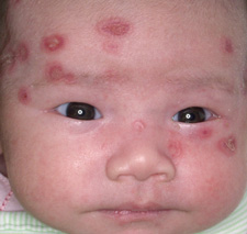
FIGURE 2
…and on the scalp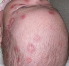
What is the differential diagnosis?
What tests should be done to make the diagnosis?
Diagnosis: Neonatal lupus
This infant has neonatal lupus erythematosus, a rare syndrome in which maternal autoantibodies are passively transferred to the baby and cause cutaneous lesions or isolated congenital heart block. The skin rash generally appears a few days to weeks after birth, typically after sun exposure, and shows well-demarcated erythematous, scaling patches that are often annular and predominately on the scalp, neck, or face. It is self-limited and generally resolves without scarring by 6 to 7 months of age.
Differential diagnosis
The differential diagnosis of neonatal lupus syndrome includes annular urticaria, tinea corporis, seborrheic dermatitis, erythema annulare centrifugum, familial annular erythema, erythema multiforme, systemic lupus erythematosus, pityrosporum infection, and photosensitive genodermatoses.1 These can be differentiated from neonatal lupus by several defining characteristics:
- Annular urticaria is transient and pruritic
- In tinea corporis, a KOH prep reveals numerous branching hyphae
- Seborrheic dermatitis is characterized by greasy scales over red, inflamed skin and distributed in facial and body folds
- Erythema annulare centrifugum usually affects the trunks and legs; the characteristic rings enlarge daily
- Familial annular erythema is generally found on the back and shoulders and accompanied by a family history of similar lesions in infancy
- Erythema multiforme is more often on extensor surfaces and is uncommon in infancy
- Systemic lupus erythematosus should be suspected if the infant has the clinical manifestations or positive serological tests while autoantibodies are absent in the mother
- Pityrosporum infection can be diagnosed by hyphae and spores (“spaghetti and meatballs”) on KOH preparation
- Photosensitive genodermatoses are a rare group of disorders that are characterized by chronicity without autoantibodies.
Causes and pathophysiology of neonatal lupus
Neonatal lupus is caused by the IgG autoantibodies anti-Ro/SSA and anti-La/SSB, which pass from the mother to the fetus via the placenta, although anti-U1-RNP antibodies can sometimes cause the cutaneous manifestations of neonatal lupus.2 The mother may or may not demonstrate features of connective tissue disease at the time of birth. Lupus has a higher incidence in Asian women than in the overall US population.
Possible symptoms in the neonate include a characteristic skin eruption or congenital heart block. Neonatal lupus accounts for 85% of cases of congenital complete heart block diagnosed in utero or in the neonatal period.3 The incidence of complete heart block in offspring of women with anti-Ro/SSA or anti-La/SSB antibodies is about 2%. The incidence of complete heart block in infants with cutaneous neonatal lupus is 15% to 30%. Incomplete heart block, which may progress to complete heart block, may also occur. A less common and self-limited sinus bradycardia has also been described in fetuses.4
Other manifestations of neonatal lupus include thrombocytopenia, aplastic anemia, hepatobiliary dysfunction, and central nervous system vasculopathy.
Diagnosis and treatment
Diagnosis is based on physical findings in an infant aged <6 months and detection of anti-Ro/SSA and anti-La/SSB antibodies in the child or mother. It should be suspected in children with congenital heart block as it is responsible for over 85% of cases.
All infants diagnosed with neonatal lupus erythematosus or born to women with anti-Ro/SSA or anti-La/SSB antibodies should have an ECG to detect heart block. If this is abnormal, referral to a pediatric cardiologist is warranted. All pregnant women with anti-Ro/SSA or anti-La/SSB antibodies should undergo regular fetal echocardiography to detect heart block. The initiation of dexamethasone or plasmapheresis as a preventative measure against congenital heart block in high-risk pregnant women is currently under consideration but is not yet justified.3
Treatment includes photoprotection (avoiding sunlight using clothing and other protective measures) and time, as nearly all cases resolve within 6 months without scarring. Mild topical steroids may be helpful. It is important to note that children with neonatal lupus may be at higher risk of developing autoimmune disorders or rheumatic disease later in life.2
Outcome
For this patient, the skin lesions were scraped with the side of a microscope slide and KOH with DMSO was used to dissolve the epithelial cells. The preparation was negative for fungal elements.This infant had anti-Ro, anti-La, and anti-RNP levels of 128.3, 150.3, and 128, respectively. Normal range is 0 to 19 for each autoantibody. Her mother’s autoantibody levels were similarly abnormal at 138.1, 158.8, and 169.1.
The patient was referred to a pediatric cardiologist for follow-up; results of a second echocardiogram and ECG were normal. The infant was seen again at 7 weeks; cutaneous lesions were present but somewhat improved, and she was otherwise doing well. Given the high probability of lupus occurring in future pregnancies, the mother received appropriate education.
CORRESPONDENCE
Keith A. Frey, MD, Department of Family Medicine, Mayo Clinic Arizona, 13737 North 92nd Street, Scottsdale, AZ 85260. E-mail: frey.keith@mayo.edu
1. Silverman ED. Neonatal lupus. In Cassidy JT, Pett RE (eds): Textbook of Pediatric Rheumatology. Philadelphia: WB Saunders; 2001:456.
2. Habif TP. Clinical Dermatology. Edinburgh: Mosby; 2004.
3. Buyon JP, Clancy RM. Neonatal lupus: Basic research and clinical perspective. Rheum Dis Clin North Am 2005;31:299-313.
4. Askanase AD, Friedman DM, Copel J, et al. Spectrum and progression of conduction abnormalities in infants born to mothers with anti-SSA/Ro-SSB/La anti-bodies. Rheum Dis Clin North Am 2002;11:145.
A full-term, healthy female newborn was delivered via cesarean section because the labor did not adequately progress. The mother, age 33 years and of Asian ancestry, had a significant medical and obstetrical history: chronic hepatitis B carrier without cirrhosis, cutaneous lupus erythematosus (positive anti-Ro and anti-La antibodies), and a positive group B streptococcal recto-vaginal culture at 35 weeks’ gestation. The mother received 4 doses of intravenous ampicillin during labor.
The infant’s initial hospital course was complicated by a transient and otherwise asymptomatic bradycardia. An electrocardiogram (ECG) confirmed a heart rate of 96 with normal interval parameters, but there were changes suggestive of left ventricular hypertrophy. An echocardiogram was normal.
Follow-up office visits for common newborn feeding problems demonstrated consistent weight gain and normal vital signs, including heart rate and facial milia. However, by age 4 weeks an erythematous eruption extending from the frontal scalp and forehead to the cheek area had developed (FIGURES 1 AND 2).
FIGURE 1
Rash on a newborn’s face …
FIGURE 2
…and on the scalp
What is the differential diagnosis?
What tests should be done to make the diagnosis?
Diagnosis: Neonatal lupus
This infant has neonatal lupus erythematosus, a rare syndrome in which maternal autoantibodies are passively transferred to the baby and cause cutaneous lesions or isolated congenital heart block. The skin rash generally appears a few days to weeks after birth, typically after sun exposure, and shows well-demarcated erythematous, scaling patches that are often annular and predominately on the scalp, neck, or face. It is self-limited and generally resolves without scarring by 6 to 7 months of age.
Differential diagnosis
The differential diagnosis of neonatal lupus syndrome includes annular urticaria, tinea corporis, seborrheic dermatitis, erythema annulare centrifugum, familial annular erythema, erythema multiforme, systemic lupus erythematosus, pityrosporum infection, and photosensitive genodermatoses.1 These can be differentiated from neonatal lupus by several defining characteristics:
- Annular urticaria is transient and pruritic
- In tinea corporis, a KOH prep reveals numerous branching hyphae
- Seborrheic dermatitis is characterized by greasy scales over red, inflamed skin and distributed in facial and body folds
- Erythema annulare centrifugum usually affects the trunks and legs; the characteristic rings enlarge daily
- Familial annular erythema is generally found on the back and shoulders and accompanied by a family history of similar lesions in infancy
- Erythema multiforme is more often on extensor surfaces and is uncommon in infancy
- Systemic lupus erythematosus should be suspected if the infant has the clinical manifestations or positive serological tests while autoantibodies are absent in the mother
- Pityrosporum infection can be diagnosed by hyphae and spores (“spaghetti and meatballs”) on KOH preparation
- Photosensitive genodermatoses are a rare group of disorders that are characterized by chronicity without autoantibodies.
Causes and pathophysiology of neonatal lupus
Neonatal lupus is caused by the IgG autoantibodies anti-Ro/SSA and anti-La/SSB, which pass from the mother to the fetus via the placenta, although anti-U1-RNP antibodies can sometimes cause the cutaneous manifestations of neonatal lupus.2 The mother may or may not demonstrate features of connective tissue disease at the time of birth. Lupus has a higher incidence in Asian women than in the overall US population.
Possible symptoms in the neonate include a characteristic skin eruption or congenital heart block. Neonatal lupus accounts for 85% of cases of congenital complete heart block diagnosed in utero or in the neonatal period.3 The incidence of complete heart block in offspring of women with anti-Ro/SSA or anti-La/SSB antibodies is about 2%. The incidence of complete heart block in infants with cutaneous neonatal lupus is 15% to 30%. Incomplete heart block, which may progress to complete heart block, may also occur. A less common and self-limited sinus bradycardia has also been described in fetuses.4
Other manifestations of neonatal lupus include thrombocytopenia, aplastic anemia, hepatobiliary dysfunction, and central nervous system vasculopathy.
Diagnosis and treatment
Diagnosis is based on physical findings in an infant aged <6 months and detection of anti-Ro/SSA and anti-La/SSB antibodies in the child or mother. It should be suspected in children with congenital heart block as it is responsible for over 85% of cases.
All infants diagnosed with neonatal lupus erythematosus or born to women with anti-Ro/SSA or anti-La/SSB antibodies should have an ECG to detect heart block. If this is abnormal, referral to a pediatric cardiologist is warranted. All pregnant women with anti-Ro/SSA or anti-La/SSB antibodies should undergo regular fetal echocardiography to detect heart block. The initiation of dexamethasone or plasmapheresis as a preventative measure against congenital heart block in high-risk pregnant women is currently under consideration but is not yet justified.3
Treatment includes photoprotection (avoiding sunlight using clothing and other protective measures) and time, as nearly all cases resolve within 6 months without scarring. Mild topical steroids may be helpful. It is important to note that children with neonatal lupus may be at higher risk of developing autoimmune disorders or rheumatic disease later in life.2
Outcome
For this patient, the skin lesions were scraped with the side of a microscope slide and KOH with DMSO was used to dissolve the epithelial cells. The preparation was negative for fungal elements.This infant had anti-Ro, anti-La, and anti-RNP levels of 128.3, 150.3, and 128, respectively. Normal range is 0 to 19 for each autoantibody. Her mother’s autoantibody levels were similarly abnormal at 138.1, 158.8, and 169.1.
The patient was referred to a pediatric cardiologist for follow-up; results of a second echocardiogram and ECG were normal. The infant was seen again at 7 weeks; cutaneous lesions were present but somewhat improved, and she was otherwise doing well. Given the high probability of lupus occurring in future pregnancies, the mother received appropriate education.
CORRESPONDENCE
Keith A. Frey, MD, Department of Family Medicine, Mayo Clinic Arizona, 13737 North 92nd Street, Scottsdale, AZ 85260. E-mail: frey.keith@mayo.edu
1. Silverman ED. Neonatal lupus. In Cassidy JT, Pett RE (eds): Textbook of Pediatric Rheumatology. Philadelphia: WB Saunders; 2001:456.
2. Habif TP. Clinical Dermatology. Edinburgh: Mosby; 2004.
3. Buyon JP, Clancy RM. Neonatal lupus: Basic research and clinical perspective. Rheum Dis Clin North Am 2005;31:299-313.
4. Askanase AD, Friedman DM, Copel J, et al. Spectrum and progression of conduction abnormalities in infants born to mothers with anti-SSA/Ro-SSB/La anti-bodies. Rheum Dis Clin North Am 2002;11:145.