User login
A 3-year-old girl is brought to dermatology by her mother, who requests evaluation of a lesion that manifested shortly after the child’s birth. The family’s primary care provider has always dismissed her lesion as a birthmark, but her parents are concerned by its tendency to abruptly change for no apparent reason. It swells up, becomes itchy and red, and then returns to normal within minutes.
According to her parents, the patient is otherwise quite healthy. She has never experienced any breathing problems and is not atopic.
EXAMINATION
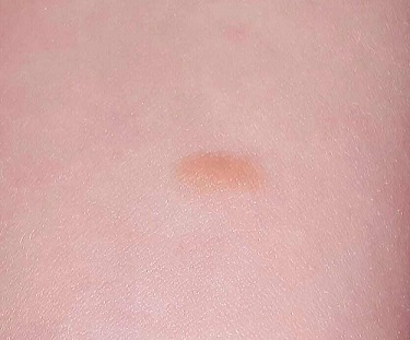
The lesion—an orange, 2-cm, round nodule with a smooth surface—is located on the volar aspect of her forearm. When forcefully stroked, it immediately begins to swell and redden, resembling a wheal. The patient verifies that the lesion itches when touched but is not tender. Before the examination ends, the lesion returns to normal.
Her type I skin is otherwise unremarkable.
What is the diagnosis?
DISCUSSION
Mastocytoma is the term for this localized accumulation of mast cells. When traumatized, histamine (among other active components) is released, causing sudden swelling and itching.
Mast cells are normal white cells involved in the natural function of the immune system, but they can be involved in rare but serious pathologic processes. For example, some children develop dozens of lesions (a condition called urticarial pigmentosa); under extreme circumstances, they can release enough histamine to induce shock and even respiratory distress.
Rarely, mast cells can undergo malignant transformation in the bone marrow, leading to mast cell leukemia. Another rare complication is mastocytosis, in which organs and tissue are invaded by mast cells, destroying the function of the organs and causing problems related to histamine release.
While simple, benign mastocytomas are common and easy to identify visually, skin biopsy is the key to diagnosing this family of diseases. With a small, stable lesion such as this patient’s, spontaneous resolution alleviates the need for treatment. Surgical removal does serve as a permanent cure, however, and as an accurate way to distinguish mastocytoma from mast cell tumors and juvenile xanthogranuloma—the other items in the differential.
TAKE-HOME LEARNING POINTS
• Mastocytomas are benign lesions composed of mast cells.
• Present at or soon after birth, mastocytomas are usually orangish brown and round to oval, and measure, on average, 1 to 3 cm.
• When traumatized by friction, they swell due to the release of histamine from the mast cells. They return to normal within minutes to hours.
• Mastocytomas only rarely require removal, since they are benign and resolve on their own.
A 3-year-old girl is brought to dermatology by her mother, who requests evaluation of a lesion that manifested shortly after the child’s birth. The family’s primary care provider has always dismissed her lesion as a birthmark, but her parents are concerned by its tendency to abruptly change for no apparent reason. It swells up, becomes itchy and red, and then returns to normal within minutes.
According to her parents, the patient is otherwise quite healthy. She has never experienced any breathing problems and is not atopic.
EXAMINATION
The lesion—an orange, 2-cm, round nodule with a smooth surface—is located on the volar aspect of her forearm. When forcefully stroked, it immediately begins to swell and redden, resembling a wheal. The patient verifies that the lesion itches when touched but is not tender. Before the examination ends, the lesion returns to normal.
Her type I skin is otherwise unremarkable.
What is the diagnosis?
DISCUSSION
Mastocytoma is the term for this localized accumulation of mast cells. When traumatized, histamine (among other active components) is released, causing sudden swelling and itching.
Mast cells are normal white cells involved in the natural function of the immune system, but they can be involved in rare but serious pathologic processes. For example, some children develop dozens of lesions (a condition called urticarial pigmentosa); under extreme circumstances, they can release enough histamine to induce shock and even respiratory distress.
Rarely, mast cells can undergo malignant transformation in the bone marrow, leading to mast cell leukemia. Another rare complication is mastocytosis, in which organs and tissue are invaded by mast cells, destroying the function of the organs and causing problems related to histamine release.
While simple, benign mastocytomas are common and easy to identify visually, skin biopsy is the key to diagnosing this family of diseases. With a small, stable lesion such as this patient’s, spontaneous resolution alleviates the need for treatment. Surgical removal does serve as a permanent cure, however, and as an accurate way to distinguish mastocytoma from mast cell tumors and juvenile xanthogranuloma—the other items in the differential.
TAKE-HOME LEARNING POINTS
• Mastocytomas are benign lesions composed of mast cells.
• Present at or soon after birth, mastocytomas are usually orangish brown and round to oval, and measure, on average, 1 to 3 cm.
• When traumatized by friction, they swell due to the release of histamine from the mast cells. They return to normal within minutes to hours.
• Mastocytomas only rarely require removal, since they are benign and resolve on their own.
