User login
A 54-year-old woman self-refers to dermatology for evaluation of asymptomatic lesions on her legs. Over the past several years, they have slowly grown larger, redder, and shinier. She denies any other skin problems. Medical history is significant for type 2 diabetes, which, according to the patient, is under good control.
The patient has sought care from a variety of professionals, including her primary care provider, her endocrinologist, and her gynecologist. Various diagnoses, including “fungal infection,” have been posited; treatment with antifungal cream and oral medications produced no effect.
She also consulted the proprietor of her local health food store, who actually examined her before diagnosing “yeast infection” and advising her to change her diet.
EXAMINATION
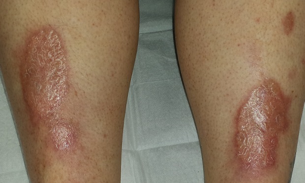
There are large, oval, reddish brown shiny plaques, measuring about 8 x 4 cm, on both anterior tibial areas. On closer inspection, the affected skin is yellowish pink and appears quite atrophic. This effect is more pronounced toward the centers of the lesions, which also display distinct telangiectasias. The borders of the plaques are slightly raised.
Examination of the rest of the patient’s skin reveals no other abnormalities.
What is the diagnosis?
DISCUSSION
Necrobiosis lipoidica (NL), though uncommon, is far from rare; it is seen somewhat regularly in dermatology practices. As this case demonstrates, it is one of hundreds of dermatologic diagnoses that you either know about or you don’t. Providers in the latter group invariably call it “fungal infection” because of its rounded borders—and because they simply have nothing else to call it.
True fungal infections are usually very superficial, involving only the outer layer of skin. This means they will manifest with scaling, a feature notably absent in this case. But more to the point, what was needed was a specific diagnosis, rather than another blind attempt at empiric treatment. Correct diagnosis dictates correct treatment.
NL was first described by Oppenhein in 1929, when he saw it in a diabetic patient. But it got its modern name in 1932 from Urbach, who thought it was invariably connected to diabetes. Now we know nondiabetic persons can develop it as well.
A number of theories exist as to the origins of this condition. One pinpoints microangiopathy of the same sort seen in the kidneys and eyes of diabetic patients. Another, proposed to account for the fact that NL is not exclusively seen in diabetes, holds that an autoimmune process might be involved—an opinion bolstered by the finding of increased TNF-alpha in the sera and skin of NL patients.
In any case, these atrophic plaques can grow quite large, eventually breaking down and ulcerating focally. Although the condition is usually asymptomatic, 25% of NL patients with advanced disease report pain.
Definitive diagnosis is made by punch biopsy, though visual diagnosis is quite adequate for those who can recognize the condition. NL lesions are most commonly seen on bilateral anterior tibial areas but can occasionally develop on the arm, face, or even genitals. The lesions can also koebnerize (ie, form along lines of trauma), occasionally being seen in surgical scars or even insulin injection sites.
Treatment, though largely unsatisfactory, includes topical and intralesional steroids. Particular care is taken to avoid or at least limit the formation of ulcers. In advanced cases, patients are referred to a wound care specialist. Oddly enough, neither the severity nor the prognosis of NL seem to have anything to do with how well or poorly the patient’s diabetes is controlled.
This patient was started on topical clobetasol cream, to be applied mostly to the actively advancing peripheral margin. Her prognosis, as with all NL patients, is guarded.
TAKE-HOME LEARNING POINTS
• Necrobiosis lipoidica (NL) is a disease of collagen degeneration that induces a granulomatous response that manifests microscopically with microangiopathy.
• NL usually appears on bilateral anterior tibial areas (but can affect the arms, face, or genitalia), as pinkish brown plaques with atrophic centers that tend to be yellow and telangiectatic.
• Though NL is usually associated with diabetes, it can develop in nondiabetic persons as well.
• The most feared complication of NL is eventual ulceration, which is why treatment is directed at taking care to avoid wounds to the plaques.
A 54-year-old woman self-refers to dermatology for evaluation of asymptomatic lesions on her legs. Over the past several years, they have slowly grown larger, redder, and shinier. She denies any other skin problems. Medical history is significant for type 2 diabetes, which, according to the patient, is under good control.
The patient has sought care from a variety of professionals, including her primary care provider, her endocrinologist, and her gynecologist. Various diagnoses, including “fungal infection,” have been posited; treatment with antifungal cream and oral medications produced no effect.
She also consulted the proprietor of her local health food store, who actually examined her before diagnosing “yeast infection” and advising her to change her diet.
EXAMINATION
There are large, oval, reddish brown shiny plaques, measuring about 8 x 4 cm, on both anterior tibial areas. On closer inspection, the affected skin is yellowish pink and appears quite atrophic. This effect is more pronounced toward the centers of the lesions, which also display distinct telangiectasias. The borders of the plaques are slightly raised.
Examination of the rest of the patient’s skin reveals no other abnormalities.
What is the diagnosis?
DISCUSSION
Necrobiosis lipoidica (NL), though uncommon, is far from rare; it is seen somewhat regularly in dermatology practices. As this case demonstrates, it is one of hundreds of dermatologic diagnoses that you either know about or you don’t. Providers in the latter group invariably call it “fungal infection” because of its rounded borders—and because they simply have nothing else to call it.
True fungal infections are usually very superficial, involving only the outer layer of skin. This means they will manifest with scaling, a feature notably absent in this case. But more to the point, what was needed was a specific diagnosis, rather than another blind attempt at empiric treatment. Correct diagnosis dictates correct treatment.
NL was first described by Oppenhein in 1929, when he saw it in a diabetic patient. But it got its modern name in 1932 from Urbach, who thought it was invariably connected to diabetes. Now we know nondiabetic persons can develop it as well.
A number of theories exist as to the origins of this condition. One pinpoints microangiopathy of the same sort seen in the kidneys and eyes of diabetic patients. Another, proposed to account for the fact that NL is not exclusively seen in diabetes, holds that an autoimmune process might be involved—an opinion bolstered by the finding of increased TNF-alpha in the sera and skin of NL patients.
In any case, these atrophic plaques can grow quite large, eventually breaking down and ulcerating focally. Although the condition is usually asymptomatic, 25% of NL patients with advanced disease report pain.
Definitive diagnosis is made by punch biopsy, though visual diagnosis is quite adequate for those who can recognize the condition. NL lesions are most commonly seen on bilateral anterior tibial areas but can occasionally develop on the arm, face, or even genitals. The lesions can also koebnerize (ie, form along lines of trauma), occasionally being seen in surgical scars or even insulin injection sites.
Treatment, though largely unsatisfactory, includes topical and intralesional steroids. Particular care is taken to avoid or at least limit the formation of ulcers. In advanced cases, patients are referred to a wound care specialist. Oddly enough, neither the severity nor the prognosis of NL seem to have anything to do with how well or poorly the patient’s diabetes is controlled.
This patient was started on topical clobetasol cream, to be applied mostly to the actively advancing peripheral margin. Her prognosis, as with all NL patients, is guarded.
TAKE-HOME LEARNING POINTS
• Necrobiosis lipoidica (NL) is a disease of collagen degeneration that induces a granulomatous response that manifests microscopically with microangiopathy.
• NL usually appears on bilateral anterior tibial areas (but can affect the arms, face, or genitalia), as pinkish brown plaques with atrophic centers that tend to be yellow and telangiectatic.
• Though NL is usually associated with diabetes, it can develop in nondiabetic persons as well.
• The most feared complication of NL is eventual ulceration, which is why treatment is directed at taking care to avoid wounds to the plaques.
