User login
An 84-year-old man is seeing an audiologist for hearing problems when the medical assistant notices a lesion on the patient’s right ear. When asked about it, the patient acknowledges that it has been bothering him. It itches and won't heal, no matter how much "medicine" the patient applies. Fortunately, it is not painful. But the patient is greatly annoyed that the lesion sometimes bleeds, leaving spots on his pillowcase and clothing.
The audiologist requests a consultation by the dermatology PA at her clinic.
EXAMINATION
The patient is an elderly white man in a wheelchair who is able to communicate readily. He is adamant that the lesion has only been present for about two weeks. However, his wife, who is with him, is sure it has been there for more than a year.
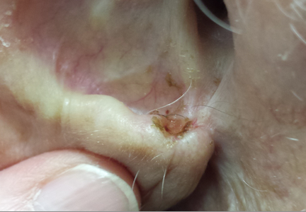
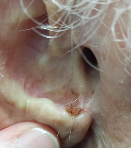
The lesion is a 1.6-cm ulcer with a telangiectatic, rolled pearly border. It is located between the tragus and antitragus on the outer floor of the patient’s external auditory meatus. It is seen in the context of the patient’s very fair and sun-damaged skin, which is also marked by numerous actinic keratoses on his face.
Under local anesthesia, shave biopsy is performed. A 5-mm slice is removed from the periphery of the lesion.
Continue for the outcome and discussion >>
DISCUSSION
The lesion proved to be the expected basal cell carcinoma (BCC), probably caused by sun exposure when the patient was younger. Patients are often incredulous about this connection, but the nuclei of skin cells have a long memory, often taking decades for cancers to actually develop. In a way, it doesn’t matter, since the damage is long since done even if the patient gets almost no sun at the present time.
But it does matter that this lesion is large and located in a difficult spot, one that does not lend itself to an easy fix. Primary closure would not be possible, given the complete lack of slack in the skin. A Mohs surgeon will need to remove the lesion, to ensure complete excision as well as perform the special procedures required to graft the site for closure.
Even though the lesion proved to be “just" a BCC (ie, not a melanoma), it still had to be dealt with lest it continue to spread. More than a few patients have had to have their entire external ear removed for this reason. Although it’s unusual, BCCs can metastasize to local lymph nodes and even to the brain and lung. More than a century ago, these were known as “rodent ulcers” that caused serious problems. If they could be caught in time, local cautery was the only chance at a cure.
The differential in this case included squamous cell carcinoma, which might have presented a more serious problem, given the location (near an orifice). Another diagnostic possibility was sebaceous carcinoma, a merkel cell cancer. Benign lesions, such as chondrodermatitis nodularis chronica helicis, occasionally present as ulcerated lesions on the ear.
BCCs are, far and away, the most common type of skin cancer, with more than a million new cases diagnosed each year in United States. They occur more commonly on fair-skinned patients and on prominently sun-exposed area (eg, ears, noses, and cheeks) but can also appear in areas where the sun seldom shines.
Several types of BCC have been described, but the nonhealing erosive papule (so-called noduloulcerative type) is the most common presentation. BCCs can also resemble scars or rashes. Though they are typically very slow to grow, not all BCCs are indolent. Some are quite aggressive both in terms of morphologic appearance and internal growth. And, as noted above, there is a differential for new, growing lesions in sun-exposed skin, making biopsy of such lesions mandatory.
Continue for Joe Monroe's take home learning points >>
TAKE-HOME LEARNING POINTS
• Nonhealing lesions on sun-exposed skin are considered to be skin cancers until proven otherwise by biopsy.
• Basal cell carcinomas (BCCs) are the most common skin cancer.
• BCCs typically grow very slowly.
• BCCs, if allowed to remain in place, can become extensive, invasive, destructive, and—in exceptional circumstances—can even metastasize.
• The differential for BCC includes squamous cell carcinoma, which has considerably more potential for metastasis than BCC, especially when it’s near an orifice.
• Quite frequently, it’s next to impossible to differentiate visually between BCC and SCC.
• BCCs and SCCs can take on several different morphologies, so any changing lesion needs to be biopsied to establish its identity, particularly since both are so common.
An 84-year-old man is seeing an audiologist for hearing problems when the medical assistant notices a lesion on the patient’s right ear. When asked about it, the patient acknowledges that it has been bothering him. It itches and won't heal, no matter how much "medicine" the patient applies. Fortunately, it is not painful. But the patient is greatly annoyed that the lesion sometimes bleeds, leaving spots on his pillowcase and clothing.
The audiologist requests a consultation by the dermatology PA at her clinic.
EXAMINATION
The patient is an elderly white man in a wheelchair who is able to communicate readily. He is adamant that the lesion has only been present for about two weeks. However, his wife, who is with him, is sure it has been there for more than a year.
The lesion is a 1.6-cm ulcer with a telangiectatic, rolled pearly border. It is located between the tragus and antitragus on the outer floor of the patient’s external auditory meatus. It is seen in the context of the patient’s very fair and sun-damaged skin, which is also marked by numerous actinic keratoses on his face.
Under local anesthesia, shave biopsy is performed. A 5-mm slice is removed from the periphery of the lesion.
Continue for the outcome and discussion >>
DISCUSSION
The lesion proved to be the expected basal cell carcinoma (BCC), probably caused by sun exposure when the patient was younger. Patients are often incredulous about this connection, but the nuclei of skin cells have a long memory, often taking decades for cancers to actually develop. In a way, it doesn’t matter, since the damage is long since done even if the patient gets almost no sun at the present time.
But it does matter that this lesion is large and located in a difficult spot, one that does not lend itself to an easy fix. Primary closure would not be possible, given the complete lack of slack in the skin. A Mohs surgeon will need to remove the lesion, to ensure complete excision as well as perform the special procedures required to graft the site for closure.
Even though the lesion proved to be “just" a BCC (ie, not a melanoma), it still had to be dealt with lest it continue to spread. More than a few patients have had to have their entire external ear removed for this reason. Although it’s unusual, BCCs can metastasize to local lymph nodes and even to the brain and lung. More than a century ago, these were known as “rodent ulcers” that caused serious problems. If they could be caught in time, local cautery was the only chance at a cure.
The differential in this case included squamous cell carcinoma, which might have presented a more serious problem, given the location (near an orifice). Another diagnostic possibility was sebaceous carcinoma, a merkel cell cancer. Benign lesions, such as chondrodermatitis nodularis chronica helicis, occasionally present as ulcerated lesions on the ear.
BCCs are, far and away, the most common type of skin cancer, with more than a million new cases diagnosed each year in United States. They occur more commonly on fair-skinned patients and on prominently sun-exposed area (eg, ears, noses, and cheeks) but can also appear in areas where the sun seldom shines.
Several types of BCC have been described, but the nonhealing erosive papule (so-called noduloulcerative type) is the most common presentation. BCCs can also resemble scars or rashes. Though they are typically very slow to grow, not all BCCs are indolent. Some are quite aggressive both in terms of morphologic appearance and internal growth. And, as noted above, there is a differential for new, growing lesions in sun-exposed skin, making biopsy of such lesions mandatory.
Continue for Joe Monroe's take home learning points >>
TAKE-HOME LEARNING POINTS
• Nonhealing lesions on sun-exposed skin are considered to be skin cancers until proven otherwise by biopsy.
• Basal cell carcinomas (BCCs) are the most common skin cancer.
• BCCs typically grow very slowly.
• BCCs, if allowed to remain in place, can become extensive, invasive, destructive, and—in exceptional circumstances—can even metastasize.
• The differential for BCC includes squamous cell carcinoma, which has considerably more potential for metastasis than BCC, especially when it’s near an orifice.
• Quite frequently, it’s next to impossible to differentiate visually between BCC and SCC.
• BCCs and SCCs can take on several different morphologies, so any changing lesion needs to be biopsied to establish its identity, particularly since both are so common.