User login
A 70-year-old man self-refers to dermatology for evaluation of a “risin’ in my finger,” which has existed for “at least 40 years.” While the lesion doesn’t really hurt, the patient wants it gone because he traumatizes it almost daily while working on his farm.
He has tried innumerable removal methods, including acids, blood root, and duct tape. Most recently, his primary care provider attempted treatment with cryotherapy.
The patient’s health is excellent in other respects, with no history of similar lesions elsewhere.
EXAMINATION
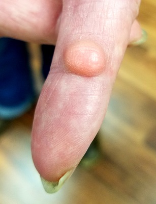
A dome-like pink nodule is seen on the palmar surface of the patient’s left index finger. The 1-cm lesion is smooth, moderately firm, and nontender. Although the majority of the lesion protrudes above the skin’s surface, there is an intradermal component. Normal skin lines on the surface are preserved, and there is no surface punctum. Palpation of the epitrochlear and axillary areas above the hand reveals no masses.
What is the diagnosis?
Traumatic puncture—especially of the palms and fingers—can invaginate the surface of skin, effectively burying the surface follicular infundibulum and associated sebaceous gland as the wound heals. These structures can continue to produce sebum and epidermal cells, which then accumulate in a space delineated by the lining of the infundibulum and form a sac called an epidermal inclusion cyst (EIC).
EICs differ from the more common epidermoid (or epidermal) cyst for several reasons. For one, epidermoid cysts are confined almost exclusively to oily skin (usually above the waist, with the back being the most common location). Unlike EICs, epidermoid cysts almost always have at least one central comedone, which acts as a plug, preventing the discharge of its contents.
The differential also includes pilar cysts—another common type, popularly known as “wens.” But these occur almost exclusively in the scalp and only rarely display a central punctum.
All of these cysts have an organized wall that contains their characteristic cheesy contents, and all can be removed surgically—preferably in one piece—under local anesthesia. Once excised, the lesion should always be submitted for pathologic examination, since cystic lesions can (albeit rarely) undergo malignant transformation.
In this case, excision was performed with anesthesia via digital block, supplemented with small amounts of lidocaine and epinephrine for hemostasis. The lesion was removed in one transverse elliptical piece and with special care to avoid trauma to underlying structures (visualized with the aid of a tourniquet). The wound was closed with interrupted sutures.
The pathology report confirmed the preoperative diagnosis, and the patient recovered uneventfully.
TAKE-HOME LEARNING POINTS
- Epidermal inclusion cysts (EICs) result from a puncture wound that buries surface follicular infundibula and their sebaceous glands under the skin, where they continue to produce material that collects in a sac.
- Unlike the more common epidermoid (epidermal) cysts, which affect the oily areas of the body (above the waist), EICs don’t have a central punctum.
- EICs can be left alone or excised, but removal must be done in one piece, lest they recur. The same is true for all aforementioned cysts.
A 70-year-old man self-refers to dermatology for evaluation of a “risin’ in my finger,” which has existed for “at least 40 years.” While the lesion doesn’t really hurt, the patient wants it gone because he traumatizes it almost daily while working on his farm.
He has tried innumerable removal methods, including acids, blood root, and duct tape. Most recently, his primary care provider attempted treatment with cryotherapy.
The patient’s health is excellent in other respects, with no history of similar lesions elsewhere.
EXAMINATION
A dome-like pink nodule is seen on the palmar surface of the patient’s left index finger. The 1-cm lesion is smooth, moderately firm, and nontender. Although the majority of the lesion protrudes above the skin’s surface, there is an intradermal component. Normal skin lines on the surface are preserved, and there is no surface punctum. Palpation of the epitrochlear and axillary areas above the hand reveals no masses.
What is the diagnosis?
Traumatic puncture—especially of the palms and fingers—can invaginate the surface of skin, effectively burying the surface follicular infundibulum and associated sebaceous gland as the wound heals. These structures can continue to produce sebum and epidermal cells, which then accumulate in a space delineated by the lining of the infundibulum and form a sac called an epidermal inclusion cyst (EIC).
EICs differ from the more common epidermoid (or epidermal) cyst for several reasons. For one, epidermoid cysts are confined almost exclusively to oily skin (usually above the waist, with the back being the most common location). Unlike EICs, epidermoid cysts almost always have at least one central comedone, which acts as a plug, preventing the discharge of its contents.
The differential also includes pilar cysts—another common type, popularly known as “wens.” But these occur almost exclusively in the scalp and only rarely display a central punctum.
All of these cysts have an organized wall that contains their characteristic cheesy contents, and all can be removed surgically—preferably in one piece—under local anesthesia. Once excised, the lesion should always be submitted for pathologic examination, since cystic lesions can (albeit rarely) undergo malignant transformation.
In this case, excision was performed with anesthesia via digital block, supplemented with small amounts of lidocaine and epinephrine for hemostasis. The lesion was removed in one transverse elliptical piece and with special care to avoid trauma to underlying structures (visualized with the aid of a tourniquet). The wound was closed with interrupted sutures.
The pathology report confirmed the preoperative diagnosis, and the patient recovered uneventfully.
TAKE-HOME LEARNING POINTS
- Epidermal inclusion cysts (EICs) result from a puncture wound that buries surface follicular infundibula and their sebaceous glands under the skin, where they continue to produce material that collects in a sac.
- Unlike the more common epidermoid (epidermal) cysts, which affect the oily areas of the body (above the waist), EICs don’t have a central punctum.
- EICs can be left alone or excised, but removal must be done in one piece, lest they recur. The same is true for all aforementioned cysts.