User login
A 67-year-old man has had a small lesion on his index finger for several years. Recently, it has grown large enough to start interfering with his normal activities. The lesion is not painful unless the patient bumps it—but as it grows, avoiding such incidents is becoming more difficult.
The diagnosis has thus far been a mystery: What is it? The patient’s primary care provider didn’t know and, per the patient’s report, had no interest in trying to remove the lesion. Thus, the patient finds himself referred to dermatology.
The patient can recall no specific injury. He does, however, report that he worked for years as an upholsterer, shaping, sewing, and stapling heavy fabric onto chairs and sofas—often puncturing his fingers in the process.
His health is otherwise “decent.” He is taking medication for hypertension and an a-blocker due to “problems” with his prostate.
EXAMINATION
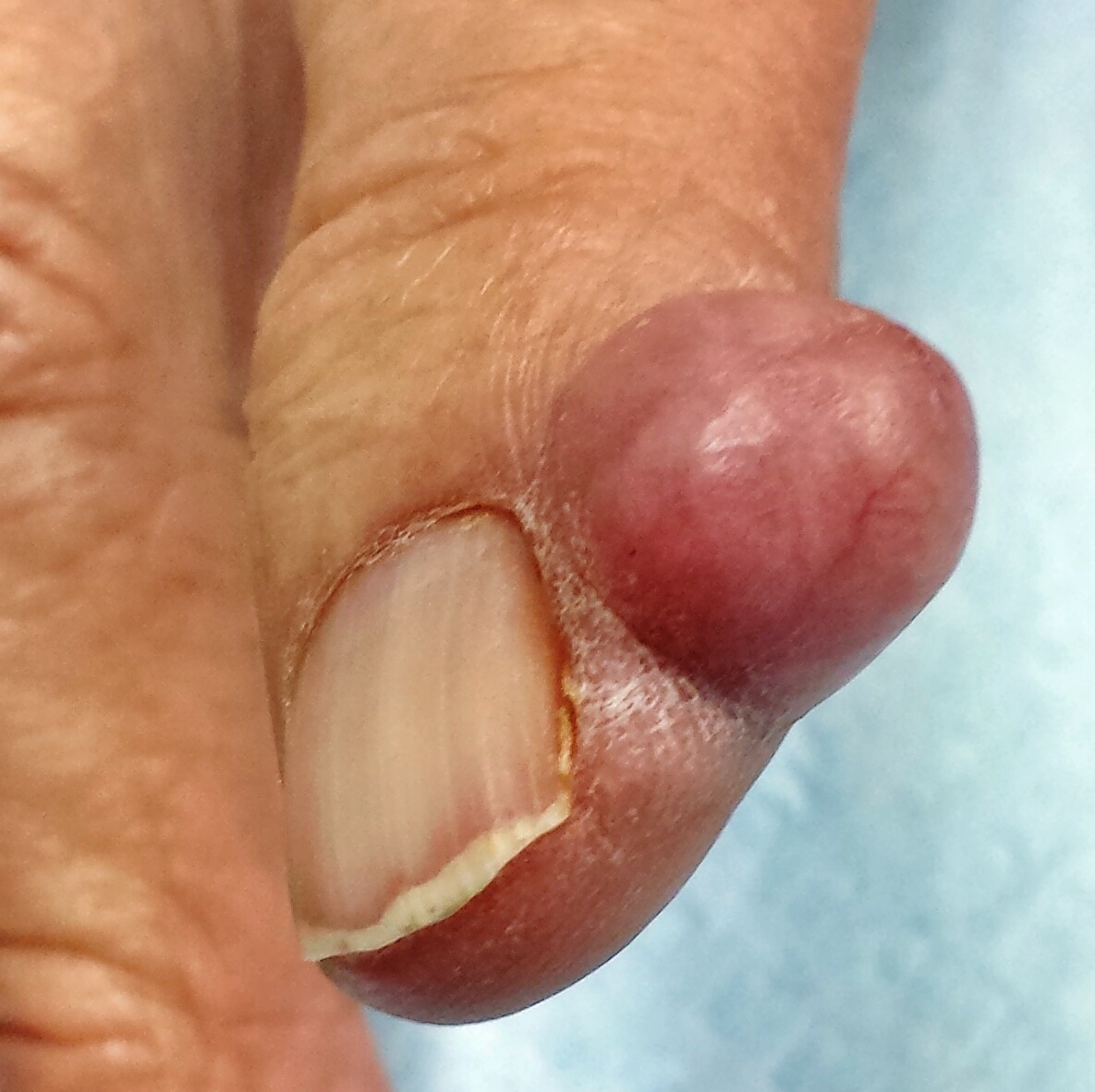
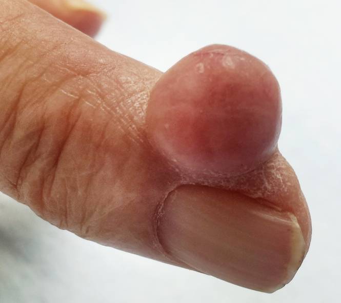
The finger lesion is a 2-cm, round, firm nodule with a smooth, telangiectatic surface. It is located on the perionychial area of patient’s index finger (lateral aspect). The lesion is attached to the finger by a broad base that rises seamlessly from the skin. It is not translucent or tender to touch. No similar lesions are seen on either hand, and no nodes are felt in the epitrochlear or axillary locations.
Within a few weeks, the patient returns to have the lesion excised. This is done with a combination of a digital block and local anesthesia, with a tourniquet to control bleeding. After primary closure, the lesion is submitted to pathology. The report indicates the lesion is a cyst, full of cheesy material.
What is the diagnosis?
DISCUSSION
This type of cyst is sometimes referred to as an implantation cyst and occurs when epidermal tissue, including sebaceous glands, is implanted into deeper tissue by a penetrating injury and continues to grow. Although this is an utterly common and benign diagnosis, the differential for a “fingeroma” includes digital mucous cyst, pyogenic granuloma, acquired digital fibrokeratoma, calcinosis cutis, tophaceous gout, wart, and giant cell tumor. Had the lesion been of bony origin, it could have represented connective tissue cancer (sarcoma) or even metastatic cancer.
Implantation cysts must be removed completely to prevent recurrence. The size and location of this particular cyst made it conducive to simple excision and primary closure. Larger lesions in more difficult locations often require the expertise of a hand surgeon for disposition.
TAKE-HOME LEARNING POINTS
• “Fingeromas” are quite common but have an extensive differential diagnosis—mostly benign, but occasionally malignant.
• Regarding malignancy, it is helpful to remember that any tissue in the body can undergo malignant transformation and that many types of primary cancers can metastasize to distant areas (including the hands).
• Removal, or at least biopsy, is the best way to differentiate between types of fingeroma lesions.
• Implantation cysts, as seen in this case, result from traumatic implantation of epidermal structures (including sebaceous glands) into deeper tissue, where they continue to grow.
• When in doubt, consider referring affected patients to dermatology or to a hand surgeon.
A 67-year-old man has had a small lesion on his index finger for several years. Recently, it has grown large enough to start interfering with his normal activities. The lesion is not painful unless the patient bumps it—but as it grows, avoiding such incidents is becoming more difficult.
The diagnosis has thus far been a mystery: What is it? The patient’s primary care provider didn’t know and, per the patient’s report, had no interest in trying to remove the lesion. Thus, the patient finds himself referred to dermatology.
The patient can recall no specific injury. He does, however, report that he worked for years as an upholsterer, shaping, sewing, and stapling heavy fabric onto chairs and sofas—often puncturing his fingers in the process.
His health is otherwise “decent.” He is taking medication for hypertension and an a-blocker due to “problems” with his prostate.
EXAMINATION
The finger lesion is a 2-cm, round, firm nodule with a smooth, telangiectatic surface. It is located on the perionychial area of patient’s index finger (lateral aspect). The lesion is attached to the finger by a broad base that rises seamlessly from the skin. It is not translucent or tender to touch. No similar lesions are seen on either hand, and no nodes are felt in the epitrochlear or axillary locations.
Within a few weeks, the patient returns to have the lesion excised. This is done with a combination of a digital block and local anesthesia, with a tourniquet to control bleeding. After primary closure, the lesion is submitted to pathology. The report indicates the lesion is a cyst, full of cheesy material.
What is the diagnosis?
DISCUSSION
This type of cyst is sometimes referred to as an implantation cyst and occurs when epidermal tissue, including sebaceous glands, is implanted into deeper tissue by a penetrating injury and continues to grow. Although this is an utterly common and benign diagnosis, the differential for a “fingeroma” includes digital mucous cyst, pyogenic granuloma, acquired digital fibrokeratoma, calcinosis cutis, tophaceous gout, wart, and giant cell tumor. Had the lesion been of bony origin, it could have represented connective tissue cancer (sarcoma) or even metastatic cancer.
Implantation cysts must be removed completely to prevent recurrence. The size and location of this particular cyst made it conducive to simple excision and primary closure. Larger lesions in more difficult locations often require the expertise of a hand surgeon for disposition.
TAKE-HOME LEARNING POINTS
• “Fingeromas” are quite common but have an extensive differential diagnosis—mostly benign, but occasionally malignant.
• Regarding malignancy, it is helpful to remember that any tissue in the body can undergo malignant transformation and that many types of primary cancers can metastasize to distant areas (including the hands).
• Removal, or at least biopsy, is the best way to differentiate between types of fingeroma lesions.
• Implantation cysts, as seen in this case, result from traumatic implantation of epidermal structures (including sebaceous glands) into deeper tissue, where they continue to grow.
• When in doubt, consider referring affected patients to dermatology or to a hand surgeon.