User login
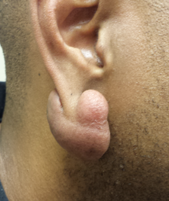
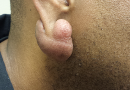
A 39-year-old African-American man has had a keloid on his right earlobe for several years. It was triggered by ear-piercing and has grown slowly to its current size. The patient denies any symptoms with the lesion.
EXAMINATION
The 5 x 2-cm lobulated mass is consistent in every way with a keloid: firm and pedunculated, with no epidermal changes. It hangs from his right earlobe. The patient has type IV skin, of African-American ancestry. After an extended discussion regarding treatment options, the patient decides to have the lesion excised.
PROCEDURE
Under sterile conditions and with local anesthesia (1% lidocaine with epinephrine), the entire keloid is removed. The wound edges are shaped to enhance the final appearance, and the wound is closed in one layer.
Since the risk for reformation is all too real, at one week post-op, the wound edges are injected with 3 mg/cc of triamcinolone, with a total of 1.5 cc used. The injections continue for three months as the wound heals. The outcome is totally acceptable to the patient, representing a huge improvement from his pre-operative appearance.
Continue reading for Joe Monroe's discussion...
DISCUSSION
Although the exact cause is still unknown, there appears to be an inheritable tendency to develop keloids. They are far more common, and grow to larger size, in patients with darker skin. Certain anatomical locations are especially at risk, such as the sternum, earlobes, shoulders (especially the deltoid areas), neck, and trunk. Keloids of the central face are almost unknown.
Keloids lie at the extreme end of the scarring continuum, with normal scars at the other end and so-called hypertrophic scars in the middle. The latter often resolve spontaneously within months of their initial formation, while true keloids never do.
Keloids are irregularly shaped, fibrous hyperpigmented (red to brown) excresences covered by thin, shiny skin. They often send out clawlike projections that ultimately obscure the original causative trauma. These tendencies help to distinguish them from mere hypertrophic scars. Biopsy, when necessary, can also help, as well as eliminate other items in the differential, including cancer (eg, dermatofibrosarcoma protuberans or metastatic cancer).
Early on, keloids are often red and uncomfortably tender. As they mature, they become less red and stabilize in size. They can become huge, especially on the chest, arms, and earlobes.
A case for surgical excision can be made, since large keloids are so cosmetically disfiguring and obvious. This patient was willing to accept the attendant risk for an even bigger postoperative keloid. With excision, primary closure, good wound care, and periodic injection of the wound edges with steroid suspension, however, he has an excellent chance to avoid this outcome.
Prior to his surgery, we reviewed alternate treatment choices, which include postexcision radiation treatment of the site, injection of the wound edges with 5-fluorouracil, or treatment of the keloid with flashlamp pulsed dye laser.
As with virtually everything we remove surgically, the keloid was sent for pathologic examination. It revealed the expected dense collection of myofibroblasts arranged in a whorled pattern, as well as a notable lack of elastic tissue.
Continue reading for Joe Monroe's take home learning points...
TAKE-HOME LEARNING POINTS
• The size and shape of keloids obscure the original triggering insult, a fact that serves to distinguish them from normal and hypertrophic scars.
• Keloids never resolve spontaneously, whereas hypertrophic scars can and do.
• Areas most prone to keloid formation include the sternum, deltoids, shoulders, earlobes, and trunk; they are almost unknown on the central face.
• In general, the darker the patient’s skin, the higher the risk for keloid formation.
• Elective surgery (eg, cyst removal) on high-risk areas and in keloid-prone patients should be avoided.
• Keloids can be injected intralesionally with glucocorticoid solutions, such as triamcinolone, or can be excised with proper postop follow-up.