User login
A 33-year-old woman presents with a lesion on the dorsum of her hand. Though it manifested several years ago, it was not problematic until recently, when it became irritated and downright painful. It has not changed in size and has never been red or swollen.
The patient denies ever having similar lesions elsewhere. She also denies any other serious health problems, specifically ophthalmologic problems.

EXAMINATION
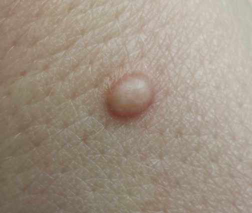
The lesion, a 5-mm ovoid tan-orange papule, is located on the dorsum of her left hand. The surface is smooth, with a very firm feel. No skin changes are noted on the surrounding skin, and there are no palpable nodes in the arm or adjacent axilla.
In light of the patient’s concern, the lesion is excised, using an elliptical incision and minimal margins. The incision is carried down into superficial adipose tissue to ensure complete removal. Bleeding is controlled and the defect closed with simple interrupted sutures.
The pathology report shows numerous densely distributed polyhedral histiocytes. Many contain a large amount of cytoplasm.
What is the diagnosis?
DISCUSSION
The clinical presentation and pathology report were both consistent with an entity called juvenile xanthogranuloma (JXG). It usually presents, as in this case, as a solitary, firm, tan to orange papule or nodule. It is more commonly seen on the head and neck of young children, although it is by no means limited to that population (despite its name, which represents anachronistic derm terminology).
Based on the pathologic picture (typified by the biopsy report in this case), JXG may represent a disordered macrophage response to an unknown tissue injury that eventuates in a granulomatous reaction.
JXG is one of the more common members of a spectrum of histiocytic disorders that includes Langerhans cell histiocytosis. In addition to its more common cutaneous distribution, JXG has been seen in almost every internal organ as well. It is rarely associated with systemic manifestations.
The most significant extracutaneous manifestation of JXG is in the eye, where it is the most common cause of spontaneous hyphema in children (usually those younger than 2). This, in turn, can lead to a secondary glaucoma and eventual blindness. This is the main caveat with solitary cutaneous JXGs: They can be associated with ocular involvement, which would manifest as changes in the color of the iris or in the size of the globe itself. Fortunately, this is a rare complication.
Beyond the potential for ocular involvement, since the vast majority of cutaneous JXG lesions are easy to diagnose and self-limiting, there is no reason to remove or otherwise treat them.
The differential for cutaneous JXG includes Spitz tumor, molluscum, and intradermal nevus.
TAKE-HOME LEARNING POINTS
• Juvenile xanthogranuloma (JXG) lesions usually resolve on their own, without treatment.
• In rare instances, cutaneous JXG can be associated with ocular involvement, which manifests with either changes in iris color or in the size of the globe itself.
• JXG lesions are commonly seen on the head and neck of young children, usually as a solitary tan to orange firm papule.
• The differential for JXG includes molluscum, Spitz tumor, and nevus. Removal of JXG lesions is usually not necessary, but pathologic examination shows pathognomic changes that effectively distinguish it from its lookalikes.
A 33-year-old woman presents with a lesion on the dorsum of her hand. Though it manifested several years ago, it was not problematic until recently, when it became irritated and downright painful. It has not changed in size and has never been red or swollen.
The patient denies ever having similar lesions elsewhere. She also denies any other serious health problems, specifically ophthalmologic problems.
EXAMINATION
The lesion, a 5-mm ovoid tan-orange papule, is located on the dorsum of her left hand. The surface is smooth, with a very firm feel. No skin changes are noted on the surrounding skin, and there are no palpable nodes in the arm or adjacent axilla.
In light of the patient’s concern, the lesion is excised, using an elliptical incision and minimal margins. The incision is carried down into superficial adipose tissue to ensure complete removal. Bleeding is controlled and the defect closed with simple interrupted sutures.
The pathology report shows numerous densely distributed polyhedral histiocytes. Many contain a large amount of cytoplasm.
What is the diagnosis?
DISCUSSION
The clinical presentation and pathology report were both consistent with an entity called juvenile xanthogranuloma (JXG). It usually presents, as in this case, as a solitary, firm, tan to orange papule or nodule. It is more commonly seen on the head and neck of young children, although it is by no means limited to that population (despite its name, which represents anachronistic derm terminology).
Based on the pathologic picture (typified by the biopsy report in this case), JXG may represent a disordered macrophage response to an unknown tissue injury that eventuates in a granulomatous reaction.
JXG is one of the more common members of a spectrum of histiocytic disorders that includes Langerhans cell histiocytosis. In addition to its more common cutaneous distribution, JXG has been seen in almost every internal organ as well. It is rarely associated with systemic manifestations.
The most significant extracutaneous manifestation of JXG is in the eye, where it is the most common cause of spontaneous hyphema in children (usually those younger than 2). This, in turn, can lead to a secondary glaucoma and eventual blindness. This is the main caveat with solitary cutaneous JXGs: They can be associated with ocular involvement, which would manifest as changes in the color of the iris or in the size of the globe itself. Fortunately, this is a rare complication.
Beyond the potential for ocular involvement, since the vast majority of cutaneous JXG lesions are easy to diagnose and self-limiting, there is no reason to remove or otherwise treat them.
The differential for cutaneous JXG includes Spitz tumor, molluscum, and intradermal nevus.
TAKE-HOME LEARNING POINTS
• Juvenile xanthogranuloma (JXG) lesions usually resolve on their own, without treatment.
• In rare instances, cutaneous JXG can be associated with ocular involvement, which manifests with either changes in iris color or in the size of the globe itself.
• JXG lesions are commonly seen on the head and neck of young children, usually as a solitary tan to orange firm papule.
• The differential for JXG includes molluscum, Spitz tumor, and nevus. Removal of JXG lesions is usually not necessary, but pathologic examination shows pathognomic changes that effectively distinguish it from its lookalikes.