User login
LOS ANGELES – Can brain imaging surpass the clock for identifying acute ischemic stroke patients who will benefit from thrombolytic or thrombectomy treatment?
That’s what experts now envision, based on early findings from several studies. Although the evidence is not yet definitive enough to justify using imaging as a replacement for time-from-stroke-onset in routine practice, the results so far are encouraging and have led to the start or planning of several phase III trials that will try to nail down a role for imaging, either CT or MR, to identify acute ischemic stroke patients who qualify for reperfusion therapy.

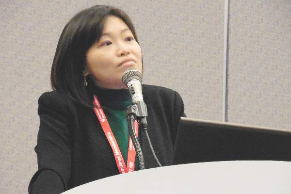
“What we’re trying to do is move away from using the clock as a surrogate marker and instead use imaging as the surrogate,” Dr. Jenny P. Tsai said in an interview at the International Stroke Conference.
The linchpin of this new approach is that clocking time elapsed from the onset of stroke symptoms to initiation of thrombolytic or endovascular treatment makes no allowance for patient-to-patient variations in collateral cerebral circulation, a factor that appears to make a big difference in whether patients can be many more hours removed from the start of their stroke and still have salvageable brain tissue. And relying on time since stroke symptom onset gives a seriously flawed estimate of a stroke’s duration when patients have an unwitnessed stroke.
The alternative is to use either CT perfusion imaging or a combination of MR perfusion and diffusion-weighted imaging “to get effective reperfusion treatments to patients who present at later time windows,” said Dr. Gregory W. Albers, professor of neurology at Stanford (Calif.) University and director of the Stanford Stroke Center. He called these two new approaches to gauging a patient’s suitability for reperfusion therapy “the next big thing in imaging” for stroke.
Using CT perfusion to assess target mismatch
Dr. Tsai presented an analysis of thrombectomy reperfusion done in 181 patients enrolled in the CT Perfusion to Predict Response to Recanalization in Ischemic Stroke Project (CRISP), which included a total of 201 acute ischemic stroke patients with large cerebral-artery occlusions treated at six U.S. centers. Her analysis excluded nine patients who presented more than 18 hours after their stroke onset, six patients who did not have successful CT perfusion assessment, and five additional patients excluded for other reasons. The 181 patients analyzed included 125 with a target mismatch in CT perfusion, indicating that salvageable tissue remained in the area of their stroke. Among these 125 patients, 111 underwent successful reperfusion by thrombectomy.
The researchers identified good treatment outcomes as patients with a modified Rankin Scale score of 2 or less 90 days after treatment. A multivariate analysis showed that among these 111 patients, achievement of a good 90-day outcome had no statistically significant relationship with time-to-treatment out to at least the first 8 hours following stroke onset, reported Dr. Tsai, a neurologist at the Stanford Stroke Center. The data also showed a nonsignificant relationship between good outcomes and time from stroke onset to treatment beyond 8 hours, but the confidence interval for this nonsignificant relationship became very wide at later times as the analysis focused on fewer and fewer patients.
“In patients with large-artery occlusions who have a target mismatch profile [on perfusion CT] and achieve successful reperfusion there is no significant association between onset-to-reperfusion-time and the probability of a good functional outcome,” suggesting that “CT perfusion is a biomarker of good outcomes beyond 6 hours” after stroke onset, Dr. Tsai concluded. CT perfusion has the advantage of being more widely available than MRI is, she added. Last year, her associates at Stanford reported similar findings using perfusion-diffusion mismatch in the cerebral area around a stroke visualized with MRI (Neurology. 2015 Aug 25;85[8]:708-15).
“MRI is not readily available” at all U.S. centers that treat stroke patients, while CT perfusion imaging is much more ubiquitous, but the findings reported by Dr. Tsai require confirmation in the several prospective, randomized trials now underway and testing this approach, Dr. Albers said.
Using MRI in unwitnessed strokes
A second challenge for using reperfusion therapy in stroke patients are patients who have unwitnessed strokes, with a completely unknown elapsed time from onset to presentation. The ability of MRI to help identify patients with unwitnessed stroke who can safely receive intravenous thrombolytic therapy with alteplase (tissue plasminogen activator, Activase) underwent testing in the MR WITNESS (Study of Intravenous Thrombolysis With Alteplase in MRI-Selected Patients) trial.
The study enrolled 80 patients with an unwitnessed acute, ischemic stroke at 10 U.S. centers. Patients had to be in a position to receive alteplase within 4.5 hours of when their stroke was first identified; 57 (71%) of the participants had wake-up strokes. All patients underwent two types of MRI to identify them as potential candidates for safe administration of alteplase: diffusion weighted imaging, to identify that a stroke had occurred, and fluid-attenuated inversion recovery (FLAIR) assessment, to identify strokes that had occurred during the prior 4 hours.
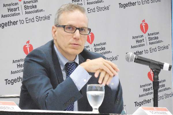
This phase II study’s primary safety endpoint was the incidence of symptomatic intracerebral hemorrhage following alteplase treatment, which occurred in 1 of the 80 patients (1.25%), not a statistically significant difference when compared with the historical standard, the 2.4% rate seen in stroke patients treated with intravenous alteplase 3 to 4.5 hours after their known stroke onset in the ECASS III (European Cooperative Acute Stroke Study) (N Engl J Med. 2008 Sept 25;359[13]:1317-29). The rate of asymptomatic intracerebral hemorrhage was not significantly different between the new study and ECASS III, reported Dr. Lee H. Schwamm, director of TeleStroke & Acute Stroke Services at the Massachusetts General Hospital in Boston.
Based on this result, “we know this approach is safe, and we saw a signal of efficacy, but we really don’t know how effective it will be” until this approach to assessing unwitnessed-stroke patients by imaging undergoes testing in a phase III trial, cautioned Dr. Schwamm at the meeting, sponsored by the American Heart Association.
Future work will also examine whether similar results can be obtained by CT imaging, which would “open this approach to every U.S. emergency department,” Dr. Schwamm said. Although the MR diffusion-weighted imaging and FLAIR analyses used in the current study do not require anything more than standard MRI equipment and software, it remains less available than CT imaging at most U.S. hospitals, he said.
The video associated with this article is no longer available on this site. Please view all of our videos on the MDedge YouTube channel
On Twitter @mitchelzoler
Traditionally, we have used a clock to identify patients who are eligible to receive reperfusion therapy, but now researchers are trying to extend patient eligibility with imaging. The Stanford (Calif.) team is trying to identify good candidates for reperfusion treatment who present with acute ischemic strokes beyond the traditional time window of 6 hours. The researchers at Massachusetts General Hospital, Boston, and their collaborators are trying to apply a similar approach to patients who have unwitnessed strokes and so have an unknown elapsed time from the start of their stroke.
The rationale behind the Stanford work is that some patients will have salvageable neurons beyond the traditional treatment time window and that this tissue can be identified by MR and CT perfusion imaging.
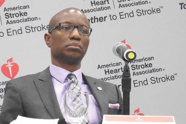 |
| Mitchel L. Zoler/Frontline Medical News Dr. Bruce I. Ovbiagele |
The workers in Boston and their collaborators used MR diffusion weighted imaging to confirm that a patient has had a stroke, and then use MR fluid attenuated inversion recovery (FLAIR) to determine if the stroke had occurred within the previous 4 hours. If a stroke has not been going on long enough to produce a positive FLAIR image, it means that the patient is still eligible for thrombolytic therapy. This could be huge for U.S. clinical practice because so many patients have unwitnessed strokes. We definitely need a larger efficacy study, but the results Dr. Schwamm reported are highly encouraging.
CT is more widely available right now in U.S. practice than is MRI, so ideally we would like to be able to use CT imaging.
Dr. Bruce I. Ovbiagele is professor and chairman of neurology at the Medical University of South Carolina in Charleston. He had no disclosures. He made these comments in an interview.
Traditionally, we have used a clock to identify patients who are eligible to receive reperfusion therapy, but now researchers are trying to extend patient eligibility with imaging. The Stanford (Calif.) team is trying to identify good candidates for reperfusion treatment who present with acute ischemic strokes beyond the traditional time window of 6 hours. The researchers at Massachusetts General Hospital, Boston, and their collaborators are trying to apply a similar approach to patients who have unwitnessed strokes and so have an unknown elapsed time from the start of their stroke.
The rationale behind the Stanford work is that some patients will have salvageable neurons beyond the traditional treatment time window and that this tissue can be identified by MR and CT perfusion imaging.
|
| Mitchel L. Zoler/Frontline Medical News Dr. Bruce I. Ovbiagele |
The workers in Boston and their collaborators used MR diffusion weighted imaging to confirm that a patient has had a stroke, and then use MR fluid attenuated inversion recovery (FLAIR) to determine if the stroke had occurred within the previous 4 hours. If a stroke has not been going on long enough to produce a positive FLAIR image, it means that the patient is still eligible for thrombolytic therapy. This could be huge for U.S. clinical practice because so many patients have unwitnessed strokes. We definitely need a larger efficacy study, but the results Dr. Schwamm reported are highly encouraging.
CT is more widely available right now in U.S. practice than is MRI, so ideally we would like to be able to use CT imaging.
Dr. Bruce I. Ovbiagele is professor and chairman of neurology at the Medical University of South Carolina in Charleston. He had no disclosures. He made these comments in an interview.
LOS ANGELES – Can brain imaging surpass the clock for identifying acute ischemic stroke patients who will benefit from thrombolytic or thrombectomy treatment?
That’s what experts now envision, based on early findings from several studies. Although the evidence is not yet definitive enough to justify using imaging as a replacement for time-from-stroke-onset in routine practice, the results so far are encouraging and have led to the start or planning of several phase III trials that will try to nail down a role for imaging, either CT or MR, to identify acute ischemic stroke patients who qualify for reperfusion therapy.
“What we’re trying to do is move away from using the clock as a surrogate marker and instead use imaging as the surrogate,” Dr. Jenny P. Tsai said in an interview at the International Stroke Conference.
The linchpin of this new approach is that clocking time elapsed from the onset of stroke symptoms to initiation of thrombolytic or endovascular treatment makes no allowance for patient-to-patient variations in collateral cerebral circulation, a factor that appears to make a big difference in whether patients can be many more hours removed from the start of their stroke and still have salvageable brain tissue. And relying on time since stroke symptom onset gives a seriously flawed estimate of a stroke’s duration when patients have an unwitnessed stroke.
The alternative is to use either CT perfusion imaging or a combination of MR perfusion and diffusion-weighted imaging “to get effective reperfusion treatments to patients who present at later time windows,” said Dr. Gregory W. Albers, professor of neurology at Stanford (Calif.) University and director of the Stanford Stroke Center. He called these two new approaches to gauging a patient’s suitability for reperfusion therapy “the next big thing in imaging” for stroke.
Using CT perfusion to assess target mismatch
Dr. Tsai presented an analysis of thrombectomy reperfusion done in 181 patients enrolled in the CT Perfusion to Predict Response to Recanalization in Ischemic Stroke Project (CRISP), which included a total of 201 acute ischemic stroke patients with large cerebral-artery occlusions treated at six U.S. centers. Her analysis excluded nine patients who presented more than 18 hours after their stroke onset, six patients who did not have successful CT perfusion assessment, and five additional patients excluded for other reasons. The 181 patients analyzed included 125 with a target mismatch in CT perfusion, indicating that salvageable tissue remained in the area of their stroke. Among these 125 patients, 111 underwent successful reperfusion by thrombectomy.
The researchers identified good treatment outcomes as patients with a modified Rankin Scale score of 2 or less 90 days after treatment. A multivariate analysis showed that among these 111 patients, achievement of a good 90-day outcome had no statistically significant relationship with time-to-treatment out to at least the first 8 hours following stroke onset, reported Dr. Tsai, a neurologist at the Stanford Stroke Center. The data also showed a nonsignificant relationship between good outcomes and time from stroke onset to treatment beyond 8 hours, but the confidence interval for this nonsignificant relationship became very wide at later times as the analysis focused on fewer and fewer patients.
“In patients with large-artery occlusions who have a target mismatch profile [on perfusion CT] and achieve successful reperfusion there is no significant association between onset-to-reperfusion-time and the probability of a good functional outcome,” suggesting that “CT perfusion is a biomarker of good outcomes beyond 6 hours” after stroke onset, Dr. Tsai concluded. CT perfusion has the advantage of being more widely available than MRI is, she added. Last year, her associates at Stanford reported similar findings using perfusion-diffusion mismatch in the cerebral area around a stroke visualized with MRI (Neurology. 2015 Aug 25;85[8]:708-15).
“MRI is not readily available” at all U.S. centers that treat stroke patients, while CT perfusion imaging is much more ubiquitous, but the findings reported by Dr. Tsai require confirmation in the several prospective, randomized trials now underway and testing this approach, Dr. Albers said.
Using MRI in unwitnessed strokes
A second challenge for using reperfusion therapy in stroke patients are patients who have unwitnessed strokes, with a completely unknown elapsed time from onset to presentation. The ability of MRI to help identify patients with unwitnessed stroke who can safely receive intravenous thrombolytic therapy with alteplase (tissue plasminogen activator, Activase) underwent testing in the MR WITNESS (Study of Intravenous Thrombolysis With Alteplase in MRI-Selected Patients) trial.
The study enrolled 80 patients with an unwitnessed acute, ischemic stroke at 10 U.S. centers. Patients had to be in a position to receive alteplase within 4.5 hours of when their stroke was first identified; 57 (71%) of the participants had wake-up strokes. All patients underwent two types of MRI to identify them as potential candidates for safe administration of alteplase: diffusion weighted imaging, to identify that a stroke had occurred, and fluid-attenuated inversion recovery (FLAIR) assessment, to identify strokes that had occurred during the prior 4 hours.
This phase II study’s primary safety endpoint was the incidence of symptomatic intracerebral hemorrhage following alteplase treatment, which occurred in 1 of the 80 patients (1.25%), not a statistically significant difference when compared with the historical standard, the 2.4% rate seen in stroke patients treated with intravenous alteplase 3 to 4.5 hours after their known stroke onset in the ECASS III (European Cooperative Acute Stroke Study) (N Engl J Med. 2008 Sept 25;359[13]:1317-29). The rate of asymptomatic intracerebral hemorrhage was not significantly different between the new study and ECASS III, reported Dr. Lee H. Schwamm, director of TeleStroke & Acute Stroke Services at the Massachusetts General Hospital in Boston.
Based on this result, “we know this approach is safe, and we saw a signal of efficacy, but we really don’t know how effective it will be” until this approach to assessing unwitnessed-stroke patients by imaging undergoes testing in a phase III trial, cautioned Dr. Schwamm at the meeting, sponsored by the American Heart Association.
Future work will also examine whether similar results can be obtained by CT imaging, which would “open this approach to every U.S. emergency department,” Dr. Schwamm said. Although the MR diffusion-weighted imaging and FLAIR analyses used in the current study do not require anything more than standard MRI equipment and software, it remains less available than CT imaging at most U.S. hospitals, he said.
The video associated with this article is no longer available on this site. Please view all of our videos on the MDedge YouTube channel
On Twitter @mitchelzoler
AT THE INTERNATIONAL STROKE CONFERENCE
Key clinical point: New CT and MR techniques provide alternatives to time-from-stroke-onset to select acute ischemic stroke patients for reperfusion therapies.
Major finding: CT perfusion identified ischemic stroke patients who could undergo effective thrombectomy more than 6 hours after their stroke onset.
Data source: The CRISP study, which enrolled 201 patients at six U.S. centers.
Disclosures: Dr. Tsai and Dr. Schwamm had no disclosures. Dr. Albers has been a consultant to iSchemaView and Covidien/Medtronic and has an ownership interest in iSchemaView.