User login
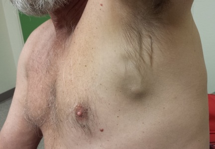
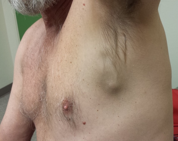
A 60-year-old man presents to a free clinic for evaluation of a “knot” in his left lower axilla. He first noticed it several months ago. Even though the lesion grew, the patient reasoned that it couldn’t be too serious since “it didn’t hurt.” But in the past month, the lesion has become much more firm and darkened.
The patient denies any other health problems. He has an 80-pack-year history of smoking. He has never had health insurance and as a result has carefully avoided any interaction with the health care establishment.
Additional history-taking reveals that he has become markedly short of breath in the past few months, which he attributes to his smoking. He denies any abdominal pain or headache. As a young man, he spent 20 years working as a roofer, often going shirtless in hot weather.
EXAMINATION
The patient is in no distress. He is accompanied by his wife, who participates in the history-taking process. Both call attention to the lesion in question, a 5.5-cm dark subcutaneous mass in the left lower axilla. The overlying skin is dark as well, and on palpation, the mass is found to be fixed and firm. Palpation elsewhere in the axillae, as well as in the head and neck, fails to detect any other lesions.
The rest of the patient's type III skin (easily tanned, seldom burned) shows limited evidence of sun damage. A few minor solar lentigines are seen on the trunk, but none stand out. Then the patient's wife points out a barely discernible, depigmented, poorly defined macule on the left lateral back. It measures about 2 cm. This lesion, she reports, "used to be real black. Then it turned scabby, and then it lost all its color. It's been this way ever since."
Continue reading for the diagnostic studies...
DIAGNOSTIC STUDIES
An incisional biopsy of the residual lesion on his back is performed. Two-thirds of the macule is removed, the wound is closed with sutures, and the specimen is submitted for pathologic examination. The resulting report indicates a superficial spreading melanoma, Breslow depth (vertical thickness) of 1.6 mm, with evidence of significant regression and a brisk mitotic rate─all predictors of metastatic potential.
Accordingly, the patient is referred to the appropriate surgical specialist, who confirms the micrometastatic nature of the axillary node and refers the patient to oncology for imaging studies that will determine the extent of metastasis. The residual primary tumor is excised with 2-cm margins.
Continue reading for the discussion...
DISCUSSION
This case illustrates the phenomenon—known as regression—in which part or all of a cutaneous melanoma is rendered invisible over time by the reaction of the immune system (although not before it has a chance to spread). Not uncommonly, these patients are diagnosed with metastatic melanoma and no primary lesion is ever discovered.
A variation on this theme is the so-called amelanotic melanoma, which can present initially in a wide variety of forms—papules, nodules, or macules; in shades of pink, blue, white, or "flesh." But unlike our patient, these cases do not involve regression.
This patient and his wife assumed that because the primary lesion had essentially disappeared, it must have been safe. And of course, they failed to connect it with the new axillary nodule.
At this writing, the extent of the patient’s disease remains to be defined by imaging studies (PET and CT scans). But given the facts of the case, his prognosis at best is guarded. A more hopeful view is that he is living in a time of meaningful discovery of better treatments for advanced melanomas.
Continue reading for Joe Monroe's take home learning points...
TAKE-HOME LEARNING POINTS
• Although the classic melanoma is black and essentially flat, it can present in many other forms, including an amelanotic version, the color of which can be pink, blue, white or even skin-colored.
• Infrequently, melanoma presents initially in metastatic form, having spread from an unknown or obscure primary lesion.
• Immune response to a cutaneous melanoma can result in partial or even total destruction of the lesion, a process termed regression.
• Predictors of a relatively poor prognosis for melanoma include: the aforementioned regression, vertical thickness (Breslow depth), mitotic rate, vascular invasion, and the presence of ulceration.
• New masses in known nodal locations require examination of surrounding skin to detect or rule out suspicious lesions.