User login
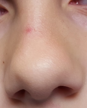
No one in the family is certain when this 6-year-old boy first developed the red spot on his nose, but it is increasingly noticeable. And with school picture day approaching, they would like the redness to resolve.
Their primary care provider reassured them, at length, that it was benign and would eventually resolve without treatment. The lesion causes no symptoms and is purely a cosmetic concern.
The child is otherwise healthy and has been since birth.
EXAMINATION
A pinpoint red dot can be seen on the upper nasal bridge, just to the right of the midline. Tiny linear red “legs” extend from the central dot, like spokes on a wheel. In aggregate, the lesion measures about 3 mm in diameter. There is no palpable component.
However, the entire lesion is blanchable: Pinpoint pressure on the central dot causes it to blanch, and as the pressure is released, the legs of the lesion refill immediately from the center outward. When a glass slide is pressed against the lesion (a process called diascopy) and then released, the same process occurs.

What is the diagnosis?
Spider angiomas (SAs), originally called nevus araneus, are actually neither angiomas nor nevi. Instead, they are telangiectasias formed from a superficial arteriole whose surrounding sphincter muscle has failed. The “legs” are tiny veins that carry blood away from the central lesion; this is why they blanch so readily with central pressure and refill from the center outward.
SAs affect 10% to 15% of children and occur only in areas along the path of the superior vena cava. Besides the face, they can be found on the trunk, arms, and dorsal hands.
When solitary, these lesions are benign and can be either left to clear on their own or removed by laser or electrodessication. The presence of three or more lesions warrants further investigation, since multiple SAs can signify underlying disease (eg, cirrhosis of the liver, thyrotoxicosis, or systemic sclerosis).
This patient was not inclined to let us treat the lesion; within a few years, a teasing comment from a classmate or two may change his mind! But with a little luck, it will disappear on its own eventually.
TAKE-HOME LEARNING POINTS
- Spider angiomas (SAs) are actually dilated telangiectatic arterioles manifesting as blanchable, bright red, pinpoint papules with venous “legs” that extend from the center.
- SAs affect 10% to 15% of all children and are confined to areas drained by the superior vena cava.
- Pressure on the lesion with a glass slide (a process called diascopy) causes total blanching; this, along with the clinical findings, confirms the diagnosis.
- Most SAs resolve on their own eventually, but they can be destroyed by laser or electrodessication.
- The presence of more than three SAs should prompt a search for potential causes, such as liver disease, thyrotoxicosis, or systemic sclerosis.
No one in the family is certain when this 6-year-old boy first developed the red spot on his nose, but it is increasingly noticeable. And with school picture day approaching, they would like the redness to resolve.
Their primary care provider reassured them, at length, that it was benign and would eventually resolve without treatment. The lesion causes no symptoms and is purely a cosmetic concern.
The child is otherwise healthy and has been since birth.
EXAMINATION
A pinpoint red dot can be seen on the upper nasal bridge, just to the right of the midline. Tiny linear red “legs” extend from the central dot, like spokes on a wheel. In aggregate, the lesion measures about 3 mm in diameter. There is no palpable component.
However, the entire lesion is blanchable: Pinpoint pressure on the central dot causes it to blanch, and as the pressure is released, the legs of the lesion refill immediately from the center outward. When a glass slide is pressed against the lesion (a process called diascopy) and then released, the same process occurs.
What is the diagnosis?
Spider angiomas (SAs), originally called nevus araneus, are actually neither angiomas nor nevi. Instead, they are telangiectasias formed from a superficial arteriole whose surrounding sphincter muscle has failed. The “legs” are tiny veins that carry blood away from the central lesion; this is why they blanch so readily with central pressure and refill from the center outward.
SAs affect 10% to 15% of children and occur only in areas along the path of the superior vena cava. Besides the face, they can be found on the trunk, arms, and dorsal hands.
When solitary, these lesions are benign and can be either left to clear on their own or removed by laser or electrodessication. The presence of three or more lesions warrants further investigation, since multiple SAs can signify underlying disease (eg, cirrhosis of the liver, thyrotoxicosis, or systemic sclerosis).
This patient was not inclined to let us treat the lesion; within a few years, a teasing comment from a classmate or two may change his mind! But with a little luck, it will disappear on its own eventually.
TAKE-HOME LEARNING POINTS
- Spider angiomas (SAs) are actually dilated telangiectatic arterioles manifesting as blanchable, bright red, pinpoint papules with venous “legs” that extend from the center.
- SAs affect 10% to 15% of all children and are confined to areas drained by the superior vena cava.
- Pressure on the lesion with a glass slide (a process called diascopy) causes total blanching; this, along with the clinical findings, confirms the diagnosis.
- Most SAs resolve on their own eventually, but they can be destroyed by laser or electrodessication.
- The presence of more than three SAs should prompt a search for potential causes, such as liver disease, thyrotoxicosis, or systemic sclerosis.