User login
A 68-year-old man is seen for evaluation of a facial sore, which has been present for “at least a month.” It concerns him because, while the lesion is not painful, it won’t heal. The patient denies any preceding trauma to the area but does admit to having several skin cancers removed in his lifetime.
His lesion has been previously diagnosed as “pyoderma” and treated unsuccessfully with topical mupirocin and oral cephalexin.
He has held a number of jobs throughout his life, all of which involved being out in the sun all day—often seven days a week. And it wasn’t until he turned 45 that he finally started wearing a hat.
EXAMINATION
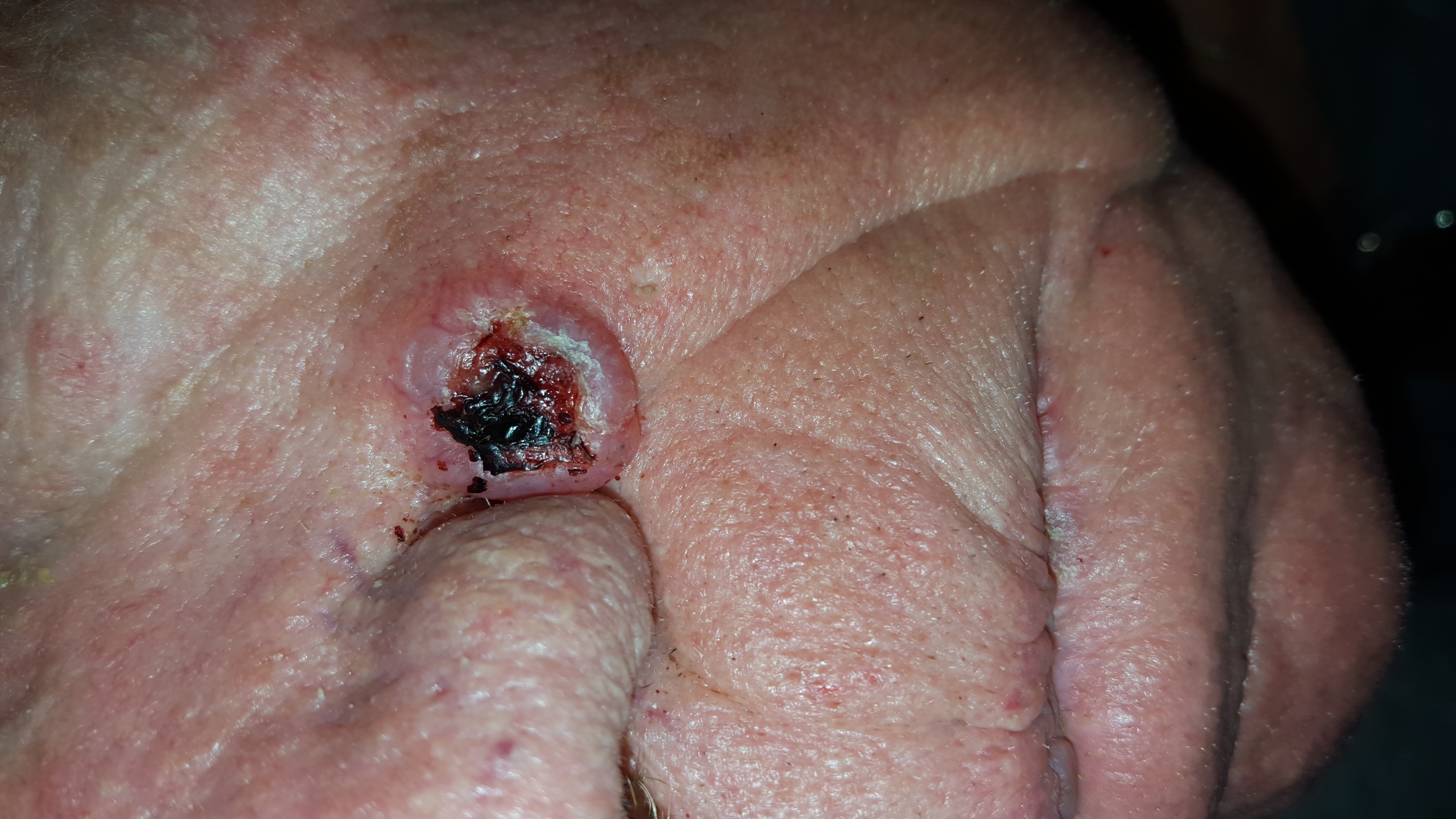
The lesion is an impressive, round, 1.2-cm nodule with extensive central ulceration. It has a rolled, translucent border and is tucked into the upper left nasolabial fold, directly adjacent to the alar bulb. It appears in the context of his fair, sun-damaged skin; he has several scars consistent with his history of skin cancer removal.

His skin elsewhere, while quite sun-damaged, is free of worrisome lesions.
A simple shave biopsy of the lesion is performed.
What is the diagnosis?
DISCUSSION
The biopsy showed changes consistent with basal cell carcinoma (BCC). It would be difficult to find a more typical example of BCC, the most common of all the sun-caused skin cancers (which include squamous cell, melanoma, and Merkel cell carcinoma). And yet, as this case illustrates, classic signs of BCC are often missed or overlooked, leading to “infection” or “pyoderma” diagnoses by providers who are unfamiliar with BCC.
I often tell medical providers that “it will be a rare day when you don’t see a BCC” because they are that common. But what I probably should say is “it will be a rare day when you don’t have a chance to see a BCC.” By that, I mean that you do have to look for them—not all are as obvious as this man’s—and you do need to have an understanding of who is susceptible and what the lesion will look like.
Older, sun-damaged individuals (mostly men) are most at risk for BBCs. Fair skin, freckles, red hair, and blue eyes are additional high-risk characteristics. This varies from melanoma patients, who tend to be much younger and whose lesions look nothing like a basal cell.
For BCC identification, focus on the areas directly and chronically exposed to the sun, such as the tops of the ears, the face, nose, chest, back, and arms.
BCCs can take on many appearances; they look as though they would hurt, but rarely do—a fact that should be noted as significant. Typically, they break down, scab, and bleed as they grow larger. They are usually very slow-growing and often take years to become noticeable. In other words, you have to think of it first, and look for it next. The fact that almost 1.5 million new BCCs are diagnosed each year in the US should heighten your suspicions considerably.
In this case, the patient will need extensive surgery, which may be deforming. Given his history of sun exposure, he will likely develop more skin cancers in the future.
TAKE-HOME LEARNING POINTS
• Basal cell carcinoma (BCC) is by far the most common type of sun-caused skin cancer; it will be seen frequently if properly looked for.
• Identification of BCCs will be more successful if the search is concentrated on the patients most likely to develop one and the areas where they are most likely to appear.
• This patient’s noduloulcerative BCC is the most common type and developed in a classic location. There is no type of infection that presents in this fashion.
• A simple shave biopsy confirms the diagnosis of BCC.
A 68-year-old man is seen for evaluation of a facial sore, which has been present for “at least a month.” It concerns him because, while the lesion is not painful, it won’t heal. The patient denies any preceding trauma to the area but does admit to having several skin cancers removed in his lifetime.
His lesion has been previously diagnosed as “pyoderma” and treated unsuccessfully with topical mupirocin and oral cephalexin.
He has held a number of jobs throughout his life, all of which involved being out in the sun all day—often seven days a week. And it wasn’t until he turned 45 that he finally started wearing a hat.
EXAMINATION
The lesion is an impressive, round, 1.2-cm nodule with extensive central ulceration. It has a rolled, translucent border and is tucked into the upper left nasolabial fold, directly adjacent to the alar bulb. It appears in the context of his fair, sun-damaged skin; he has several scars consistent with his history of skin cancer removal.
His skin elsewhere, while quite sun-damaged, is free of worrisome lesions.
A simple shave biopsy of the lesion is performed.
What is the diagnosis?
DISCUSSION
The biopsy showed changes consistent with basal cell carcinoma (BCC). It would be difficult to find a more typical example of BCC, the most common of all the sun-caused skin cancers (which include squamous cell, melanoma, and Merkel cell carcinoma). And yet, as this case illustrates, classic signs of BCC are often missed or overlooked, leading to “infection” or “pyoderma” diagnoses by providers who are unfamiliar with BCC.
I often tell medical providers that “it will be a rare day when you don’t see a BCC” because they are that common. But what I probably should say is “it will be a rare day when you don’t have a chance to see a BCC.” By that, I mean that you do have to look for them—not all are as obvious as this man’s—and you do need to have an understanding of who is susceptible and what the lesion will look like.
Older, sun-damaged individuals (mostly men) are most at risk for BBCs. Fair skin, freckles, red hair, and blue eyes are additional high-risk characteristics. This varies from melanoma patients, who tend to be much younger and whose lesions look nothing like a basal cell.
For BCC identification, focus on the areas directly and chronically exposed to the sun, such as the tops of the ears, the face, nose, chest, back, and arms.
BCCs can take on many appearances; they look as though they would hurt, but rarely do—a fact that should be noted as significant. Typically, they break down, scab, and bleed as they grow larger. They are usually very slow-growing and often take years to become noticeable. In other words, you have to think of it first, and look for it next. The fact that almost 1.5 million new BCCs are diagnosed each year in the US should heighten your suspicions considerably.
In this case, the patient will need extensive surgery, which may be deforming. Given his history of sun exposure, he will likely develop more skin cancers in the future.
TAKE-HOME LEARNING POINTS
• Basal cell carcinoma (BCC) is by far the most common type of sun-caused skin cancer; it will be seen frequently if properly looked for.
• Identification of BCCs will be more successful if the search is concentrated on the patients most likely to develop one and the areas where they are most likely to appear.
• This patient’s noduloulcerative BCC is the most common type and developed in a classic location. There is no type of infection that presents in this fashion.
• A simple shave biopsy confirms the diagnosis of BCC.