User login
A 25-year-old woman is concerned about lesions on the dorsum of her right foot that first appeared about four months ago. Initially, there was one red lesion; several similarly colored lesions manifested within a few weeks. The original lesion darkened from red to brown before gradually fading to its current appearance. All the lesions are asymptomatic.
The patient denies injury to the foot. She denies any history of skin problems, including any similar lesions elsewhere on her body. She describes her general health as “quite good,” aside from mild seasonal allergies. She is taking no medications of any kind.
EXAMINATION
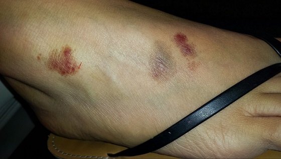
Three macular patches—each about 2.4 cm and roughly round, with sharp margins—are seen on the dorsum of her right foot. There is no palpable component to any of them, nor increased warmth or tenderness. Two are dark reddish brown, with nonblanchable redness. The middle lesion looks quite different—brownish gray—but it too is totally macular. Elsewhere, her type IV Hispanic skin is free of any notable lesions.
A shave biopsy is taken from one of the active (red) lesions. The pathology results indicate capillaritis.
What is the diagnosis?
DISCUSSION
When red inflammatory processes fail to blanch with digital pressure, we know that red blood cells (RBCs) have likely leaked from damaged blood vessels and stained the interstitial spaces. Generically, we call this purpura. On occasion, purpura can be an ominous sign, indicating a vasculitis such as leukocytoclastic vasculitis (LCV), in which an immune complex damages the walls of blood vessels that supply vital organs. This can occur in the context of a connective tissue disease, such as lupus, or an allergic reaction to an ingestant (food, drink, or medicine).
But there are specific histologic criteria (eg, nuclear dust from white blood cells that have expended themselves in attacking blood vessel walls and extravasated RBCs from these same leaky walls) for the diagnosis of LCV that were missing in this case. Significantly, the affected blood vessels in this case were capillaries, which are not affected by LCV.
The histologic and clinical findings put us squarely into a relatively benign group of conditions called capillaritis. Collectively, these are known as pigmented purpuris dermatoses; the most common is Schamberg disease (progressive pigmentary dermatosis). These dermatoses are characterized by extravasation of RBCs with marked hemosiderin deposition.
In this case, however, we’re looking at another common form, lichen aureus (LA). The lesions of LA are round to polygonal, sometimes shiny, and brownish red (which can look darker in patients with darker skin).
Schamberg lesions are said to resemble sprinkled cayenne pepper. They typically start on the lower legs and move upward to just below the knee, then slowly descend and resolve. This entire process will occur within a period of several months.
The cause of this family of dermatoses is unknown, but, thankfully, they are all asymptomatic and self-limited. Topical and systemic treatment does not help.
The differential also includes granulomatous disease (eg, granuloma annulare) and fixed drug eruption. Biopsy, as in this case, is often warranted.
TAKE-HOME LEARNING POINTS
• Lichen aureus and Schamberg disease are the most common forms of capillaritis, a benign condition usually affecting the legs and feet.
• These forms of capillaritis are caused by extravasation of red blood cells (RBCs) from damaged capillaries.
• They typically manifest with brownish red macules and patches that do not blanch with digital pressure.
• The cause of capillaritis is unknown, although it is clear that it results from damage to capillary vessel walls that allows RBCs to leak into the interstitial tissues.
• The main item in the differential is leukocytoclastic vasculitis (LCV), which is of more acute onset and usually has a petichial appearance. Since it has systemic implications, suspected LCV must be confirmed or ruled out with biopsy.
A 25-year-old woman is concerned about lesions on the dorsum of her right foot that first appeared about four months ago. Initially, there was one red lesion; several similarly colored lesions manifested within a few weeks. The original lesion darkened from red to brown before gradually fading to its current appearance. All the lesions are asymptomatic.
The patient denies injury to the foot. She denies any history of skin problems, including any similar lesions elsewhere on her body. She describes her general health as “quite good,” aside from mild seasonal allergies. She is taking no medications of any kind.
EXAMINATION
Three macular patches—each about 2.4 cm and roughly round, with sharp margins—are seen on the dorsum of her right foot. There is no palpable component to any of them, nor increased warmth or tenderness. Two are dark reddish brown, with nonblanchable redness. The middle lesion looks quite different—brownish gray—but it too is totally macular. Elsewhere, her type IV Hispanic skin is free of any notable lesions.
A shave biopsy is taken from one of the active (red) lesions. The pathology results indicate capillaritis.
What is the diagnosis?
DISCUSSION
When red inflammatory processes fail to blanch with digital pressure, we know that red blood cells (RBCs) have likely leaked from damaged blood vessels and stained the interstitial spaces. Generically, we call this purpura. On occasion, purpura can be an ominous sign, indicating a vasculitis such as leukocytoclastic vasculitis (LCV), in which an immune complex damages the walls of blood vessels that supply vital organs. This can occur in the context of a connective tissue disease, such as lupus, or an allergic reaction to an ingestant (food, drink, or medicine).
But there are specific histologic criteria (eg, nuclear dust from white blood cells that have expended themselves in attacking blood vessel walls and extravasated RBCs from these same leaky walls) for the diagnosis of LCV that were missing in this case. Significantly, the affected blood vessels in this case were capillaries, which are not affected by LCV.
The histologic and clinical findings put us squarely into a relatively benign group of conditions called capillaritis. Collectively, these are known as pigmented purpuris dermatoses; the most common is Schamberg disease (progressive pigmentary dermatosis). These dermatoses are characterized by extravasation of RBCs with marked hemosiderin deposition.
In this case, however, we’re looking at another common form, lichen aureus (LA). The lesions of LA are round to polygonal, sometimes shiny, and brownish red (which can look darker in patients with darker skin).
Schamberg lesions are said to resemble sprinkled cayenne pepper. They typically start on the lower legs and move upward to just below the knee, then slowly descend and resolve. This entire process will occur within a period of several months.
The cause of this family of dermatoses is unknown, but, thankfully, they are all asymptomatic and self-limited. Topical and systemic treatment does not help.
The differential also includes granulomatous disease (eg, granuloma annulare) and fixed drug eruption. Biopsy, as in this case, is often warranted.
TAKE-HOME LEARNING POINTS
• Lichen aureus and Schamberg disease are the most common forms of capillaritis, a benign condition usually affecting the legs and feet.
• These forms of capillaritis are caused by extravasation of red blood cells (RBCs) from damaged capillaries.
• They typically manifest with brownish red macules and patches that do not blanch with digital pressure.
• The cause of capillaritis is unknown, although it is clear that it results from damage to capillary vessel walls that allows RBCs to leak into the interstitial tissues.
• The main item in the differential is leukocytoclastic vasculitis (LCV), which is of more acute onset and usually has a petichial appearance. Since it has systemic implications, suspected LCV must be confirmed or ruled out with biopsy.
