User login
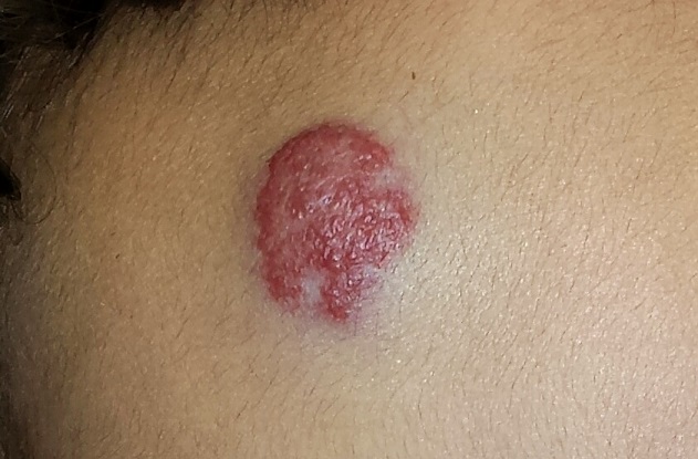
A 4-month-old boy is brought in by his mother for evaluation of a “birthmark” that has become more prominent with time. The child complains a bit when the lesion is touched.
His mother gives a history of a normal full-term pregnancy, with an uneventful delivery. Other than the skin lesion, there have been no other known problems with the child’s health.

EXAMINATION
The lesion in question is a 2-cm bright red nodule in the middle of the child’s forehead. There is a faint blue halo around it, but no other abnormalities are seen in the area or elsewhere on the child’s body. The patient otherwise appears well developed and well nourished; he is quite alert and reactive to verbal stimuli.
What is the diagnosis?
DISCUSSION
Most first-year medical students could tell you this was a case of an infantile hemangioma—but within the past five years, the categorization and treatment of hemangiomas have changed rapidly.
Hemangiomas are benign and usually self-involuting vascular tumors; they are distinct from the family of permanent congenital vascular lesions, such as port wine stains. About 80% occur on the face or neck, and they more commonly occur in female patients. Those that occur near the skin’s surface tend to be bright red, while those of deeper origin are more bluish. Hemangiomas can also occur in extracutaneous areas (eg, the liver); these are usually detected via imaging.
Most infantile hemangiomas appear in the first few weeks of life but begin to involute shortly after they reach maximal size (usually by age 12 months). Involution is complete by age 5 in about 50% of cases and by age 7 in 70% of them. The remainder usually resolve by age 12.
Until recently, the parents of an affected child would be told that the lesions were benign and would likely disappear on their own by the time the child was ages 7 to 10—all true enough. These lesions were treated only when they were large or symptomatic, or if their location blocked or otherwise interfered with the function of the eyes, ears, mouth, anus, or vagina. Unfortunately, this approach ignored the psychosocial aspects of having a prominent and fire-engine red lesion, which often becomes the object of unwanted attention, and even ridicule, at a crucial developmental age.
In the past, the main treatment options were destructive in nature (laser, excision) and often caused as many problems as they solved. Pharmacologic treatment with oral prednisone, and later with interferon, was also tried with mixed success.
In 2008, in a serendipitous observation, a French team came up with a groundbreaking approach: giving systemic propranolol multiple times over a 12 to 18–month period. This method proved to be both safe and effective when used in the right setting by specialists (pediatric dermatologists and/or pediatricians) experienced in this modality. It was found that the lesions responded best if treated before age 6 months.
This has worked so well and so safely that it has quickly become routine. But it will take time before it is sufficiently well known to become the actual standard of care for these common lesions.
TAKE-HOME LEARNING POINTS
• Infantile hemangiomas (IH) usually resolve (involute) by age 12 at the latest.
• IH, by definition, is different from permanent congenital vascular malformations, such as port wine stains.
• About 80% of IHs occur on the head and neck; they are often the object of unwanted attention or even ridicule.
• A new treatment approach, systemic propranolol, is now being used routinely and successfully. It works best when the patient is 6 months old or younger.
• Ideally, this new treatment approach should be overseen by the relevant specialist, be it in a pediatric or dermatologic setting.
A 4-month-old boy is brought in by his mother for evaluation of a “birthmark” that has become more prominent with time. The child complains a bit when the lesion is touched.
His mother gives a history of a normal full-term pregnancy, with an uneventful delivery. Other than the skin lesion, there have been no other known problems with the child’s health.
EXAMINATION
The lesion in question is a 2-cm bright red nodule in the middle of the child’s forehead. There is a faint blue halo around it, but no other abnormalities are seen in the area or elsewhere on the child’s body. The patient otherwise appears well developed and well nourished; he is quite alert and reactive to verbal stimuli.
What is the diagnosis?
DISCUSSION
Most first-year medical students could tell you this was a case of an infantile hemangioma—but within the past five years, the categorization and treatment of hemangiomas have changed rapidly.
Hemangiomas are benign and usually self-involuting vascular tumors; they are distinct from the family of permanent congenital vascular lesions, such as port wine stains. About 80% occur on the face or neck, and they more commonly occur in female patients. Those that occur near the skin’s surface tend to be bright red, while those of deeper origin are more bluish. Hemangiomas can also occur in extracutaneous areas (eg, the liver); these are usually detected via imaging.
Most infantile hemangiomas appear in the first few weeks of life but begin to involute shortly after they reach maximal size (usually by age 12 months). Involution is complete by age 5 in about 50% of cases and by age 7 in 70% of them. The remainder usually resolve by age 12.
Until recently, the parents of an affected child would be told that the lesions were benign and would likely disappear on their own by the time the child was ages 7 to 10—all true enough. These lesions were treated only when they were large or symptomatic, or if their location blocked or otherwise interfered with the function of the eyes, ears, mouth, anus, or vagina. Unfortunately, this approach ignored the psychosocial aspects of having a prominent and fire-engine red lesion, which often becomes the object of unwanted attention, and even ridicule, at a crucial developmental age.
In the past, the main treatment options were destructive in nature (laser, excision) and often caused as many problems as they solved. Pharmacologic treatment with oral prednisone, and later with interferon, was also tried with mixed success.
In 2008, in a serendipitous observation, a French team came up with a groundbreaking approach: giving systemic propranolol multiple times over a 12 to 18–month period. This method proved to be both safe and effective when used in the right setting by specialists (pediatric dermatologists and/or pediatricians) experienced in this modality. It was found that the lesions responded best if treated before age 6 months.
This has worked so well and so safely that it has quickly become routine. But it will take time before it is sufficiently well known to become the actual standard of care for these common lesions.
TAKE-HOME LEARNING POINTS
• Infantile hemangiomas (IH) usually resolve (involute) by age 12 at the latest.
• IH, by definition, is different from permanent congenital vascular malformations, such as port wine stains.
• About 80% of IHs occur on the head and neck; they are often the object of unwanted attention or even ridicule.
• A new treatment approach, systemic propranolol, is now being used routinely and successfully. It works best when the patient is 6 months old or younger.
• Ideally, this new treatment approach should be overseen by the relevant specialist, be it in a pediatric or dermatologic setting.