User login
Dr Bechtel, editor of “Derm Dilemma,” is a professor of medicine and director of the division of dermatology at the Ohio State University College of Medicine in Columbus. He is also a member of the EMERGENCY MEDICINE editorial board. Ms Bechtel is a nurse educational consultant in Columbus.
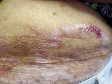
Case 1
Skin biopsy revealed epidermal atrophy, dermal edema, and a band-like lymphocytic infiltrate below the epidermis, consistent with lichen sclerosis. Lichen sclerosis often manifests as ivory-white plaques with a cigarette-paper texture in the anogenital region. As noted in this patient, nongenital sites can be involved. Lichen sclerosis can affect both adult women and female pediatric patients. Symptoms include a burning feeling during urination and purpura within skin lesions in the anogenital region. The initial treatment of choice for lichen sclerosis involves moderate-to-potent topical steroids, which are gradually tapered based on clinical response. It is important for the health care provider to monitor for thinning of skin secondary to topical steroid use.
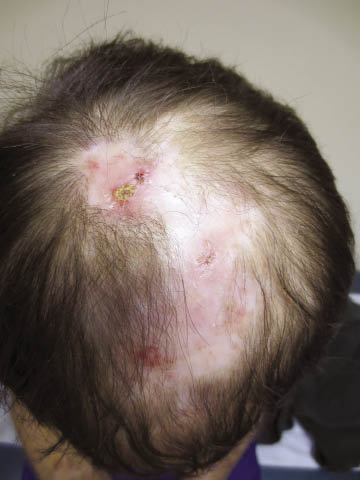
Case 2
The dermatology consult and biopsy confirm a diagnosis of discoid lupus erythematosus (DLE). The clinical clues to recognizing DLE are erythematous rashes on sun-exposed areas, often with cutaneous scarring and follicular plugging; individual lesions demonstrating central hypopigmentation and hyperpigmentation of the margins; and scarring alopecia. DLE can be complicated by arthralgias, scarring alopecia, and squamous cell cancer developing within the scars. Approximately 5% to 15% of patients with DLE develop systemic lupus erythematosus; patients with widespread lesions are more likely to develop systemic disease. Treatment options include topical steroids, antimalarial agents, and avoiding sun exposure (eg, use of sunscreen and protective clothing). Patients must be monitored for development of cutaneous squamous cell cancer in scar tissue.
Dr Bechtel, editor of “Derm Dilemma,” is a professor of medicine and director of the division of dermatology at the Ohio State University College of Medicine in Columbus. He is also a member of the EMERGENCY MEDICINE editorial board. Ms Bechtel is a nurse educational consultant in Columbus.
Case 1
Skin biopsy revealed epidermal atrophy, dermal edema, and a band-like lymphocytic infiltrate below the epidermis, consistent with lichen sclerosis. Lichen sclerosis often manifests as ivory-white plaques with a cigarette-paper texture in the anogenital region. As noted in this patient, nongenital sites can be involved. Lichen sclerosis can affect both adult women and female pediatric patients. Symptoms include a burning feeling during urination and purpura within skin lesions in the anogenital region. The initial treatment of choice for lichen sclerosis involves moderate-to-potent topical steroids, which are gradually tapered based on clinical response. It is important for the health care provider to monitor for thinning of skin secondary to topical steroid use.
Case 2
The dermatology consult and biopsy confirm a diagnosis of discoid lupus erythematosus (DLE). The clinical clues to recognizing DLE are erythematous rashes on sun-exposed areas, often with cutaneous scarring and follicular plugging; individual lesions demonstrating central hypopigmentation and hyperpigmentation of the margins; and scarring alopecia. DLE can be complicated by arthralgias, scarring alopecia, and squamous cell cancer developing within the scars. Approximately 5% to 15% of patients with DLE develop systemic lupus erythematosus; patients with widespread lesions are more likely to develop systemic disease. Treatment options include topical steroids, antimalarial agents, and avoiding sun exposure (eg, use of sunscreen and protective clothing). Patients must be monitored for development of cutaneous squamous cell cancer in scar tissue.