User login
HISTORY
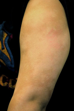
Pigment loss is no joke, especially when it appears annually in the same locations. Although it eventually clears up, this 30-year-old-woman has been worried about her condition ever since her (no doubt, well-meaning) sister-in-law suggested it might be vitiligo.
The patient had no idea what vitiligo was but soon found out when she looked it up. To add to that angst, she has been seen by any number of providers over the years and has been given about as many different diagnoses, including the ever-popular “ringworm,” psoriasis, and mere dry skin.
The patient knows she has always had exceptionally dry skin, as well as seasonal allergies. These are problems almost everyone on her mother’s side of the family has had (as well as the patient’s two siblings).
The patient’s pigment loss seems to peak in the summer, resolving almost completely by Christmas.
DISCUSSION
In general, change in pigment is called dyschromia, but when one adds or loses the normal color of the skin, it’s called either hyperchromia or hypochromia. The distinction is not merely academic, since dyschromic patients can turn a variety of colors: blue (with ingestion of silver salts or minocycline, for example), bronze (seen with Wilson’s and Addison’s diseases and hemochromatosis), brown (as in melasma), or gray (seen with administration of gold salts).
One unusual form of hypochromia is vitiligo, an autoimmune disease eventuating in sharply demarcated areas of complete pigment loss, leaving perfectly white skin in its wake. Vitiligo seldom, if ever, waxes and wanes and never involves associated scaling. But it can certainly be a cosmetic problem, often affecting the face, hands, arms, and legs.
Vitilgo can be treated with varying degrees of success, depending on how early the treatment is instituted and how aggressive the disease is. Quite often, it progresses unnoticed until it has become permanent.
Fortunately, our patient did not have vitiligo and instead had the extremely common pityriasis alba (PA). Osler, the most famous physician of his time (1900), once quipped that he could ”forgive dermatology its complexity, but never its terminology.” Pityriasis alba (PA) sounds imposing. But in fact, it represents a simple but important concept: As cutaneous inflammation subsides, the epidermis can add pigment (called postinflammatory hyperpigmentation) or lose it (postinflammatory hypopigmentation).
PA manifests with patchy partial pigment loss as a postinflammatory consequence of antecedent eczema. The latter, originating most commonly in winter, is almost always part of an overarching diagnosis of atopic dermatitis, minor diagnostic criteria for which also include dry, sensitive skin, seasonal allergies, and asthma (all inherited traits).
The usual progression is thus: Patients with xerosis (dry skin), which is worsened by the low humidity of winter and the appeal of long, hot showers, begin to develop faintly defined, round, slightly scaly patches of eczema on the sides of the face and on the arms. These are so faint that they are often missed by the patient and family until later, in the spring, when the postinflammatory loss of pigment is made more obvious by tanning of the surrounding skin. As one might expect, this is especially obvious in darker-skinned patients whose eczema has long since faded, leaving an exceedingly fine scale (if any).
When the eczema is especially active and inflamed, the annular shape and scaly surface put the inexperienced provider in mind of “ringworm” (tinea corporis). This can be ruled out with a KOH prep and brief history-taking to identify whether a potential source for fungal organisms (new pet, playmates, siblings) exists. Keep in mind that pityriasis alba is far more common than tinea corporis.
The differential diagnosis for patchy hypopigmentation also includes tinea versicolor and (in older patients) cutaneous T-cell lymphoma, which would be progressive (albeit slowly) and not seasonal. The organism responsible for tinea versicolor (the commensal yeast Malassezia furfur) needs, among other things, much sebum in order to thrive. As this is an ingredient missing in prepubescent children, it is an important point in ruling out tinea versicolor, which is almost always seasonal as well, in young patients.
On a practical level, the “treatment” of PA predominantly involves easing patients’ (and parents’) minds by giving them a firm diagnosis that does not involve “ringworm,” and a clear idea of the self-limiting nature of the problem. See below for other treatment ideas.
LEARNING POINTS
Pityriasis alba (PA) favors the sides of the face and triceps areas of both arms; it is especially common in darker-skinned children.
PA is nearly always part of atopic dermatitis, the presence of which can help to corroborate the diagnosis.
The use of sunscreen helps to prevent the darkening of surrounding skin, making PA less obvious.
Preventing the eczema in the first place, by taking short showers, using emollients soaps, and moisturizing daily, may be beneficial.
Treating the antecedent eczema early on with group IV or V steroid creams (such as desonide) is useful in decreasing hypopigmentation later on.
HISTORY
Pigment loss is no joke, especially when it appears annually in the same locations. Although it eventually clears up, this 30-year-old-woman has been worried about her condition ever since her (no doubt, well-meaning) sister-in-law suggested it might be vitiligo.
The patient had no idea what vitiligo was but soon found out when she looked it up. To add to that angst, she has been seen by any number of providers over the years and has been given about as many different diagnoses, including the ever-popular “ringworm,” psoriasis, and mere dry skin.
The patient knows she has always had exceptionally dry skin, as well as seasonal allergies. These are problems almost everyone on her mother’s side of the family has had (as well as the patient’s two siblings).
The patient’s pigment loss seems to peak in the summer, resolving almost completely by Christmas.
DISCUSSION
In general, change in pigment is called dyschromia, but when one adds or loses the normal color of the skin, it’s called either hyperchromia or hypochromia. The distinction is not merely academic, since dyschromic patients can turn a variety of colors: blue (with ingestion of silver salts or minocycline, for example), bronze (seen with Wilson’s and Addison’s diseases and hemochromatosis), brown (as in melasma), or gray (seen with administration of gold salts).
One unusual form of hypochromia is vitiligo, an autoimmune disease eventuating in sharply demarcated areas of complete pigment loss, leaving perfectly white skin in its wake. Vitiligo seldom, if ever, waxes and wanes and never involves associated scaling. But it can certainly be a cosmetic problem, often affecting the face, hands, arms, and legs.
Vitilgo can be treated with varying degrees of success, depending on how early the treatment is instituted and how aggressive the disease is. Quite often, it progresses unnoticed until it has become permanent.
Fortunately, our patient did not have vitiligo and instead had the extremely common pityriasis alba (PA). Osler, the most famous physician of his time (1900), once quipped that he could ”forgive dermatology its complexity, but never its terminology.” Pityriasis alba (PA) sounds imposing. But in fact, it represents a simple but important concept: As cutaneous inflammation subsides, the epidermis can add pigment (called postinflammatory hyperpigmentation) or lose it (postinflammatory hypopigmentation).
PA manifests with patchy partial pigment loss as a postinflammatory consequence of antecedent eczema. The latter, originating most commonly in winter, is almost always part of an overarching diagnosis of atopic dermatitis, minor diagnostic criteria for which also include dry, sensitive skin, seasonal allergies, and asthma (all inherited traits).
The usual progression is thus: Patients with xerosis (dry skin), which is worsened by the low humidity of winter and the appeal of long, hot showers, begin to develop faintly defined, round, slightly scaly patches of eczema on the sides of the face and on the arms. These are so faint that they are often missed by the patient and family until later, in the spring, when the postinflammatory loss of pigment is made more obvious by tanning of the surrounding skin. As one might expect, this is especially obvious in darker-skinned patients whose eczema has long since faded, leaving an exceedingly fine scale (if any).
When the eczema is especially active and inflamed, the annular shape and scaly surface put the inexperienced provider in mind of “ringworm” (tinea corporis). This can be ruled out with a KOH prep and brief history-taking to identify whether a potential source for fungal organisms (new pet, playmates, siblings) exists. Keep in mind that pityriasis alba is far more common than tinea corporis.
The differential diagnosis for patchy hypopigmentation also includes tinea versicolor and (in older patients) cutaneous T-cell lymphoma, which would be progressive (albeit slowly) and not seasonal. The organism responsible for tinea versicolor (the commensal yeast Malassezia furfur) needs, among other things, much sebum in order to thrive. As this is an ingredient missing in prepubescent children, it is an important point in ruling out tinea versicolor, which is almost always seasonal as well, in young patients.
On a practical level, the “treatment” of PA predominantly involves easing patients’ (and parents’) minds by giving them a firm diagnosis that does not involve “ringworm,” and a clear idea of the self-limiting nature of the problem. See below for other treatment ideas.
LEARNING POINTS
Pityriasis alba (PA) favors the sides of the face and triceps areas of both arms; it is especially common in darker-skinned children.
PA is nearly always part of atopic dermatitis, the presence of which can help to corroborate the diagnosis.
The use of sunscreen helps to prevent the darkening of surrounding skin, making PA less obvious.
Preventing the eczema in the first place, by taking short showers, using emollients soaps, and moisturizing daily, may be beneficial.
Treating the antecedent eczema early on with group IV or V steroid creams (such as desonide) is useful in decreasing hypopigmentation later on.
