User login
Six month ago, this 36-year-old man’s left fourth finger began to bother him. He’s tried topical antibiotics (triple-antibiotic cream and mupirocin), hot soaks in solutions of Epsom salts, application of colloidal silver solution, and courses of two different oral antibiotics (cephalexin and ciprofloxacin). None have provided any relief from the pain.
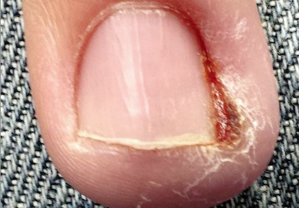
The patient admits removing a hangnail from the nail in question, causing a little bleeding. Then followed the slow onset of chronic pain and swelling, which has never been severe but is painful enough to interfere with normal activities—particularly his job, which requires extensive computer time.
EXAMINATION
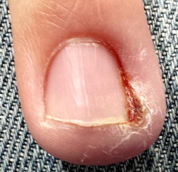
The medial perionychial area of his left fourth finger is modestly swollen and red and markedly tender to touch. No pus can be expressed from the area, and there are no palpable lymph nodes in either the epitrochlear or axillary locations of this arm. No lymphangiitic streaking is seen on the hand or arm. The affected area is a small section of the nail fold, where glistening, friable tissue is seen in the proximal invaginated area.
After a brief discussion of treatment alternatives, the decision is made to anesthetize the digit by means of a digital block, using 2 cc of 2% plain lidocaine. Once anesthesia is achieved, the hand is elevated above the chest and a tourniquet applied to minimize bleeding. The back of the patient’s hand is placed on his chest, then the swollen nail fold is pulled back. A 3-mm remnant of hangnail, still attached proximally, is revealed. It is removed and the area curetted. The site is cleaned and dressed, then the tourniquet is released.
Within 2 weeks, the finger is no longer swollen or painful.
Continue for Joe Monroe's discussion and take-home learning points >>
DISCUSSION
“Infection” is only one potential cause of redness, swelling, increased warmth, and localized pain. Classically termed rubor, tumor, calor, and dolor, these are indicators of inflammation, which can occur in many conditions besides “infection.” One tragic example is inflammatory breast cancer, which is all too often mistreated as mastitis until the lack of response to treatment finally forces—often belatedly—consideration of other items in the differential.
A far less dramatic example is the case described, in which a simple hangnail is incompletely removed, leaving a shard of nail that then digs into the perionychial skin as it grows out. This sets into motion a healing process that cannot proceed to resolution, because the tissue is re-injured every time the finger strikes the computer keyboard. This not only causes the wound to get stuck in a certain phase of healing (angioneogenesis) but also prevents completion of the process. The tissue’s response is what we see with this patient, invariably (and erroneously) called “infection.”
This is basically the identical process we see with ingrown toenails, except for the unfortunate fact that we stand upright, compress the toe with a shoe, and walk on it—all of which greatly magnify the pain, redness, and swelling. With toes more than fingers, we also tend to see the production of a button of granulated tissue at the site. This results from ongoing, inappropriate angioneogenesis. Sometimes termed pyogenic granuloma (or sclerosing hemangioma), this tissue is quite friable and bleeds copiously with any amount of trauma.
Ironically, acute bacterial paronychia of the fingers, usually caused by ordinary staph, can start in much the same way (without, of course, the retained nail shard). However, it presents with more focal concentration of redness and swelling, a collection of thick, green pus, and exquisite tenderness, all of which is relieved by simple incision and drainage.
With this patient, and those with ingrown toenails, it’s quite compelling to prescribe oral antibiotics. But these never help, and for good reason: The problem is intolerance of the “foreign body,” not infection. The solution is to “reboot” the healing process by removing the offending nail shard.
TAKE-HOME LEARNING POINTS
• While “ingrown fingernails” are far less common than ingrown toenails, both are caused by nail fragments slicing into live tissue.
• The cure is to remove the offending fragment, which allows the wound to heal.
• For ingrown toenails, an extra step is often necessary: destroying the offending segment of nail matrix with curettement and/or application of phenol.
• Anesthesia for digits should never be accomplished by local infiltration of the affected area for these kinds of procedures. Instead, employ a digital block technique, which is far less painful and provides complete anesthesia when done properly.
• Resist the urge to reflexively diagnose “infection” when confronted with redness, swelling, etc. Instead, consider other potential causes for inflammation first.
Six month ago, this 36-year-old man’s left fourth finger began to bother him. He’s tried topical antibiotics (triple-antibiotic cream and mupirocin), hot soaks in solutions of Epsom salts, application of colloidal silver solution, and courses of two different oral antibiotics (cephalexin and ciprofloxacin). None have provided any relief from the pain.
The patient admits removing a hangnail from the nail in question, causing a little bleeding. Then followed the slow onset of chronic pain and swelling, which has never been severe but is painful enough to interfere with normal activities—particularly his job, which requires extensive computer time.
EXAMINATION
The medial perionychial area of his left fourth finger is modestly swollen and red and markedly tender to touch. No pus can be expressed from the area, and there are no palpable lymph nodes in either the epitrochlear or axillary locations of this arm. No lymphangiitic streaking is seen on the hand or arm. The affected area is a small section of the nail fold, where glistening, friable tissue is seen in the proximal invaginated area.
After a brief discussion of treatment alternatives, the decision is made to anesthetize the digit by means of a digital block, using 2 cc of 2% plain lidocaine. Once anesthesia is achieved, the hand is elevated above the chest and a tourniquet applied to minimize bleeding. The back of the patient’s hand is placed on his chest, then the swollen nail fold is pulled back. A 3-mm remnant of hangnail, still attached proximally, is revealed. It is removed and the area curetted. The site is cleaned and dressed, then the tourniquet is released.
Within 2 weeks, the finger is no longer swollen or painful.
Continue for Joe Monroe's discussion and take-home learning points >>
DISCUSSION
“Infection” is only one potential cause of redness, swelling, increased warmth, and localized pain. Classically termed rubor, tumor, calor, and dolor, these are indicators of inflammation, which can occur in many conditions besides “infection.” One tragic example is inflammatory breast cancer, which is all too often mistreated as mastitis until the lack of response to treatment finally forces—often belatedly—consideration of other items in the differential.
A far less dramatic example is the case described, in which a simple hangnail is incompletely removed, leaving a shard of nail that then digs into the perionychial skin as it grows out. This sets into motion a healing process that cannot proceed to resolution, because the tissue is re-injured every time the finger strikes the computer keyboard. This not only causes the wound to get stuck in a certain phase of healing (angioneogenesis) but also prevents completion of the process. The tissue’s response is what we see with this patient, invariably (and erroneously) called “infection.”
This is basically the identical process we see with ingrown toenails, except for the unfortunate fact that we stand upright, compress the toe with a shoe, and walk on it—all of which greatly magnify the pain, redness, and swelling. With toes more than fingers, we also tend to see the production of a button of granulated tissue at the site. This results from ongoing, inappropriate angioneogenesis. Sometimes termed pyogenic granuloma (or sclerosing hemangioma), this tissue is quite friable and bleeds copiously with any amount of trauma.
Ironically, acute bacterial paronychia of the fingers, usually caused by ordinary staph, can start in much the same way (without, of course, the retained nail shard). However, it presents with more focal concentration of redness and swelling, a collection of thick, green pus, and exquisite tenderness, all of which is relieved by simple incision and drainage.
With this patient, and those with ingrown toenails, it’s quite compelling to prescribe oral antibiotics. But these never help, and for good reason: The problem is intolerance of the “foreign body,” not infection. The solution is to “reboot” the healing process by removing the offending nail shard.
TAKE-HOME LEARNING POINTS
• While “ingrown fingernails” are far less common than ingrown toenails, both are caused by nail fragments slicing into live tissue.
• The cure is to remove the offending fragment, which allows the wound to heal.
• For ingrown toenails, an extra step is often necessary: destroying the offending segment of nail matrix with curettement and/or application of phenol.
• Anesthesia for digits should never be accomplished by local infiltration of the affected area for these kinds of procedures. Instead, employ a digital block technique, which is far less painful and provides complete anesthesia when done properly.
• Resist the urge to reflexively diagnose “infection” when confronted with redness, swelling, etc. Instead, consider other potential causes for inflammation first.