User login
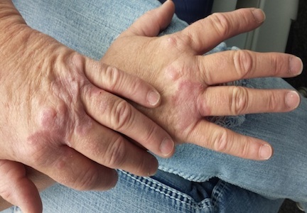
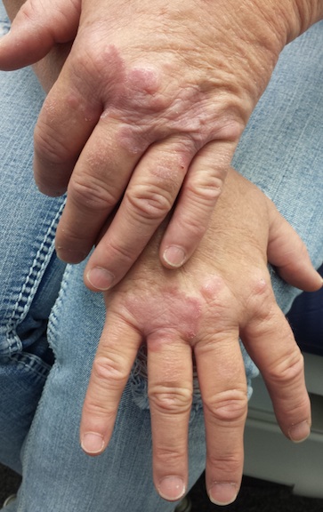
Several months ago, lesions appeared on both of this 48-year-old woman’s hands. They have been completely unresponsive to any of the treatments prescribed by her primary care provider, including oral (terbinafine) and topical antifungal medications.
The lesions, which are asymptomatic, manifested slowly. They first appeared on the dorsa of her hands and then gradually spread until they covered the central portion of both hands. The lesions wax and wane in terms of thickness and extent but never disappear.
The patient denies similar lesions elsewhere. She also denies contact with animals or children. She has never been immunosuppressed and is quite healthy aside from her skin problems. She has a family history but no personal history of diabetes.
EXAMINATION
The condition affects both hands equally. It is composed of intradermal reddish brown papules and plaques that cover the metacarpal areas and extend onto the proximal interphalangeal skin and the distal dorsa. No epidermal component (scale or other broken skin surface) is seen.
The margins of the lesions are arciform, slightly raised, and smooth. The centers of several are concave.
What is the diagnosis?
DISCUSSION
This is the classic presentation of granuloma annulare (GA), in terms of morphology, distribution, and patient gender. Though this common condition can manifest in a number of locations and in men, it tends to favor the extensor surfaces of the extremities of women. The dorsum of the foot, anterior tibial skin, and elbows are typical locations that make it a relatively easy condition to recognize.
GA can be more difficult to identify, however, when it is generalized or deeper in the dermis. The latter, termed subcutaneous GA, is most common in children, who present with large dark annular patches of discoloration too deep in the dermis to produce any palpable surface changes. The more rare generalized GA can be difficult to diagnose and treat. Biopsy of suspected generalized or subcutaneous GA is necessary to differentiate it from its lookalikes, such as secondary syphilis, lichen planus, sarcoidosis, and tinea.
The annular borders of GA lesions are notorious for deceiving the unwary practitioner into diagnosing fungal infection. But the latter is, by definition, an infection of the outermost layers of the skin on which fungal organisms feed. That process almost invariably produces significant scaling, especially on the periphery of the lesions. Such scaling is completely absent on GA lesions, which also have a papularity and induration rarely seen with superficial fungal infections. In this particular case, the patient denied any potential source for a fungal infection (animals, children) and denied the itching we would expect with one.
The cause of GA is unknown, but it appears to represent a reaction to an unknown antigen. One previous hypothesis held that it was connected to diabetes, but this was proved false.
Ordinary GA resolves on its own (though this can take months)—which is fortunate, since there is no uniformly successful treatment. Many things have been tried, including topical or intralesional steroids and cryotherapy, with varying levels of success. Generalized GA is even more difficult to treat, can linger for years, and can be highly pruritic. Treatments have included hydroxychloroquine, dapsone, potassium iodide, isotretinoin, and PUVA. More recently, biologics are being used with some success.
The bigger problem with GA is simply to consider it in the differential for odd, annular, or generalized lesions.
TAKE-HOME LEARNING POINTS
• Granuloma annulare (GA) is often mistaken for fungal infection, but it is an intradermal rather than epidermal process.
• The extensor surfaces of the extremities of women are favored sites for GA lesions.
• Biopsy of suspected GA lesions confirms the diagnosis while ruling out other items in the differential (eg, sarcoidosis and lichen planus).
• GA has no connection to diabetes.