User login
Several months ago, this 8-year-old boy began losing hair from his scalp. Other than mild itching, there are no associated symptoms. The patient has no pets at home, but he spends his after-school hours with his cousin, who does.
The child is allergy prone but otherwise healthy. No one else in the family (ie, his two younger siblings) has been similarly affected.
EXAMINATION
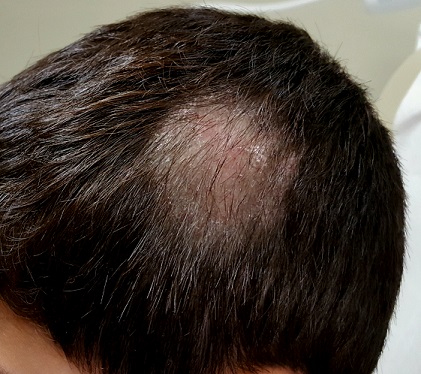
About three-quarters of the hair is missing from a 6-cm oval patch on the parietal scalp. A few short hairs remain. The skin in this area is slightly edematous, with focal areas of broken, scaly skin.
Palpation of the head and neck reveals adenopathy in the nuchal area of the affected side. A KOH prep is performed with a #10 blade; the sample includes hairs as well as skin.
What is the diagnosis?
Examination of the sample revealed endothrix, in which fungal spores and hyphae are found inside the broken-off hairs, especially near the roots. A fungal culture confirmed the presence of Trychophyton tonsurans.
T tonsurans is the most common culprit in tinea captitis cases in the United States. This dermatophytic infection of the scalp is a common diagnosis in children, who typically contract it from other children. (Some causative species—such as Microsporum audouinii—spread via contact with animals, but these organisms are generally rare in the US.) Tinea capitis is seen more frequently in boys than in girls, and African-American patients are especially at risk.
Tinea capitis infects the deep hair shaft but spares the skin. Diagnosis requires a combination of clinical signs and identification of the organism in the hair shaft; the latter will also help to guide treatment. In contrast, tinea corporis is diagnosed by clinical features and KOH examination of external scales where the organism resides. Traditionally, infected hairs have needed to be removed for KOH exam—but practical experience has shown that a vigorous scrape that captures infected hairs can accomplish the same thing.
The results of fungal culture may take a month or more to finalize; in the interim, patients such as this one may be treated with griseofulvin (10 mg/kg/d) and application of topical ciclopirox cream bid to reduce infectivity. Total clearance will take at least two months.
Tinea capitis has several forms including inflammatory (which manifests with a large, swollen, inflamed mass) and black dot (named for the tips of broken hair shafts that remain in the affected areas). The differential includes psoriasis, alopecia areata, and seborrhea.
TAKE-HOME LEARNING POINTS
- Tinea capitis is a dermatophytic infection of the scalp usually caused by the dermatophytes Trychophyton tonsurans or T rubrum.
- These infections involve the hair shaft below the skin line, rather than the surface of the skin.
- The organisms that commonly cause tinea capitis in the US typically spread through contact with another person.
- Diagnosis can be made from clinical findings only, including reactive adenopathy. KOH and culture can be necessary in questionable cases, and because of the length of treatment.
Several months ago, this 8-year-old boy began losing hair from his scalp. Other than mild itching, there are no associated symptoms. The patient has no pets at home, but he spends his after-school hours with his cousin, who does.
The child is allergy prone but otherwise healthy. No one else in the family (ie, his two younger siblings) has been similarly affected.
EXAMINATION
About three-quarters of the hair is missing from a 6-cm oval patch on the parietal scalp. A few short hairs remain. The skin in this area is slightly edematous, with focal areas of broken, scaly skin.
Palpation of the head and neck reveals adenopathy in the nuchal area of the affected side. A KOH prep is performed with a #10 blade; the sample includes hairs as well as skin.
What is the diagnosis?
Examination of the sample revealed endothrix, in which fungal spores and hyphae are found inside the broken-off hairs, especially near the roots. A fungal culture confirmed the presence of Trychophyton tonsurans.
T tonsurans is the most common culprit in tinea captitis cases in the United States. This dermatophytic infection of the scalp is a common diagnosis in children, who typically contract it from other children. (Some causative species—such as Microsporum audouinii—spread via contact with animals, but these organisms are generally rare in the US.) Tinea capitis is seen more frequently in boys than in girls, and African-American patients are especially at risk.
Tinea capitis infects the deep hair shaft but spares the skin. Diagnosis requires a combination of clinical signs and identification of the organism in the hair shaft; the latter will also help to guide treatment. In contrast, tinea corporis is diagnosed by clinical features and KOH examination of external scales where the organism resides. Traditionally, infected hairs have needed to be removed for KOH exam—but practical experience has shown that a vigorous scrape that captures infected hairs can accomplish the same thing.
The results of fungal culture may take a month or more to finalize; in the interim, patients such as this one may be treated with griseofulvin (10 mg/kg/d) and application of topical ciclopirox cream bid to reduce infectivity. Total clearance will take at least two months.
Tinea capitis has several forms including inflammatory (which manifests with a large, swollen, inflamed mass) and black dot (named for the tips of broken hair shafts that remain in the affected areas). The differential includes psoriasis, alopecia areata, and seborrhea.
TAKE-HOME LEARNING POINTS
- Tinea capitis is a dermatophytic infection of the scalp usually caused by the dermatophytes Trychophyton tonsurans or T rubrum.
- These infections involve the hair shaft below the skin line, rather than the surface of the skin.
- The organisms that commonly cause tinea capitis in the US typically spread through contact with another person.
- Diagnosis can be made from clinical findings only, including reactive adenopathy. KOH and culture can be necessary in questionable cases, and because of the length of treatment.