User login
A 79-year-old man presents for a routine skin check, in the context of his 40-year history of nonmelanoma skin cancer. He grew up on a farm and then became a farmer himself, spending almost every day in the sun (usually without a hat). His skin burned easily but would take on a “tan” by the start of summer.
In the succeeding years, he developed so many skin cancers (and had them removed) that he lost count. All were basal cell or squamous cell carcinomas, predominately manifesting on his face, arms, and ears. Several required Mohs surgery for removal.

EXAMINATION
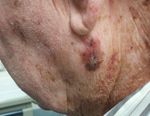
Abundant evidence of excessive sun exposure is seen on the patient’s skin: actinic keratoses on the forehead, ears, and neck, and multiple solar lentigines on the face, neck, and arms. On his left neck, below the ear, is a large, oddly pigmented, dark macular patch. Dermatoscopic examination reveals focal pigmentary clumping and streaming, which prompts the decision to perform an incisional biopsy. The darkest and most irregular part of the lesion is taken as a sample. The pathology report shows lentigo maligna.
What is lentigo maligna?
DISCUSSION
Lentigo maligna (LM), also known as Hutchinson freckle, is a type of melanoma in situ that is typically seen on sun-exposed skin. It has indistinct margins with predominantly brown and black coloration. LM is usually seen on older, mostly fair-skinned patients who have a history of extensive sun exposure. Its preferred sites include the face, ears, neck, and upper extremities.
LM is, by definition, entirely superficial and therefore safe. Its main significance is that it can become focally invasive to the dermis, a phenomenon termed lentigo maligna melanoma (LMM). When that occurs, the clusters of spindle-shaped atypical cells can then progress to a vertical growth phase, resulting in intravascular invasion that eventuates in metastasis.
LMs grow so slowly that they often escape detection; they are sometimes mistaken for solar lentigines (SL). They can “collide” with SLs or other benign lesions (eg, seborrheic keratoses), which can effectively camouflage them. Suspicion is usually triggered by change (color, size) in a lesion.
Biopsy is the only method of detection. In this case, the central portion of this large, oddly pigmented and bordered, multicolored patch was excised and the darkest, most irregular part of the lesion collected. This is the gold standard for biopsy of a potential melanoma. A large deep shave biopsy (“saucerization”) would have accomplished the same thing; studies show that neither process will cause metastasis. The main mistake to avoid is performing a single punch biopsy, which risks a false-negative result.
The definitive surgical approach is to remove the lesion with a 1-cm margin around it and into the underlying adipose layer. The wound can either be left to heal by secondary intention or closed primarily. Either way, this patient’s prognosis is excellent, unless the final pathology report indicates focal invasion. With a patient of this age, the LM could have been left alone—although, once discovered, LM is irresistibly compelling to treat.
TAKE-HOME LEARNING POINTS
• Lentigo maligna (LM) is a type of melanoma in situ with the potential to progress to lentigo maligna melanoma, an invasive form of early melanoma.
• LM is common on sun-exposed skin of older patients with a history of excessive sun exposure.
• Faces, ears, arms, and necks are common areas for LM to manifest.
• Biopsy of suspected melanoma should incorporate a significant portion of the darkest, most irregular part of the lesion; it can be done by incisional technique or deep saucerization.
• LM is also known as Hutchinson freckle, since it often resembles a large, irregularly bordered and pigmented freckle.
A 79-year-old man presents for a routine skin check, in the context of his 40-year history of nonmelanoma skin cancer. He grew up on a farm and then became a farmer himself, spending almost every day in the sun (usually without a hat). His skin burned easily but would take on a “tan” by the start of summer.
In the succeeding years, he developed so many skin cancers (and had them removed) that he lost count. All were basal cell or squamous cell carcinomas, predominately manifesting on his face, arms, and ears. Several required Mohs surgery for removal.
EXAMINATION
Abundant evidence of excessive sun exposure is seen on the patient’s skin: actinic keratoses on the forehead, ears, and neck, and multiple solar lentigines on the face, neck, and arms. On his left neck, below the ear, is a large, oddly pigmented, dark macular patch. Dermatoscopic examination reveals focal pigmentary clumping and streaming, which prompts the decision to perform an incisional biopsy. The darkest and most irregular part of the lesion is taken as a sample. The pathology report shows lentigo maligna.
What is lentigo maligna?
DISCUSSION
Lentigo maligna (LM), also known as Hutchinson freckle, is a type of melanoma in situ that is typically seen on sun-exposed skin. It has indistinct margins with predominantly brown and black coloration. LM is usually seen on older, mostly fair-skinned patients who have a history of extensive sun exposure. Its preferred sites include the face, ears, neck, and upper extremities.
LM is, by definition, entirely superficial and therefore safe. Its main significance is that it can become focally invasive to the dermis, a phenomenon termed lentigo maligna melanoma (LMM). When that occurs, the clusters of spindle-shaped atypical cells can then progress to a vertical growth phase, resulting in intravascular invasion that eventuates in metastasis.
LMs grow so slowly that they often escape detection; they are sometimes mistaken for solar lentigines (SL). They can “collide” with SLs or other benign lesions (eg, seborrheic keratoses), which can effectively camouflage them. Suspicion is usually triggered by change (color, size) in a lesion.
Biopsy is the only method of detection. In this case, the central portion of this large, oddly pigmented and bordered, multicolored patch was excised and the darkest, most irregular part of the lesion collected. This is the gold standard for biopsy of a potential melanoma. A large deep shave biopsy (“saucerization”) would have accomplished the same thing; studies show that neither process will cause metastasis. The main mistake to avoid is performing a single punch biopsy, which risks a false-negative result.
The definitive surgical approach is to remove the lesion with a 1-cm margin around it and into the underlying adipose layer. The wound can either be left to heal by secondary intention or closed primarily. Either way, this patient’s prognosis is excellent, unless the final pathology report indicates focal invasion. With a patient of this age, the LM could have been left alone—although, once discovered, LM is irresistibly compelling to treat.
TAKE-HOME LEARNING POINTS
• Lentigo maligna (LM) is a type of melanoma in situ with the potential to progress to lentigo maligna melanoma, an invasive form of early melanoma.
• LM is common on sun-exposed skin of older patients with a history of excessive sun exposure.
• Faces, ears, arms, and necks are common areas for LM to manifest.
• Biopsy of suspected melanoma should incorporate a significant portion of the darkest, most irregular part of the lesion; it can be done by incisional technique or deep saucerization.
• LM is also known as Hutchinson freckle, since it often resembles a large, irregularly bordered and pigmented freckle.