User login
Wellness vacations
It’s best practice to not set an alarm when on vacation. The point of vacation, after all, is to escape the rock-hard constraints of the daily grind. But the melody pulling me from slumber wasn’t coming from my phone. It was the ethereal chant of Fajr, morning prayer rising from surrounding mosques. I was in the medina of Marrakesh sleeping in a hotel that was once a home, called a riad. Some parts of the building date from the medieval period. Fajr occurs at dawn, before morning light. Getting from the bed to the toilet was treacherous – you must traverse cold, uneven steps to get there. Yet I had to get dressed: Morning yoga on our riad rooftop would start with sunrise. I guess even my vacations have agendas: I was in Morocco not only to holiday, but also to improve mind, body, and spirit.

For those who can afford them, your travel is fully arranged and activities such as yoga, cooking classes, hikes, and meditation are scheduled. This was my first wellness trip and it was transformative. Many times have I tried to disconnect from the distractions of life, but there is nothing so purifying as having no phone or Internet access. When I made peace with the reality that I couldn’t access EPIC or email, it was like a ringing in the ears had lifted: I could hear silence again.
This trip took us to three locations: Marrakesh, the Atlas Mountains, and the edge of the Sahara Desert. Yoga was prescribed twice a day. Morning practice was 90 minutes of shedding layers as the sun rose and our bodies warmed to increasingly difficult sequences. This was followed by Moroccan breakfast with fellow travelers from around the world. All were professionals and I wasn’t surprised to learn that burnout is common to many. I was surprised to realize that sharing stories with strangers about the vicissitudes of life was deeply bonding. (Or perhaps it was doing yoga inversions together.)
Also surprising was how easy it is to get lost in the maze that is Marrakesh. And yet, it was rewarding. Finding our way back through the mass of people, donkeys, and motorbikes along dark, unmarked alleys – without Waze – was intensely clarifying. Few things help you be present “in the moment” as being adrift and disoriented in a foreign city.
There was relaxation too. We made Khobz, traditional Moroccan bread by mixing just the right amounts of flour, yeast, sugar, oil, water, and salt. Knead, add, knead, add, and stop when done. We then walked a half mile to give our doughy creations to a baker who, with blackened calloused hands, worked an ancient communal oven. Then we waited patiently for the sardines ahead of us to finish baking first. I’ve no idea how long it all took – I had nowhere else to be.
The next day we hiked to a village in the Ourika Valley. There we had lunch at the home of a local Berber family. They served us their best tea, vegetable couscous, and lamb tagine while their chickens and donkeys watched us curiously. It was Thanksgiving (not on the Berber calendar of course) and sharing a meal prepared by a faraway stranger who doesn’t speak English makes you feel thankful in a refreshing way. Way more alike than different we are, I learned.
We finished our trip with a little desert “glamping.” The vast expanse of desert, interrupted by swirling winds and camel bellows quiets your mind, opens you to the immensity of life. That night we sat close to a bonfire and watched the Milky Way drift across the true black sky. I woke the next morning to the best night’s sleep I’ve had all year. My last wellness activity was unplanned, but meaningful nonetheless. As it happens, there’s no hot water in the desert and a bracingly cold shower marked the end of my treatment/vacation.
If the opposite of burned out is repleted, then I am. Also grateful to have such a transformative experience, for friends new and old who love me, and for hot water. Prescribe yourself one if you can.
Dr. Benabio is director of Healthcare Transformation and chief of dermatology at Kaiser Permanente San Diego. The opinions expressed in this column are his own and do not represent those of Kaiser Permanente. Dr. Benabio is @Dermdoc on Twitter. Write to him at dermnews@mdedge.com.
It’s best practice to not set an alarm when on vacation. The point of vacation, after all, is to escape the rock-hard constraints of the daily grind. But the melody pulling me from slumber wasn’t coming from my phone. It was the ethereal chant of Fajr, morning prayer rising from surrounding mosques. I was in the medina of Marrakesh sleeping in a hotel that was once a home, called a riad. Some parts of the building date from the medieval period. Fajr occurs at dawn, before morning light. Getting from the bed to the toilet was treacherous – you must traverse cold, uneven steps to get there. Yet I had to get dressed: Morning yoga on our riad rooftop would start with sunrise. I guess even my vacations have agendas: I was in Morocco not only to holiday, but also to improve mind, body, and spirit.
For those who can afford them, your travel is fully arranged and activities such as yoga, cooking classes, hikes, and meditation are scheduled. This was my first wellness trip and it was transformative. Many times have I tried to disconnect from the distractions of life, but there is nothing so purifying as having no phone or Internet access. When I made peace with the reality that I couldn’t access EPIC or email, it was like a ringing in the ears had lifted: I could hear silence again.
This trip took us to three locations: Marrakesh, the Atlas Mountains, and the edge of the Sahara Desert. Yoga was prescribed twice a day. Morning practice was 90 minutes of shedding layers as the sun rose and our bodies warmed to increasingly difficult sequences. This was followed by Moroccan breakfast with fellow travelers from around the world. All were professionals and I wasn’t surprised to learn that burnout is common to many. I was surprised to realize that sharing stories with strangers about the vicissitudes of life was deeply bonding. (Or perhaps it was doing yoga inversions together.)
Also surprising was how easy it is to get lost in the maze that is Marrakesh. And yet, it was rewarding. Finding our way back through the mass of people, donkeys, and motorbikes along dark, unmarked alleys – without Waze – was intensely clarifying. Few things help you be present “in the moment” as being adrift and disoriented in a foreign city.
There was relaxation too. We made Khobz, traditional Moroccan bread by mixing just the right amounts of flour, yeast, sugar, oil, water, and salt. Knead, add, knead, add, and stop when done. We then walked a half mile to give our doughy creations to a baker who, with blackened calloused hands, worked an ancient communal oven. Then we waited patiently for the sardines ahead of us to finish baking first. I’ve no idea how long it all took – I had nowhere else to be.
The next day we hiked to a village in the Ourika Valley. There we had lunch at the home of a local Berber family. They served us their best tea, vegetable couscous, and lamb tagine while their chickens and donkeys watched us curiously. It was Thanksgiving (not on the Berber calendar of course) and sharing a meal prepared by a faraway stranger who doesn’t speak English makes you feel thankful in a refreshing way. Way more alike than different we are, I learned.
We finished our trip with a little desert “glamping.” The vast expanse of desert, interrupted by swirling winds and camel bellows quiets your mind, opens you to the immensity of life. That night we sat close to a bonfire and watched the Milky Way drift across the true black sky. I woke the next morning to the best night’s sleep I’ve had all year. My last wellness activity was unplanned, but meaningful nonetheless. As it happens, there’s no hot water in the desert and a bracingly cold shower marked the end of my treatment/vacation.
If the opposite of burned out is repleted, then I am. Also grateful to have such a transformative experience, for friends new and old who love me, and for hot water. Prescribe yourself one if you can.
Dr. Benabio is director of Healthcare Transformation and chief of dermatology at Kaiser Permanente San Diego. The opinions expressed in this column are his own and do not represent those of Kaiser Permanente. Dr. Benabio is @Dermdoc on Twitter. Write to him at dermnews@mdedge.com.
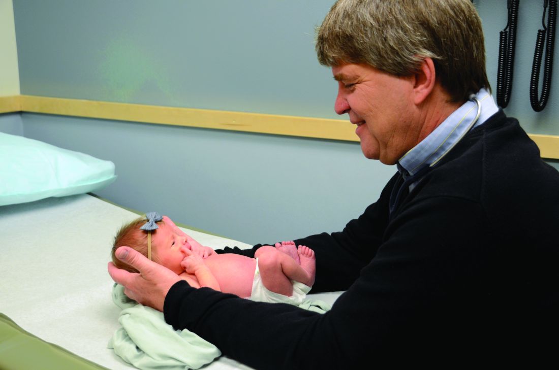
Obstetrical care in crisis
For the last 25 years I have had the privilege of caring for a rural community, practicing full-scope family medicine including obstetrics with cesarean sections. I have had a deeply rewarding career, and delivering babies and watching them grow up has been one of the most gratifying parts of my work.
My concern is that, as the number of family physicians who practice maternity care has decreased, the infant and maternal mortality rate in the United States has increased, especially in rural and minority populations. Currently, 5 million women of reproductive age have no access to maternity care.
At the same time 23% of incoming family medicine residents would like to offer maternity care and are trained to do so, but few are able to find a job where this is possible.1 This is unfortunate because family physicians have the training and expertise to provide comprehensive maternity care. Although they have lower rates of cesarean section than ob-gyns, with similar outcomes, family physicians do have surgical skills, including providing cesarean sections, that are often necessary for safe delivery.2,3
In addition, family physicians have the internal medicine and behavioral health background to care for postpartum complications, as well as substance use disorders. Because they also care for children, they see postpartum women when they come in with their children for well-child checks. These visits offer an excellent opportunity to also check on the mother for postpartum depression and other signs of postpartum illness.
Centers for Disease Control and Prevention data reveal that maternal mortality can be divided into three nearly equal parts: pregnancy, delivery, and post partum. They define delivery as the week of delivery. The 48 hours post delivery accounted for only 12% of overall mortality. This means that even if women travel to metropolitan areas, they are likely to be home when they have fatal complications. The lack of trained and experienced physicians in the communities where women live increases their risks should they have complications. Most maternal fatalities occur when conditions are not recognized in a timely fashion. Some responses require procedural skills such as dilation and curettage (D&C).
As a member of the National Advisory Committee on Rural Health and Human Services, I visited several states to evaluate their rural health systems. We looked at infant mortality by county and found an enormous disparity between counties, largely caused by lack of prenatal services and obstetrical services.
These disparities between counties are getting worse. The United States is losing critical access hospitals at a rapid pace. We have lost 117 critical access hospitals in the last 10 years, with 40 in the last year alone. According to the National Rural Health Association, 4,673 additional facilities – representing more than one-third of rural hospitals in the United States – are vulnerable and could close. The reasons are multiple, but the result has been an erosion of the rural safety net, especially with regard to maternity care.
These hospital closures force women to travel farther distances for maternity care, including cesarean sections, and this contributes to increased maternal and infant mortality.5 In a study from Canada, the complication rates increased substantially as distances increased. Women are more likely to have premature deliveries, deliver on the side of the road, or end up in inappropriate facilities.
The distance from delivery is directly related to outcomes. A study from the early 1990s showed that women did better if they received maternity care from local hospitals and physicians.6 From a family medicine perspective, this makes sense because traveling to a metropolitan area means isolation from family and social networks. Stress increases because pregnant women also are often the primary caregiver of other children and the primary wage earner of the family. Although we are unsure what impact stress has on pregnancy, we do know it does have an effect on greater risk of prematurity and poor outcomes.
Obstetricians provide excellent care, but they are not a panacea. Only half of U.S. counties have adequate ob.gyn. coverage. Moreover, in many of those counties, the ob.gyns. subspecialize in gynecologic surgery and infertility, but don’t provide obstetrical care. Another challenge: ob.gyns. cannot survive financially in smaller communities; our policies must include incentives to recruit and retain them in underserved areas.
Certified nurse midwives also provide excellent care and are an invaluable member of the patient-care team, but again, they cannot be the only solution. Obstetrical emergencies do occur, and mothers need a physician trained in providing on-site medical or surgical care. They also need a hospital with adequate staff to care for emergencies.
In communities large enough to support a multispecialty group, certified medical technicians, family physicians, and ob.gyns. would ideally work alongside each other. In small communities four family physicians can provide a high level of maternity care including surgical deliveries, while supporting themselves with caring for children and elders in clinics, hospitals, and EDs.
It is unconscionable that a country as wealthy as ours would accept rates of maternal and infant mortality that rival and are often worse than developing countries. Although the reasons are many, there is no excuse. Family physicians are an essential part of reversing this trend. We need policies that enable family physicians to help resolve the shortage of maternity care for underserved communities, to address the maternal and infant mortality rate, and to provide maternity care that is part of family medicine’s full scope of practice.
Dr. Cullen is board chair of the American Academy of Family Physicians and a practicing family physician in Valdez, Alaska.
References
1. Am Board Fam Med. 2017 Jul-Aug;30(4):405-6.
2. CMAJ. 2015 Oct 27;187:1125-32.
3. J Am Board Fam Med. 2013 Jul-Aug;26(4):366-72.
4. NRHA Save Rural Hospitals Action Center. www.ruralhealthweb.org/advocate/save-rural-hospitals.
5. BMC Health Serv Res. 2011 Jun 10;11:147.
6. Am J Public Health. 1990 Jul;80(7):814-8.
Novel analysis links insomnia to first-onset major depressive disorder
a prospective study of 768 adults with a history of depression suggests.
Insomnia has been identified as a risk factor for depression, but the impact of lifetime depression history and the role of insomnia in major depressive disorder (MDD) remains unclear, wrote Tessa Blanken, MSc, of the Netherlands Institute for Neuroscience, Amsterdam, and colleagues. Studies of this relationship have been hampered by the difficulty of isolating the impact of insomnia as an independent predictor of MDD from depression and other disorders.
In a study published in Sleep, the researchers reviewed data from 768 adults aged 18-65 years who were participants in the Netherlands Study of Depression and Anxiety, a multicenter, longitudinal study that included four assessments over 6 years. The participants had no current or prior diagnosis of MDD. The average age of the participants was 41 years, and 63% were women.
The investigators used Network Outcome Analysis to study the link between insomnia and MDD. The investigators wrote, “Network modeling techniques provide a unique framework to study the interactions among symptoms and their role in the development and maintenance of psychiatric disorders. Using network analysis we can estimate the unique association between pairs of symptoms, while controlling for the state and associations of all other symptoms.”
Over 6-years’ follow-up, 141 participants (18%) were diagnosed with first-onset MDD. Overall, insomnia severity was a significant predictor of first-onset MDD (hazard ratio 1.11, 95% confidence interval). The analysis showed that the predictive effect of insomnia on first-onset MDD was driven solely by the item “Did you have trouble falling asleep” (hazard ratio, 1.33; 95% confidence interval, 1.12-1.57; observed range, 0-4). Those individuals who had trouble falling asleep 3-4 times or more than 4 times a week were 2.3 or 3.2 times, respectively, more likely to develop first-onset MDD. None of the other sleep complaints, such as nocturnal and early morning awakening, significantly increased the risk of first-onset MDD.
The study findings were limited by several factors including the full impact of short sleep duration and lack of chronotype assessment, the researchers noted. However, “the identification of ‘difficulty initiating sleep’ as a risk factor is particularly promising because a recent meta-analysis showed that cognitive behavioural therapy, the treatment of choice for insomnia, is highly effective,” the researchers wrote. The results suggest that treating problems in sleep initiation could contribute to preventing first-onset depression and reducing the overall burden of MDD, they concluded.
The study was supported by the European Research Council. The researchers had no financial conflicts to disclose.
SOURCE: Blanken TF et al. Sleep. 2019 Dec 2. doi: 10.1093/sleep/zsz288.
FROM SLEEP
ctDNA, CTCs predict TNBC relapse – now what?
SAN ANTONIO – The presence of circulating tumor DNA (ctDNA) and to a lesser degree circulating tumor cells (CTCs) following neoadjuvant chemotherapy for triple-negative breast cancer (TNBC) augurs poor prognosis, but it’s still unclear how this information can inform therapy.

An analysis of plasma samples from 142 women enrolled in a trial that compared outcomes of genomically directed therapy with those of physician’s choice of a regimen for treatment of TNBC showed that, at a median follow-up of 17.2 months, detection of ctDNA was significantly associated with worse distant disease-free survival and the risk of relapse was especially high for patients with both detectable ctDNA and CTCs, reported Milan Radovich, PhD, from Indiana University in Indianapolis.
“Patients with TNBC at high clinical risk of relapse due to incomplete response to neoadjuvant chemotherapy can be further risk-stratified based on the presence of [minimal residual disease] as determined by ctDNA,” he said at the San Antonio Breast Cancer Symposium.
As previously reported, the presence of detectable ctDNA after surgery for breast cancer is a significant predictor of inferior disease-free survival (DFS), but the findings, while clinically valid, are not yet applicable to clinical care, Dr. Radovich acknowledged in a briefing shortly before his presentation of the data in a general session.
As part of the BRE12-158 trial, Dr. Radovich and colleagues conducted a prospective analysis of patients with early-stage TNBC who had significant residual disease at the time of surgery, despite having undergone neoadjuvant chemotherapy. The tumors were genomically sequenced and the patients were then randomized to receive either genomocially directed therapy or the treating physician’s choice of a regimen.
Of 177 evaluable patients, a total of 142 had successful ctDNA results and 123 had successful CTC testing. The ctDNA samples were sequenced using a liquid assay for 70 oncogenes commonly mutated in cancer. CTCs were isolated from peripheral blood with a positive-selection microfluidic device. CTC-positivity was defined at detection of one or more cells.
The investigators found that ctDNA was significantly associated with distant DFS, with a median of 32.5 months for ctDNA-positive patients versus median not reached for ctDNA-negative patients. The respective 2-year distant DFS rates were 56% and 81%, respectively. The hazard ratio for worse distant DFS among ctDNA-positive patients was 2.99 (P = .0055).
Similarly, DFS was significantly inferior among ctDNA-positive patients, with a median of 22.8 months versus not reached for ctDNA-negative patients, and median 2-year DFS rates of 50% and 76%. The HR for worse survival in ctDNA-positive patients was 2.67 (P = .0069).
Overall survival (OS) was also worse for ctDNA-positive patients; although the median OS was not reached in either positive or negative patients, the 2-year OS rates were 57% among ctDNA-positive patients and 80% among negative patients. The HR for death among the ctDNA-positive patients was 4.16 (P = .0024).
CTC detection, in contrast, showed a trend toward a significant association with clinical outcomes, but neither distant DFS, DFS, nor OS measures achieved statistical significance.
However, when ctDNA and CTC were combined, the results showed significantly inferior distant DFS for patients positive for both markers, compared with that of patients who were ctDNA positive but CTC negative (median, 32.5 months vs. not reached; 2 year distant DFS, 52% vs. 89%; HR, 5.29; P = .0095).
Similarly, this group of patients had worse DFS (median, 20.8 months vs. not reached; 2-year DFS, 43% vs. 80%; HR, 3.15; P = .0370) and worse OS (median, OS not reached in either group; 2-year OS, 51% vs. 76%; HR, 8.60; P = .0074).
To take advantage of these data, the investigators are preparing to launch a phase 2 trial (BRE18-334) in which patients with residual disease are randomized by ctDNA status (positive or negative) to either genetically directed therapy or therapy with no genomic target. The patients randomized to genomically directed therapy will be treated according to mutation category.
For example, capecitabine will given plus either a PARP (poly [ADP-ribose] polymerase) inhibitor for patients with mutations in DNA repair pathways, atezolizumab (Tecentriq) for patients with mutations in the immunotherapy pathway, ipatasertib for mutations in the P13K/AKT/mTOR pathway, or a PARP inhibitor plus atezolizumab for patients with mutations in the both DNA repair immunotherapy pathways.
At the briefing, moderator Virginia Kaklamani, MD, DSc, from University of Texas Health San Antonio, commented that the results of the study can inform clinical research, but “we don’t have predictive data as to how we’re going to act on this and improve patient outcomes. What we need is exactly the trial that the investigators have designed to see whether these results can then further help us in treating our patients and improving their outcomes.”
“Should we be doing this on Monday in clinic, and the answer is no,” Dr. Radovich agreed. “What we don’t know is if having this information is really going to improve outcomes.”
In an interview, senior author Bryan P. Schneider, MD from Indiana University School of Medicine put it this way: “I’ve always believed that you don’t order a test until you know what to do with it,” he said.
The study was funded by the Vera Bradley Foundation for Breast Cancer and the Indiana University Grand Challenge Precision Health Initiative. Dr. Radovich and Dr. Schneider reported no relevant disclosures. Dr. Kaklamani disclosed speakers bureau activities and consulting for multiple companies, as well as research funding from Eiasi.
SOURCE: Radovich M. SABCS 2019, Abstract GS5-02.
SAN ANTONIO – The presence of circulating tumor DNA (ctDNA) and to a lesser degree circulating tumor cells (CTCs) following neoadjuvant chemotherapy for triple-negative breast cancer (TNBC) augurs poor prognosis, but it’s still unclear how this information can inform therapy.
An analysis of plasma samples from 142 women enrolled in a trial that compared outcomes of genomically directed therapy with those of physician’s choice of a regimen for treatment of TNBC showed that, at a median follow-up of 17.2 months, detection of ctDNA was significantly associated with worse distant disease-free survival and the risk of relapse was especially high for patients with both detectable ctDNA and CTCs, reported Milan Radovich, PhD, from Indiana University in Indianapolis.
“Patients with TNBC at high clinical risk of relapse due to incomplete response to neoadjuvant chemotherapy can be further risk-stratified based on the presence of [minimal residual disease] as determined by ctDNA,” he said at the San Antonio Breast Cancer Symposium.
As previously reported, the presence of detectable ctDNA after surgery for breast cancer is a significant predictor of inferior disease-free survival (DFS), but the findings, while clinically valid, are not yet applicable to clinical care, Dr. Radovich acknowledged in a briefing shortly before his presentation of the data in a general session.
As part of the BRE12-158 trial, Dr. Radovich and colleagues conducted a prospective analysis of patients with early-stage TNBC who had significant residual disease at the time of surgery, despite having undergone neoadjuvant chemotherapy. The tumors were genomically sequenced and the patients were then randomized to receive either genomocially directed therapy or the treating physician’s choice of a regimen.
Of 177 evaluable patients, a total of 142 had successful ctDNA results and 123 had successful CTC testing. The ctDNA samples were sequenced using a liquid assay for 70 oncogenes commonly mutated in cancer. CTCs were isolated from peripheral blood with a positive-selection microfluidic device. CTC-positivity was defined at detection of one or more cells.
The investigators found that ctDNA was significantly associated with distant DFS, with a median of 32.5 months for ctDNA-positive patients versus median not reached for ctDNA-negative patients. The respective 2-year distant DFS rates were 56% and 81%, respectively. The hazard ratio for worse distant DFS among ctDNA-positive patients was 2.99 (P = .0055).
Similarly, DFS was significantly inferior among ctDNA-positive patients, with a median of 22.8 months versus not reached for ctDNA-negative patients, and median 2-year DFS rates of 50% and 76%. The HR for worse survival in ctDNA-positive patients was 2.67 (P = .0069).
Overall survival (OS) was also worse for ctDNA-positive patients; although the median OS was not reached in either positive or negative patients, the 2-year OS rates were 57% among ctDNA-positive patients and 80% among negative patients. The HR for death among the ctDNA-positive patients was 4.16 (P = .0024).
CTC detection, in contrast, showed a trend toward a significant association with clinical outcomes, but neither distant DFS, DFS, nor OS measures achieved statistical significance.
However, when ctDNA and CTC were combined, the results showed significantly inferior distant DFS for patients positive for both markers, compared with that of patients who were ctDNA positive but CTC negative (median, 32.5 months vs. not reached; 2 year distant DFS, 52% vs. 89%; HR, 5.29; P = .0095).
Similarly, this group of patients had worse DFS (median, 20.8 months vs. not reached; 2-year DFS, 43% vs. 80%; HR, 3.15; P = .0370) and worse OS (median, OS not reached in either group; 2-year OS, 51% vs. 76%; HR, 8.60; P = .0074).
To take advantage of these data, the investigators are preparing to launch a phase 2 trial (BRE18-334) in which patients with residual disease are randomized by ctDNA status (positive or negative) to either genetically directed therapy or therapy with no genomic target. The patients randomized to genomically directed therapy will be treated according to mutation category.
For example, capecitabine will given plus either a PARP (poly [ADP-ribose] polymerase) inhibitor for patients with mutations in DNA repair pathways, atezolizumab (Tecentriq) for patients with mutations in the immunotherapy pathway, ipatasertib for mutations in the P13K/AKT/mTOR pathway, or a PARP inhibitor plus atezolizumab for patients with mutations in the both DNA repair immunotherapy pathways.
At the briefing, moderator Virginia Kaklamani, MD, DSc, from University of Texas Health San Antonio, commented that the results of the study can inform clinical research, but “we don’t have predictive data as to how we’re going to act on this and improve patient outcomes. What we need is exactly the trial that the investigators have designed to see whether these results can then further help us in treating our patients and improving their outcomes.”
“Should we be doing this on Monday in clinic, and the answer is no,” Dr. Radovich agreed. “What we don’t know is if having this information is really going to improve outcomes.”
In an interview, senior author Bryan P. Schneider, MD from Indiana University School of Medicine put it this way: “I’ve always believed that you don’t order a test until you know what to do with it,” he said.
The study was funded by the Vera Bradley Foundation for Breast Cancer and the Indiana University Grand Challenge Precision Health Initiative. Dr. Radovich and Dr. Schneider reported no relevant disclosures. Dr. Kaklamani disclosed speakers bureau activities and consulting for multiple companies, as well as research funding from Eiasi.
SOURCE: Radovich M. SABCS 2019, Abstract GS5-02.
REPORTING FROM SABCS 2019
Fast, aggressive eczema treatment linked to fewer food allergies by age 2
Researchers in Japan report that
For their research published in the Journal of Allergy and Clinical Immunology: In Practice, Yumiko Miyaji, MD, PhD, of Japan’s National Center for Child Health and Development in Tokyo and colleagues looked at 3 years’ worth of records for 177 infants younger than 1 year of age seen at a hospital allergy center for eczema. Of these infants, 89 were treated with betamethasone valerate within 4 months of disease onset, and 88 were treated after 4 months of onset. Most (142) were followed-up at 22-24 months, when all were in complete remission or near remission from eczema.
At follow-up, clinicians collected information about anaphylactic reactions to food, administered specific food challenges, and tested serum immunoglobin E levels for food allergens. Dr. Miyaji and colleagues found a significant difference in the prevalence of allergies between the early-treated and late-treated groups to chicken egg, cow’s milk, wheat, peanuts, soy, or fish (25% vs. 46%, respectively; P equal to .013). For individual food allergies, only chicken egg was associated with a statistically significant difference in prevalence (15% vs 36%, P equal to .006).
“Our present study may be the first to demonstrate that early aggressive topical corticosteroid treatment to shorten the duration of eczema in infants was significantly associated with a decrease in later development of [food allergies],” Dr. Miyaji and colleagues wrote in their analysis.
The investigators acknowledged as limitations of their study some between-group differences at baseline, with characteristics such as Staphylococcus aureus infections and some inflammatory biomarkers higher in the early treatment group.
The Japan Agency for Medical Research and Development supported the study, and the investigators disclosed no conflicts of interest related to their findings.
SOURCE: Miyaji Y et al. J Allergy Clin Immunol Pract. 2019. doi: 10.1016/j.jaip.2019.11.036
FROM THE JOURNAL OF ALLERGY AND CLINICAL IMMUNOLOGY: IN PRACTICE
ENGAGE AF-TIMI: Insulin linked to greater risk for stroke, CV death, bleeding
LOS ANGELES – Patients with diabetes had significantly higher adjusted risk of bleeding, cardiovascular-related death, and poorer net outcomes, particularly those treated with insulin, a subanalysis of the ENGAGE AF-TIMI 48 trial has shown.

In addition, the pharmacokinetic and pharmacodynamic profile of the study drug, edoxaban – a novel oral anticoagulant drug and a direct factor Xa inhibitor – was generally similar in patients with and without diabetes.
“We know that atrial fibrillation is associated with a fivefold increased risk of stroke,” Anna Plitt, MD, said at the World Congress on Insulin Resistance, Diabetes & Cardiovascular Disease. “Type 2 diabetes is associated with a twofold increased risk of stroke, and longer duration of diabetes is associated with even higher ischemic event rates. The coexistence of [atrial fibrillation] and type 2 diabetes further increases thromboembolic risk.”
Dr. Plitt, a cardiology fellow at Mount Sinai Hospital, New York, noted that, although type 2 diabetes is characterized by a prothrombotic and inflammatory state, the mechanism of action by which hyperglycemia and/or insulin resistance leads to the development of atrial fibrillation (AFib) remains unknown. “Given the complex clinical interactions between AFib and type 2 diabetes, care for these patients remains challenging,” she said. “Recommendations for anticoagulation managements vary based on the presence of additional risk factors and which guidelines are followed.”
In the ENGAGE AF-TIMI 48 trial, 21,105 patients with documented AFib within the previous 12 months were randomized to standard-care warfarin or high-dose edoxaban (60 mg daily) or low-dose edoxaban (30 mg daily). The edoxaban dose was reduced by 50% if creatinine clearance reached 30-50 mL/min, patient weight reached 60 kg or less, or there was concomitant use of a P-glycoprotein inhibitor (N Engl J Med. 2013;369:2093-104). The median follow-up was 2.8 years, and the primary efficacy endpoint was stroke or systemic embolic events (SEEs). The primary safety endpoint was major bleeding, as defined by the International Society on Thrombosis and Haemostasis criteria.
The findings showed that edoxaban was noninferior to warfarin in preventing stroke/SEEs. It also significantly reduced major bleeding, cardiovascular death, and net outcomes. “Therefore, the higher dose of edoxaban was approved globally for treating patients with AFib,” Dr. Plitt said. “The lower-dose regimen was not approved because there was less protection from ischemic stroke, compared with warfarin.”
For the current subanalysis, Dr. Plitt and colleagues set out to further evaluate outcomes of patients enrolled in the ENGAGE AF-TIMI 48 trial, excluding those who were in the low-dose edoxaban group. The presence or absence of diabetes was determined by the local investigator at randomization. The investigators further stratified patients into insulin-treated and non–insulin treated groups and used multivariate Cox regression models to adjust for baseline characteristics across the groups stratified by diabetes status. Next, they analyzed edoxaban concentration, anti–factor Xa activity, and international normalized ratio data and compared outcomes of high-dose edoxaban with those of warfarin.
The primary endpoint and the primary safety endpoint of interest were the same as in the main ENGAGE AF-TIMI 48 trial. Key secondary endpoints included in the subanalysis were cardiovascular death, stroke/SEE, major adverse cardiovascular events (MACE, a composite of myocardial infarction, stroke, SEE, or death because of cardiovascular cause or bleeding), and all-cause death.
In all, 7,624 of the 21,105 patients in the ENGAGE AF-TIMI 48 trial had diabetes, for a rate of 36%. Most of the patients with diabetes did not require insulin (30%), while 6% did. There were fewer female patients with diabetes than without (37% vs. 39%, respectively). Of note was that history of prior stroke/transient ischemic attack was higher in the no-diabetes group than in the diabetes group (33% vs. 21%), as was congestive heart failure (63% vs. 48%).
The mean CHA2DS2-VASc score for predicting thromboembolic risk (0, low risk; greater than 1, high risk) was 4.6 in the diabetes group and 4.2 in the no-diabetes group. When diabetes was not included in the score, the mean CHA2DS2-VASc score was 3.6 in the diabetes group. “Because the trial entry criteria required a minimum CHADS2 score of 2, patients without diabetes were enriched with stroke risk factors other than diabetes,” Dr. Plitt said.
Adjusted outcomes from the subanalysis showed that the risk of stroke/SEE was similar between patients with and without diabetes (hazard ratio, 1.08). However, patients with diabetes were at higher adjusted risk for cardiovascular death than patients without diabetes (HR, 1.29), MACE (HR, 1.28), major bleed (HR, 1.28), and the net outcome of stroke, SEE, major bleed, or all-cause death (HR, 1.25).
The researchers also analyzed the pharmacodynamic and pharmacokinetic data of high-dose edoxaban, stratified by diabetes status. They found that the parameters were generally similar between patients with and without diabetes, including trough concentrations of edoxaban (34.3 and 37.2 ng/mL, respectively; P = .04), trough exogenous anti–factor Xa activity (0.59 and 0.68 IU/mL; P = .11), and the percentage change from baseline in the peak endogenous anti–factor Xa activity (P = .66). The percentage changes from baseline of the trough endogenous anti–factor Xa activity was slightly lower in patients with diabetes, compared with patients without diabetes (P less than .001). “However, these modest differences between the two groups are of unclear clinical significance,” Dr. Plitt said.
Results from the main ENGAGE AF-TIMI 48 showed that the rates of stroke/SEE were reduced by 13% on high-dose edoxaban. However, the subanalysis found no significant effect modification in the reduction in stroke/SEE with edoxaban, compared with warfarin, when stratified by diabetes status (reductions of 16% vs. 7% in the no-diabetes and diabetes groups, respectively; P for interaction = .54). The researchers also observed similar reductions with edoxaban in the risks of secondary outcomes when patients were stratified by diabetes status.
In another finding, patients with diabetes who were treated with insulin were at a higher adjusted risk for all outcomes, compared with those with diabetes who were not treated with insulin. This included stroke/SEE (HR, 1.44), cardiovascular-related death (HR, 1.83), MACE (HR, 1.78), major bleed (HR, 1.31), and net outcome (HR, 1.57).
Next, the researchers compared the study endpoints of high-dose edoxaban and warfarin, with and without insulin. “None of the efficacy, safety, or net outcomes demonstrated evidence of treatment effect modification related to the use of insulin among [patients with diabetes],” she said.
Dr. Plitt disclosed having received honoraria for educational activities from Bristol-Myers Squibb.
LOS ANGELES – Patients with diabetes had significantly higher adjusted risk of bleeding, cardiovascular-related death, and poorer net outcomes, particularly those treated with insulin, a subanalysis of the ENGAGE AF-TIMI 48 trial has shown.
In addition, the pharmacokinetic and pharmacodynamic profile of the study drug, edoxaban – a novel oral anticoagulant drug and a direct factor Xa inhibitor – was generally similar in patients with and without diabetes.
“We know that atrial fibrillation is associated with a fivefold increased risk of stroke,” Anna Plitt, MD, said at the World Congress on Insulin Resistance, Diabetes & Cardiovascular Disease. “Type 2 diabetes is associated with a twofold increased risk of stroke, and longer duration of diabetes is associated with even higher ischemic event rates. The coexistence of [atrial fibrillation] and type 2 diabetes further increases thromboembolic risk.”
Dr. Plitt, a cardiology fellow at Mount Sinai Hospital, New York, noted that, although type 2 diabetes is characterized by a prothrombotic and inflammatory state, the mechanism of action by which hyperglycemia and/or insulin resistance leads to the development of atrial fibrillation (AFib) remains unknown. “Given the complex clinical interactions between AFib and type 2 diabetes, care for these patients remains challenging,” she said. “Recommendations for anticoagulation managements vary based on the presence of additional risk factors and which guidelines are followed.”
In the ENGAGE AF-TIMI 48 trial, 21,105 patients with documented AFib within the previous 12 months were randomized to standard-care warfarin or high-dose edoxaban (60 mg daily) or low-dose edoxaban (30 mg daily). The edoxaban dose was reduced by 50% if creatinine clearance reached 30-50 mL/min, patient weight reached 60 kg or less, or there was concomitant use of a P-glycoprotein inhibitor (N Engl J Med. 2013;369:2093-104). The median follow-up was 2.8 years, and the primary efficacy endpoint was stroke or systemic embolic events (SEEs). The primary safety endpoint was major bleeding, as defined by the International Society on Thrombosis and Haemostasis criteria.
The findings showed that edoxaban was noninferior to warfarin in preventing stroke/SEEs. It also significantly reduced major bleeding, cardiovascular death, and net outcomes. “Therefore, the higher dose of edoxaban was approved globally for treating patients with AFib,” Dr. Plitt said. “The lower-dose regimen was not approved because there was less protection from ischemic stroke, compared with warfarin.”
For the current subanalysis, Dr. Plitt and colleagues set out to further evaluate outcomes of patients enrolled in the ENGAGE AF-TIMI 48 trial, excluding those who were in the low-dose edoxaban group. The presence or absence of diabetes was determined by the local investigator at randomization. The investigators further stratified patients into insulin-treated and non–insulin treated groups and used multivariate Cox regression models to adjust for baseline characteristics across the groups stratified by diabetes status. Next, they analyzed edoxaban concentration, anti–factor Xa activity, and international normalized ratio data and compared outcomes of high-dose edoxaban with those of warfarin.
The primary endpoint and the primary safety endpoint of interest were the same as in the main ENGAGE AF-TIMI 48 trial. Key secondary endpoints included in the subanalysis were cardiovascular death, stroke/SEE, major adverse cardiovascular events (MACE, a composite of myocardial infarction, stroke, SEE, or death because of cardiovascular cause or bleeding), and all-cause death.
In all, 7,624 of the 21,105 patients in the ENGAGE AF-TIMI 48 trial had diabetes, for a rate of 36%. Most of the patients with diabetes did not require insulin (30%), while 6% did. There were fewer female patients with diabetes than without (37% vs. 39%, respectively). Of note was that history of prior stroke/transient ischemic attack was higher in the no-diabetes group than in the diabetes group (33% vs. 21%), as was congestive heart failure (63% vs. 48%).
The mean CHA2DS2-VASc score for predicting thromboembolic risk (0, low risk; greater than 1, high risk) was 4.6 in the diabetes group and 4.2 in the no-diabetes group. When diabetes was not included in the score, the mean CHA2DS2-VASc score was 3.6 in the diabetes group. “Because the trial entry criteria required a minimum CHADS2 score of 2, patients without diabetes were enriched with stroke risk factors other than diabetes,” Dr. Plitt said.
Adjusted outcomes from the subanalysis showed that the risk of stroke/SEE was similar between patients with and without diabetes (hazard ratio, 1.08). However, patients with diabetes were at higher adjusted risk for cardiovascular death than patients without diabetes (HR, 1.29), MACE (HR, 1.28), major bleed (HR, 1.28), and the net outcome of stroke, SEE, major bleed, or all-cause death (HR, 1.25).
The researchers also analyzed the pharmacodynamic and pharmacokinetic data of high-dose edoxaban, stratified by diabetes status. They found that the parameters were generally similar between patients with and without diabetes, including trough concentrations of edoxaban (34.3 and 37.2 ng/mL, respectively; P = .04), trough exogenous anti–factor Xa activity (0.59 and 0.68 IU/mL; P = .11), and the percentage change from baseline in the peak endogenous anti–factor Xa activity (P = .66). The percentage changes from baseline of the trough endogenous anti–factor Xa activity was slightly lower in patients with diabetes, compared with patients without diabetes (P less than .001). “However, these modest differences between the two groups are of unclear clinical significance,” Dr. Plitt said.
Results from the main ENGAGE AF-TIMI 48 showed that the rates of stroke/SEE were reduced by 13% on high-dose edoxaban. However, the subanalysis found no significant effect modification in the reduction in stroke/SEE with edoxaban, compared with warfarin, when stratified by diabetes status (reductions of 16% vs. 7% in the no-diabetes and diabetes groups, respectively; P for interaction = .54). The researchers also observed similar reductions with edoxaban in the risks of secondary outcomes when patients were stratified by diabetes status.
In another finding, patients with diabetes who were treated with insulin were at a higher adjusted risk for all outcomes, compared with those with diabetes who were not treated with insulin. This included stroke/SEE (HR, 1.44), cardiovascular-related death (HR, 1.83), MACE (HR, 1.78), major bleed (HR, 1.31), and net outcome (HR, 1.57).
Next, the researchers compared the study endpoints of high-dose edoxaban and warfarin, with and without insulin. “None of the efficacy, safety, or net outcomes demonstrated evidence of treatment effect modification related to the use of insulin among [patients with diabetes],” she said.
Dr. Plitt disclosed having received honoraria for educational activities from Bristol-Myers Squibb.
REPORTING FROM THE WCIRDC 2019
CvLPRIT: Complete revascularization benefits persist long term
The greater reduction in major adverse cardiovascular events with complete revascularization for ST-segment elevation myocardial infarction, compared with target-lesion only, persists for many years after the procedure, a study has found.

In the Journal of the American College of Cardiology, researchers report the outcomes of long-term follow-up of 272 patients admitted with ST-segment elevation myocardial infarction, who were enrolled in CvLPRIT (Complete Versus Lesion-Only Primary PCI Trial).
The trial randomized patients to complete revascularization or infarct-related artery revascularization only, with a median follow-up of 5.6 years after randomization.
Anthony H. Gershlick, MD, from the University of Leicester (England) and NIHR Leicester Biomedical Research Centre, and coauthors highlighted conflicting evidence on the relative benefit of complete revascularization, compared with revascularization focused on the culprit artery only.
“The aim of this study was, for the first time, to determine if there is a sustained benefit in favor of multivessel percutaneous coronary intervention [PCI] in the longer term,” they wrote.
In the group of patients who underwent complete revascularization, the composite major adverse cardiovascular event rate at 5.6 years was 43% lower than in the infarct-related artery revascularization group (24.0 vs. 37.7%; P = .0079), according to the intention-to-treat analysis.
The complete revascularization group also showed a significantly lower rate of the secondary composite endpoint of death or MI, which was 10% in the complete revascularization group and 18.5% in the target lesion group (hazard ratio, 0.47; P = .0175).
“Our data suggest that total revascularization, known to have benefits in various cohorts with coronary artery disease, should now probably be considered the standard of care in suitable patients with STEMI with multivessel disease,” they wrote.
However they did find that the rates of ischemia-driven revascularization were not significantly different between the two groups at the long-term follow-up.
The authors also did an analysis of outcomes from the end of the original 12-month study to the final follow-up point. This showed a nonsignificant trend toward a lower rate of major adverse cardiovascular events in the group who underwent complete revascularization; 17.1%, compared with 23.3% in the infarct-related artery revascularization group. The rates of the individual components of that primary endpoint also trended toward lower rates in individuals with complete revascularization.
Similarly, the rates of ischemia-driven revascularization were similar in both groups when analyzed after the 12-month mark, and the authors noted that the need for ischemia-driven revascularization was equally spread between infarct-related arteries and non–infarct-related arteries.
The authors commented that the event rate curves for the two groups remained separated even to the median follow-up point of 5.6 years, showing that the highly significant difference in major adverse cardiovascular event rates between the two groups persists.
“All of these data suggest that lower rates of events seen within 12 months do translate into longer-term benefit, predominantly through nonattenuation of benefit,” they wrote.
They speculated that the longer-term benefit of early complete revascularization could be the result of improvement in blood flow to areas around the original site of ischemia, and because it managed lesions in nontarget vessels in patients with disease in multiple arteries.
“Certainly, given that both the MRI and nuclear medicine substudies of CvLPRIT showed no difference between the groups in infarct size (at 1 week) and no difference in ischemic burden at 6 weeks, the benefit we have demonstrated does not appear to be explained simply in terms of ischemic burden being dealt with prophylactically in the complete group,” they wrote.
Commenting on the study’s limitations, the authors noted that the overall numbers of patients were small, and that the use of all-cause mortality rather than cardiovascular mortality may affect the interpretation of results.
The CvLPRIT study was funded by the British Heart Foundation, with support from the National Institute for Health Research Comprehensive Local Research Networks. No conflicts of interest were declared.
SOURCE: Gershlick A et al. J Am Coll Cardiol. 2019 Dec 16;74:3083-9.
Multivessel coronary artery disease is present in around half of all patients presenting with STEMI and is associated with worse outcomes. However the decision about whether to revascularize beyond the culprit lesions – including lesions that may be asymptomatic and cause no ischemia – has been a matter of debate.
This longer-term follow-up from the CvLPRIT trial, along with evidence from other studies, has confirmed that complete revascularization should be considered in STEMI patients with multivessel disease. However, we suggest an individualized approach, rather than one-size-fits-all. This should also take into account factors such as the patient’s age and comorbidities, to avoid futile complex procedures in very old or frail patients. It is also important not to underestimate the importance of revascularization of the target lesion only.
There also remain questions about the best timing for complete revascularization and how to select the nonculprit lesions for revascularization.
Guillaume Cayla, MD, and Benoit Lattuca, MD, are from the Service de cardiologie, CHU de Nimes, ACTION Study Group at the Université de Montpellier, Nimes, France. These comments are adapted from an accompanying editorial (J Am Coll Cardiol. 2019; 74:3095-8. doi. org/10.1016/j.jacc.2019.10.037). Both authors declared research grants and lecture or consultancy fees from the pharmaceutical sector.
The greater reduction in major adverse cardiovascular events with complete revascularization for ST-segment elevation myocardial infarction, compared with target-lesion only, persists for many years after the procedure, a study has found.
In the Journal of the American College of Cardiology, researchers report the outcomes of long-term follow-up of 272 patients admitted with ST-segment elevation myocardial infarction, who were enrolled in CvLPRIT (Complete Versus Lesion-Only Primary PCI Trial).
The trial randomized patients to complete revascularization or infarct-related artery revascularization only, with a median follow-up of 5.6 years after randomization.
Anthony H. Gershlick, MD, from the University of Leicester (England) and NIHR Leicester Biomedical Research Centre, and coauthors highlighted conflicting evidence on the relative benefit of complete revascularization, compared with revascularization focused on the culprit artery only.
“The aim of this study was, for the first time, to determine if there is a sustained benefit in favor of multivessel percutaneous coronary intervention [PCI] in the longer term,” they wrote.
In the group of patients who underwent complete revascularization, the composite major adverse cardiovascular event rate at 5.6 years was 43% lower than in the infarct-related artery revascularization group (24.0 vs. 37.7%; P = .0079), according to the intention-to-treat analysis.
The complete revascularization group also showed a significantly lower rate of the secondary composite endpoint of death or MI, which was 10% in the complete revascularization group and 18.5% in the target lesion group (hazard ratio, 0.47; P = .0175).
“Our data suggest that total revascularization, known to have benefits in various cohorts with coronary artery disease, should now probably be considered the standard of care in suitable patients with STEMI with multivessel disease,” they wrote.
However they did find that the rates of ischemia-driven revascularization were not significantly different between the two groups at the long-term follow-up.
The authors also did an analysis of outcomes from the end of the original 12-month study to the final follow-up point. This showed a nonsignificant trend toward a lower rate of major adverse cardiovascular events in the group who underwent complete revascularization; 17.1%, compared with 23.3% in the infarct-related artery revascularization group. The rates of the individual components of that primary endpoint also trended toward lower rates in individuals with complete revascularization.
Similarly, the rates of ischemia-driven revascularization were similar in both groups when analyzed after the 12-month mark, and the authors noted that the need for ischemia-driven revascularization was equally spread between infarct-related arteries and non–infarct-related arteries.
The authors commented that the event rate curves for the two groups remained separated even to the median follow-up point of 5.6 years, showing that the highly significant difference in major adverse cardiovascular event rates between the two groups persists.
“All of these data suggest that lower rates of events seen within 12 months do translate into longer-term benefit, predominantly through nonattenuation of benefit,” they wrote.
They speculated that the longer-term benefit of early complete revascularization could be the result of improvement in blood flow to areas around the original site of ischemia, and because it managed lesions in nontarget vessels in patients with disease in multiple arteries.
“Certainly, given that both the MRI and nuclear medicine substudies of CvLPRIT showed no difference between the groups in infarct size (at 1 week) and no difference in ischemic burden at 6 weeks, the benefit we have demonstrated does not appear to be explained simply in terms of ischemic burden being dealt with prophylactically in the complete group,” they wrote.
Commenting on the study’s limitations, the authors noted that the overall numbers of patients were small, and that the use of all-cause mortality rather than cardiovascular mortality may affect the interpretation of results.
The CvLPRIT study was funded by the British Heart Foundation, with support from the National Institute for Health Research Comprehensive Local Research Networks. No conflicts of interest were declared.
SOURCE: Gershlick A et al. J Am Coll Cardiol. 2019 Dec 16;74:3083-9.
FROM THE JOURNAL OF THE AMERICAN COLLEGE OF CARDIOLOGY
Minimum transcatheter mitral valve intervention case load recommended
U.S. sites that perform transcatheter mitral valve interventions should treat a minimum of 20 such patients each year or at least 40 every 2 years, according to revised recommendations and requirements released on Dec. 16, 2019, by several U.S. cardiology and cardiac surgery societies.

The revised recommendations also for the first time address patient selection for using transcatheter mitral valve (MV) intervention in patients with mitral regurgitation (MR) secondary to heart failure and a left ventricular ejection fraction of 21%-49%, a patient target for transcatheter interventions approved by the Food and Drug Administration in March 2019. The FDA first approved the same transcatheter MV intervention system (MitraClip) in 2013 for patients with primary MR caused by an abnormality of the MV and a prohibitive risk for surgical MV repair or replacement, and the same societies had previously issued their recommendations based on that approval (J Am Coll Cardiol. 2014 Oct; 64[14]:1515-26).
The need for updated recommendations on the delivery of transcatheter MV interventions including the personnel and facilities required for a valid U.S. program was driven by “new transcatheter technology, new trial results, and the new FDA-approved indication” of secondary MR. “We did the update for patients with secondary MR,” said Robert O. Bonow, MD, professor of medicine at Northwestern University, Chicago, and chair of the committee that wrote the revised recommendations.
“Primary and secondary MR are like two different diseases. Primary MR is a disease of the MV leaflets. Secondary MR is a disease of heart failure, left ventricular dysfunction, and left ventricular enlargement.” That makes secondary MR an indication with a potentially huge upside, given how many patients have heart failure today, plus projections for steadily increasing numbers of patients as the geriatric population grows.
“Heart failure is a huge issue, and the numbers are growing, which is why we felt it was important to say which heart failure patients should be candidates. We did not endorse this for all patients, although MR is very common in heart failure,” with estimated prevalence rates as high as two-thirds of all heart failure patients, Dr. Bonow said.
The new recommendations spell out an approach to patient selection that aims to apply transcatheter MV intervention to patients who match those enrolled in the COAPT (Cardiovascular Outcomes Assessment of the MitraClip Percutaneous Therapy for Heart Failure Patients With Functional Mitral Regurgitation) trial, which randomized 614 patients with MR secondary to heart failure and found for that MV intervention cut the rate of heart failure hospitalization by about half during 2 years of follow-up, a statistically significant effect for the study’s primary endpoint (N Engl J Med. 2018 Dec 13;379[24]:2307-18). The new recommendations provide “a way to replicate the COAPT trial” in routine practice, Dr. Bonow said in an interview.”We thought it was important to try to replicate the COAPT results,” and a key step toward trying to accomplish that is to apply the multidisciplinary team concept to evaluating patients and determining their management strategy. Dr. Bonow coauthored a commentary that discussed how the COAPT results should influence routine practice (N Engl J Med. 2018 Dec 13;379[24]:2374-6).
“In this case, perhaps the most important member of the multidisciplinary team is the heart failure specialist, because secondary MR is a disease of heart failure and it’s important to also optimize medical therapy to reduce MR and prolong life,” he said. Treatment optimization before undertaking a transcatheter MV intervention involves not only drug treatment but also treatment with indicated devices, such as biventricular pacing, to optimize left ventricular size and function. The 2019 FDA approval of the transcatheter approach for treating MV disease specified that eligible patients had to continue to have significant MR despite receiving optimal drug and device treatment.
Treatment decisions for patients with MR must be highly individualized, and unlike transcatheter aortic valve replacement (TAVR), which occurs against a background of “really good results” for surgical aortic valve replacement, surgical treatment of MR “has never been shown to prolong life, although it can improve function,” Dr. Bonow said. In addition, open surgical repair or replacement of a faulty MV “is much better than the MitraClip for elimination a MV leak; usually with transcatheter intervention there is some residual leak. And other etiologies of MR, such as atrial fibrillation causing atrial enlargement and dilatation of the mitral annulus, generally have much better results with surgery than with a transcatheter intervention. For both primary and secondary MR, deciding when to use surgery and when to do a transcatheter intervention is very individualized,” concluded Dr. Bonow, and a fact that distinguishes transcatheter MV interventions from TAVR, which has been shown to have efficacy that’s comparable with surgical aortic valve replacement. The MitraClip system is currently the only device with U.S. marketing approval for transcatheter MV intervention, although other devices are in development.
Case volume requirements
The designation of a minimum transcatheter MV intervention case load of 20 procedures a year or 40 every 2 years reflected a “consensus among interventional cardiologists and cardiac surgeons of what the experience had to be for MV repair,” Dr. Bonow said. This number contrasts with a minimum case volume of 50 procedures/year or 100 every 2 years to maintain a TAVR program proposed in 2018 by a similar expert panel organized by U.S. cardiology and cardiac surgery societies (J Am Coll of Cardiol. 2019 Jan;73[3]:340-74). The new MV recommendations “follow a similar template [as the TAVR recommendations], but the numbers are what we thought would be best for optimal transcatheter MV expertise. MV interventions will likely increase, and we felt it would be best to define the transcatheter operators and are the right patients; the volume is unclear. There are a lot of heart failure patients, but we know from COAPT that not everyone is a candidate. The existing MV device does not fit all settings. We thought the numbers we selected were most appropriate, at least when we are starting.”
Dr. Bonow and coauthors who wrote the new recommendations will rely on payers, particularly the Centers for Medicare & Medicaid Services, to adopt the societal recommendations as part of their criteria for reimbursement and thereby give them teeth. In June 2019, CMS announced its Medicare coverage determination for TAVR, which included procedure minimums of 20 per year or 40 over 2 years for TAVR programs, a number that fell substantially below the 50 per year or 100 over 2 years that had been proposed by the societies. “We hope CMS will use our MV recommendations as a starting point,” but the final CMS coverage decision for transcatheter MV intervention could again differ from what the societies proposed, Dr. Bonow acknowledged.
In addition to strongly promoting a multidisciplinary team approach (and spelling out the members of the team) and shared decision making involvement of the patient with the team, the new recommendations also endorse participation of MV intervention programs in the Transcatheter Valve Therapy U.S. patient registry that’s maintained by two of the societies that helped organize the writing committee. The recommendations discuss the need to collect 30-day (and longer) outcomes data from transcatheter MV intervention programs through the registry as is now done for TAVR programs (N Engl J Med. 2019 Jun 27;380[26]:2541-50). Dr. Bonow declined to predict when 30-day outcomes data may start appearing for programs performing transcatheter MV interventions.
Dr. Bonow had no disclosures. The COAPT study was funded by Abbott, the company that markets the MitrClip clip delivery system.
SOURCE: Bonow RO et al. J Am Coll Cardiol. 2019 Dec 16. doi: 10.1016/j.jacc.2019.12.002.
U.S. sites that perform transcatheter mitral valve interventions should treat a minimum of 20 such patients each year or at least 40 every 2 years, according to revised recommendations and requirements released on Dec. 16, 2019, by several U.S. cardiology and cardiac surgery societies.
The revised recommendations also for the first time address patient selection for using transcatheter mitral valve (MV) intervention in patients with mitral regurgitation (MR) secondary to heart failure and a left ventricular ejection fraction of 21%-49%, a patient target for transcatheter interventions approved by the Food and Drug Administration in March 2019. The FDA first approved the same transcatheter MV intervention system (MitraClip) in 2013 for patients with primary MR caused by an abnormality of the MV and a prohibitive risk for surgical MV repair or replacement, and the same societies had previously issued their recommendations based on that approval (J Am Coll Cardiol. 2014 Oct; 64[14]:1515-26).
The need for updated recommendations on the delivery of transcatheter MV interventions including the personnel and facilities required for a valid U.S. program was driven by “new transcatheter technology, new trial results, and the new FDA-approved indication” of secondary MR. “We did the update for patients with secondary MR,” said Robert O. Bonow, MD, professor of medicine at Northwestern University, Chicago, and chair of the committee that wrote the revised recommendations.
“Primary and secondary MR are like two different diseases. Primary MR is a disease of the MV leaflets. Secondary MR is a disease of heart failure, left ventricular dysfunction, and left ventricular enlargement.” That makes secondary MR an indication with a potentially huge upside, given how many patients have heart failure today, plus projections for steadily increasing numbers of patients as the geriatric population grows.
“Heart failure is a huge issue, and the numbers are growing, which is why we felt it was important to say which heart failure patients should be candidates. We did not endorse this for all patients, although MR is very common in heart failure,” with estimated prevalence rates as high as two-thirds of all heart failure patients, Dr. Bonow said.
The new recommendations spell out an approach to patient selection that aims to apply transcatheter MV intervention to patients who match those enrolled in the COAPT (Cardiovascular Outcomes Assessment of the MitraClip Percutaneous Therapy for Heart Failure Patients With Functional Mitral Regurgitation) trial, which randomized 614 patients with MR secondary to heart failure and found for that MV intervention cut the rate of heart failure hospitalization by about half during 2 years of follow-up, a statistically significant effect for the study’s primary endpoint (N Engl J Med. 2018 Dec 13;379[24]:2307-18). The new recommendations provide “a way to replicate the COAPT trial” in routine practice, Dr. Bonow said in an interview.”We thought it was important to try to replicate the COAPT results,” and a key step toward trying to accomplish that is to apply the multidisciplinary team concept to evaluating patients and determining their management strategy. Dr. Bonow coauthored a commentary that discussed how the COAPT results should influence routine practice (N Engl J Med. 2018 Dec 13;379[24]:2374-6).
“In this case, perhaps the most important member of the multidisciplinary team is the heart failure specialist, because secondary MR is a disease of heart failure and it’s important to also optimize medical therapy to reduce MR and prolong life,” he said. Treatment optimization before undertaking a transcatheter MV intervention involves not only drug treatment but also treatment with indicated devices, such as biventricular pacing, to optimize left ventricular size and function. The 2019 FDA approval of the transcatheter approach for treating MV disease specified that eligible patients had to continue to have significant MR despite receiving optimal drug and device treatment.
Treatment decisions for patients with MR must be highly individualized, and unlike transcatheter aortic valve replacement (TAVR), which occurs against a background of “really good results” for surgical aortic valve replacement, surgical treatment of MR “has never been shown to prolong life, although it can improve function,” Dr. Bonow said. In addition, open surgical repair or replacement of a faulty MV “is much better than the MitraClip for elimination a MV leak; usually with transcatheter intervention there is some residual leak. And other etiologies of MR, such as atrial fibrillation causing atrial enlargement and dilatation of the mitral annulus, generally have much better results with surgery than with a transcatheter intervention. For both primary and secondary MR, deciding when to use surgery and when to do a transcatheter intervention is very individualized,” concluded Dr. Bonow, and a fact that distinguishes transcatheter MV interventions from TAVR, which has been shown to have efficacy that’s comparable with surgical aortic valve replacement. The MitraClip system is currently the only device with U.S. marketing approval for transcatheter MV intervention, although other devices are in development.
Case volume requirements
The designation of a minimum transcatheter MV intervention case load of 20 procedures a year or 40 every 2 years reflected a “consensus among interventional cardiologists and cardiac surgeons of what the experience had to be for MV repair,” Dr. Bonow said. This number contrasts with a minimum case volume of 50 procedures/year or 100 every 2 years to maintain a TAVR program proposed in 2018 by a similar expert panel organized by U.S. cardiology and cardiac surgery societies (J Am Coll of Cardiol. 2019 Jan;73[3]:340-74). The new MV recommendations “follow a similar template [as the TAVR recommendations], but the numbers are what we thought would be best for optimal transcatheter MV expertise. MV interventions will likely increase, and we felt it would be best to define the transcatheter operators and are the right patients; the volume is unclear. There are a lot of heart failure patients, but we know from COAPT that not everyone is a candidate. The existing MV device does not fit all settings. We thought the numbers we selected were most appropriate, at least when we are starting.”
Dr. Bonow and coauthors who wrote the new recommendations will rely on payers, particularly the Centers for Medicare & Medicaid Services, to adopt the societal recommendations as part of their criteria for reimbursement and thereby give them teeth. In June 2019, CMS announced its Medicare coverage determination for TAVR, which included procedure minimums of 20 per year or 40 over 2 years for TAVR programs, a number that fell substantially below the 50 per year or 100 over 2 years that had been proposed by the societies. “We hope CMS will use our MV recommendations as a starting point,” but the final CMS coverage decision for transcatheter MV intervention could again differ from what the societies proposed, Dr. Bonow acknowledged.
In addition to strongly promoting a multidisciplinary team approach (and spelling out the members of the team) and shared decision making involvement of the patient with the team, the new recommendations also endorse participation of MV intervention programs in the Transcatheter Valve Therapy U.S. patient registry that’s maintained by two of the societies that helped organize the writing committee. The recommendations discuss the need to collect 30-day (and longer) outcomes data from transcatheter MV intervention programs through the registry as is now done for TAVR programs (N Engl J Med. 2019 Jun 27;380[26]:2541-50). Dr. Bonow declined to predict when 30-day outcomes data may start appearing for programs performing transcatheter MV interventions.
Dr. Bonow had no disclosures. The COAPT study was funded by Abbott, the company that markets the MitrClip clip delivery system.
SOURCE: Bonow RO et al. J Am Coll Cardiol. 2019 Dec 16. doi: 10.1016/j.jacc.2019.12.002.
Atopic dermatitis in egg-, milk-allergic kids may up anaphylaxis risk
compared with allergic patients without atopic dermatitis, based on retrospective data from 347 individuals.
Atopic dermatitis has been associated with increased risk of food allergies, but the association and predictive factors of skin reactions to certain foods remain unclear, wrote Bryce C. Hoffman, MD, of National Jewish Health, Denver, and colleagues.
In a letter published in the Annals of Allergy, Asthma & Immunology, the researchers identified children aged 0-18 years with peanut, cow’s milk, and/or egg allergies with or without atopic dermatitis (AD) using an institutional research database and conducted a retrospective study of medical records.
Overall, children with egg and milk allergies plus AD had significantly higher rates of anaphylaxis than allergic children without AD (47% vs. 11% for egg, 50% vs. 19% for milk). Anaphylaxis rates were similar in children with peanut allergies with or without AD (27% vs. 23%).
“This finding may suggest that skin barrier dysfunction plays a role in the severity of [food allergy]. However, this is not universal to all food antigens, and other mechanisms are likely important in the association of anaphylaxis with a particular food,” the researchers noted.
Rates of tolerance for both baked egg and baked milk were similar between AD and non-AD patients (83% vs. 61% for milk; 82% vs. 67% for egg). In addition, levels of total IgE were increased in children with egg and milk allergies plus AD, compared with children without AD. However, children with peanut allergies plus AD had decreased total IgE, compared with children with peanut allergies but no AD. This “may support a link between Th2 polarization and [food allergy] severity, ” Dr. Hoffman and associates wrote.
The findings were limited by several factors, including the retrospective study design, exclusion of many patients, and lack of data on the amount of food that triggered anaphylactic reactions, the researchers noted.
Nonetheless, the results suggest that children with atopic dermatitis and allergies to eggs and milk are at increased risk and that clinicians should counsel these patients and families about the potential for more-severe reactions to oral food challenges, Dr. Hoffman and associates concluded.
The study was supported by National Jewish Health and the Edelstein Family Chair of Pediatric Allergy and Immunology. The researchers had no financial conflicts to disclose.
SOURCE: Hoffman BC et al. Ann Allergy Asthma Immunol. 2019 Sep 11. doi: 10.1016/j.anai.2019.09.008.
FROM THE ANNALS OF ALLERGY, ASTHMA & IMMUNOLOGY
Ambulatory surgery centers 101: What new GIs need to know
Almost 20 years ago, I joined Digestive Disease Specialists in Oklahoma, where I am an owner in two ambulatory endoscopy centers (AECs). Through this experience, I’ve learned a thing or two about the advantages of these centers and what to consider when joining a practice that has ownership in an endoscopy center or an ambulatory surgery center (ASC).

ASCs – or in my case AECs – are highly specialized, modern health care facilities in which physicians provide safe, high-quality procedures to millions of Americans each year, including diagnostic and preventive procedures. ASCs and AECs allow us to provide a more convenient and cost-effective alternative to performing GI procedures in a hospital. As you can imagine, these facilities are a vital part of being an independent gastroenterologist.
Quality care at a lower cost
When looking into practices with ownership in surgery centers, quality of care is one of the most critical considerations. Each center must enter into an agreement with Medicare and meet its certification requirements, which are similar to those required for hospital outpatient departments. We also undergo accreditation by the Accreditation Association for Ambulatory Healthcare. And while not everyone participates in quality registries such as GIquic – which helps us define and refine our endoscopic outcomes – our AEC does because we put patient care first.
This focus on quality leads to better care. A 2016 study of Medicare claims in the Journal of Health Economics shows that ASCs and AECs, on average, provide higher-quality care for outpatient procedures than hospitals.1
We get into medicine because we care about patients and want to provide them with the best and most convenient care possible. Because we don’t have to operate in the context of a large hospital, we can be nimbler when it comes to patient needs. As is true in other areas, the more you specialize, the more effective and efficient you can become performing certain procedures. For instance, our practice is highly specialized in endoscopy. As a result, we can be highly efficient in the turnaround time in between patient procedures. This helps people get back to their daily lives as soon as possible, whereas patients may spend a lot more time waiting to have their procedure in the hospital setting.
Performing procedures in an ASC or AEC also helps us keep costs down, and we spend a lot of time educating policymakers about the role independent physicians play in lowering health care costs. According to a recent study in Health Affairs, hospital-based outpatient care prices grew at four times the rate of physician prices from 2007 to 2014.2 And the National Institute for Health Care Reform found that the average Medicare facility payment for a basic colonoscopy was more than twice as much in a hospital setting.3
The challenges and benefits of running an ASC or AEC
With consolidation of hospitals and insurance companies, operating an independent ASC or AEC has become more challenging. The players are bigger, providing more competition. Where we practice in Oklahoma, we are the only independent gastroenterologists. The way we have kept up is by diversifying the ancillary services we offer. This includes infusion center services, pathology, anesthesia, and research alongside our standard endoscopic and office/hospital-based practice.
As doctors, we are not typically trained on the business side of medicine, but when you own an ASC, you need to learn to be your own boss. You will need to understand the details of how the business operates, from relevant state and federal regulations to the supportive services that make the center run smoothly.
Gastroenterologists who are new to the field can and should ask questions of any practice they are considering joining about the buy-in process for ASCs and the path to becoming a partner. One of the things to consider is what kind of planning has gone into determining how many investors there are in the ASC. It is possible to have too many physician partners, but having too few could also present challenges. If there are too many physician owners, ownership interests could be diluted. On the flip side, if there aren’t many physician owners, it will be more expensive to buy in and there will be greater risk.
Another question to ask is if the practice partners with a hospital for its ASC or AEC. About one in four surgery centers have a hospital partner. This can be helpful with managed care contracting and concerns physicians may have about being excluded from having hospital privileges. Partnering with a hospital is not an indication of a successful center given there are still many surgery centers with hospital partners that are not run well or underperform.
While I have outlined some of the challenges of owning an ASC or AEC, physician ownership can be very attractive under the right circumstances. There are many benefits including having more control over your life. You get to set your own schedules and determine your patient load.
When physicians own the surgery center, we can control the schedule and the environment in the operating room. In a hospital, there is not much we can do if the operating room is not running efficiently. If our cases are bumped, the care of our patients is undermined. In our own surgery center, we can ensure that these things do not happen. We are able to provide our patients the best care possible.
When I look back over my time in our AEC, I wouldn’t change anything. Even though there may be challenges in running an AEC, I would advise young gastroenterologists to consider this path. Nothing worthwhile is easy, but the ability to provide the best care to our patients is its own reward.
References
1. Munnich E et al. J Health Economics, January 2018;57:147-67. https://doi.org/10.1016/j.jhealeco.2017.11.004.
2. Cooper Z et al. https://doi.org/10.1377/hlthaff.2018.05424. Health Affairs 2019;38(2).
3. Reschovsky J et al. NIHCR Research Brief No. 16. http://nihcr.org/wp-content/uploads/2016/07/Research_Brief_No._16.pdf. National Institute for Health Care Reform; June 2014.
Dr. Stokesberry is a practicing gastroenterologist at Digestive Disease Specialists in Oklahoma City and serves as an executive committee member of the Digestive Health Physicians Association.
Almost 20 years ago, I joined Digestive Disease Specialists in Oklahoma, where I am an owner in two ambulatory endoscopy centers (AECs). Through this experience, I’ve learned a thing or two about the advantages of these centers and what to consider when joining a practice that has ownership in an endoscopy center or an ambulatory surgery center (ASC).
ASCs – or in my case AECs – are highly specialized, modern health care facilities in which physicians provide safe, high-quality procedures to millions of Americans each year, including diagnostic and preventive procedures. ASCs and AECs allow us to provide a more convenient and cost-effective alternative to performing GI procedures in a hospital. As you can imagine, these facilities are a vital part of being an independent gastroenterologist.
Quality care at a lower cost
When looking into practices with ownership in surgery centers, quality of care is one of the most critical considerations. Each center must enter into an agreement with Medicare and meet its certification requirements, which are similar to those required for hospital outpatient departments. We also undergo accreditation by the Accreditation Association for Ambulatory Healthcare. And while not everyone participates in quality registries such as GIquic – which helps us define and refine our endoscopic outcomes – our AEC does because we put patient care first.
This focus on quality leads to better care. A 2016 study of Medicare claims in the Journal of Health Economics shows that ASCs and AECs, on average, provide higher-quality care for outpatient procedures than hospitals.1
We get into medicine because we care about patients and want to provide them with the best and most convenient care possible. Because we don’t have to operate in the context of a large hospital, we can be nimbler when it comes to patient needs. As is true in other areas, the more you specialize, the more effective and efficient you can become performing certain procedures. For instance, our practice is highly specialized in endoscopy. As a result, we can be highly efficient in the turnaround time in between patient procedures. This helps people get back to their daily lives as soon as possible, whereas patients may spend a lot more time waiting to have their procedure in the hospital setting.
Performing procedures in an ASC or AEC also helps us keep costs down, and we spend a lot of time educating policymakers about the role independent physicians play in lowering health care costs. According to a recent study in Health Affairs, hospital-based outpatient care prices grew at four times the rate of physician prices from 2007 to 2014.2 And the National Institute for Health Care Reform found that the average Medicare facility payment for a basic colonoscopy was more than twice as much in a hospital setting.3
The challenges and benefits of running an ASC or AEC
With consolidation of hospitals and insurance companies, operating an independent ASC or AEC has become more challenging. The players are bigger, providing more competition. Where we practice in Oklahoma, we are the only independent gastroenterologists. The way we have kept up is by diversifying the ancillary services we offer. This includes infusion center services, pathology, anesthesia, and research alongside our standard endoscopic and office/hospital-based practice.
As doctors, we are not typically trained on the business side of medicine, but when you own an ASC, you need to learn to be your own boss. You will need to understand the details of how the business operates, from relevant state and federal regulations to the supportive services that make the center run smoothly.
Gastroenterologists who are new to the field can and should ask questions of any practice they are considering joining about the buy-in process for ASCs and the path to becoming a partner. One of the things to consider is what kind of planning has gone into determining how many investors there are in the ASC. It is possible to have too many physician partners, but having too few could also present challenges. If there are too many physician owners, ownership interests could be diluted. On the flip side, if there aren’t many physician owners, it will be more expensive to buy in and there will be greater risk.
Another question to ask is if the practice partners with a hospital for its ASC or AEC. About one in four surgery centers have a hospital partner. This can be helpful with managed care contracting and concerns physicians may have about being excluded from having hospital privileges. Partnering with a hospital is not an indication of a successful center given there are still many surgery centers with hospital partners that are not run well or underperform.
While I have outlined some of the challenges of owning an ASC or AEC, physician ownership can be very attractive under the right circumstances. There are many benefits including having more control over your life. You get to set your own schedules and determine your patient load.
When physicians own the surgery center, we can control the schedule and the environment in the operating room. In a hospital, there is not much we can do if the operating room is not running efficiently. If our cases are bumped, the care of our patients is undermined. In our own surgery center, we can ensure that these things do not happen. We are able to provide our patients the best care possible.
When I look back over my time in our AEC, I wouldn’t change anything. Even though there may be challenges in running an AEC, I would advise young gastroenterologists to consider this path. Nothing worthwhile is easy, but the ability to provide the best care to our patients is its own reward.
References
1. Munnich E et al. J Health Economics, January 2018;57:147-67. https://doi.org/10.1016/j.jhealeco.2017.11.004.
2. Cooper Z et al. https://doi.org/10.1377/hlthaff.2018.05424. Health Affairs 2019;38(2).
3. Reschovsky J et al. NIHCR Research Brief No. 16. http://nihcr.org/wp-content/uploads/2016/07/Research_Brief_No._16.pdf. National Institute for Health Care Reform; June 2014.
Dr. Stokesberry is a practicing gastroenterologist at Digestive Disease Specialists in Oklahoma City and serves as an executive committee member of the Digestive Health Physicians Association.