User login
Get Ready for Better Robotics and Virtual Reality
The future of vascular surgery – the procedures themselves and the settings in which they occur – will be showcased in “Vascular Robotics; Imaging Systems; Virtual Reality and Guidance; Hybrid Rooms,” on Thursday afternoon.
The session will be co-moderated by Dr. Mark A. Farber, professor of surgery and radiology at the University of North Carolina, Chapel Hill, and director, UNC Aortic Center, UNC Hospitals, and by Dr. Jean Bismuth, associate professor at Houston Methodist Hospital and Methodist DeBakey Heart & Vascular Center.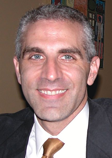
“In the new paradigm of minimally invasive vascular surgery, image fusion is an application that is essential to all endovascular surgeons,” Dr. Bismuth added. “Understanding its features promotes effective and safe complex procedures. That, along with an appreciation of the evolution in robotics and navigation technologies, has the potential to elevate the integration of the complex hybrid OR. Of great importance for the endovascular surgeon is an appreciation of what the hybrid OR is actually capable of and preparing for the revolution that will further evolve that space over the next 5 years,” Dr. Bismuth emphasized.
Presentations begin with an exploration of robotic technology for vascular surgery. Dr. Fabien Thaveau, professor of vascular surgery, Medical School of Strasbourg, will address the impact of the latest technology on abdominal aortic aneurysms in his talk, “Advances in Robotic Laparoscopic Treatment of AAAs: Value of the Gore Hybrid Graft in Dealing with Complex AAAs.” Dr. Nicholas J.W. Cheshire, professor of vascular surgery, Imperial College, and chief of vascular surgery, Aortic Centre, Royal Brompton Hospital, will continue the discussion by reviewing the current use of robotics and electromagnetic guidance in endovascular procedures, and exploring what developments clinicians might expect to see in the future.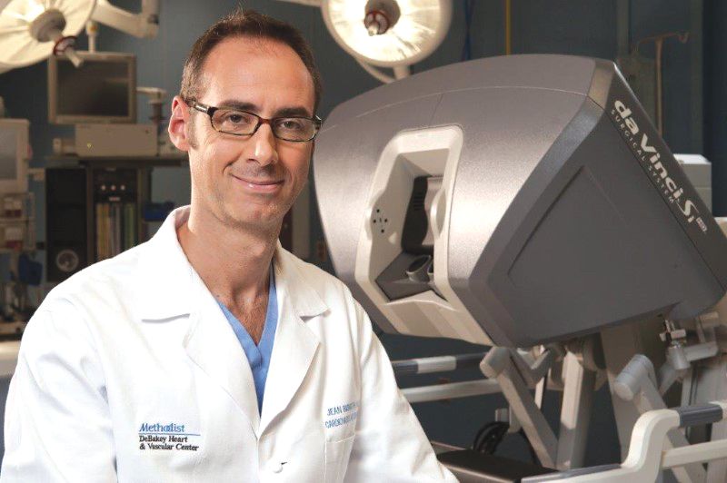
“The relevant issues are the actual imaging equipment, radiation burden, the ease of use of the equipment, a platform that is as noninvasive as possible, and promotes ideal ergonomics, and ultimately optimizes quality and efficiency of patient care,” said Dr. Bismuth.
“A core understanding of advanced imaging techniques is the foundation and the tool which will allow all other technologies to be integrated,” he added. Several speakers focus on the topic of fusion imaging. “Understanding fusion imaging should almost be a take-home message for all at the VEITHsymposium. This is an indispensable tool for all those performing endovascular procedures,” said Dr. Bismuth. Dr. Neal S. Cayne, associate professor of surgery, New York University School of Medicine, and director of endovascular surgery, New York University Medical Center, will describe the pros and cons of the technology in his talk, “Role of 3D Fusion Imaging and Guidance with the Siemens Artis Zeego System for Complex Vascular Interventions: Advantages and Limitations.”
Dr. Cayne will be followed by Dr. Stephan Haulon, professor, Université Paris Sud, and chief of Aortic Center, Hôpital Marie Lannelongue, Paris, who will discuss new developments in advanced fusion imaging and how the technology may be used for managing translumbar type 2 endoleaks and percutaneous distal branch puncture. He will also explain the potential role of fusion imaging for retrograde wiring “when prograde techniques fail during F/B/EVAR.” The fusion imaging portion of the session will continue with a presentation by Dr. Herve Rousseau, professor of radiology, Paul Sabatier University, and chief of department, CHU Rangueil Toulouse, France, on the benefits of fusion imaging for the treatment of type B aortic dissections (TBADs). Next, Dr. Klaus M. Overbeck, Newcastle University and City Hospitals Sunderland, England, will speak on the value of “fusion, re-entry devices, and a variable curvature sheath,” for the endovascular treatment of patients with complicated aorto-iliac occlusive disease. All fusion imaging is not created equal, according to Dr. Giovanni F. Torsello, a vascular surgeon, Charité Universitätsmedizin Berlin, who will address the differences between 2D-3D fusion and 3D-3D fusion, and why these differences are important in endovascular procedures.
A video presentation by Dr. Marc L. Schermerhorn, associate professor of surgery,Harvard Medical School, on “CTA Image Fusion with the Phillips Vessel Navigator to Facilitate F/EVAR” will conclude the presentations.
Go Global for Innovative Venous and Aortic Research
The 2016 Associate Faculty Global Podium session “Fascinating Venous and Aortic Topics” will be a series of presentations from experts from around the world on topics such as inflammatory aortic aneurysms and outcomes after endovascular-only repair, and emergency reconstruction of an inferior vena cava.
“There are presently multiple options for the treatment of both venous and arterial diseases. Surgeons will benefit by learning from others’ clinical experiences and operative techniques. This will help them in providing optimal therapy for each of their patients,” session co-moderator Dr. John Blebea said.
“Surgeons, by nature, are curious and always interested in learning new techniques or surgical skills,” said Dr. Blebea, professor of vascular surgery at the University of Oklahoma and immediate past president of the American Venous Forum. Co-moderating the session are Dr. Alan M. Dietzek of the University of Vermont and president-elect of the Society for Clinical Vascular Surgery; and Dr. Nick Morrison of the Morrison Vein Institute and President of the International Union of Phlebology.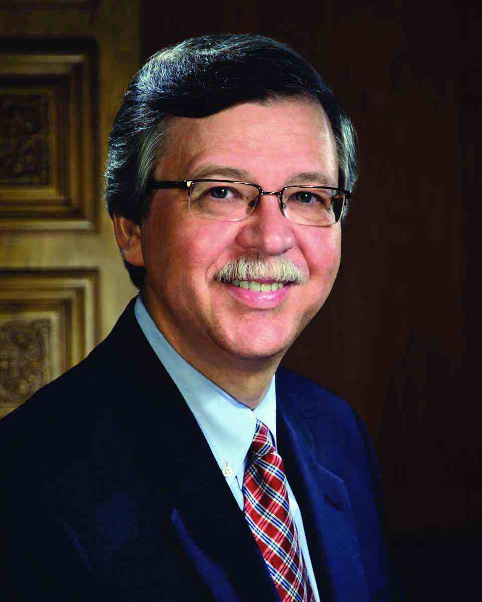
“The value of the presentations will further be enhanced by the commentary and critique of the three moderators, who are each experienced clinicians and world-recognized experts,” Dr. Blebea added.
“As the title of the session implies, this 3-hour conference will provide a fascinating glimpse into both venous and aortic arterial topics,” Dr. Blebea said. “The treatment of venous disease is an area of increasing interest and concern, on multiple levels. When one considers that an estimated 25 million Americans are affected by venous disease, ranging from cosmetic spider veins to recalcitrant non-healing ulcers, it is understandable that greater attention is being given to its treatment,” he noted.
“In addition, during the past two decades, there have been dramatic changes in the therapeutic options available. On the arterial side, advances in endovascular techniques now provide patients with interventions that are associated with less morbidity and mortality,” said Dr. Blebea. “However, there is also a need to customize interventions based on both anatomic restrictions and patient needs. This session will provide physicians with clinical insights which can be used immediately in their own practice, in a broad range of circumstances,” he emphasized.
"I think it is important to show support for the more junior members of our specialty," added Dr. Dietzek. "This is a wonderful opportunity for U.S. and international, early career vascular surgeons, residents, and fellows to present their research at what is arguably the foremost vascular conference in the world. Attending this session will heighten the experience of the presenter and will be a learning experience for the attendee – a win-win!"
Open surgery “remains a good and valid option for the treatment of inflammatory aortic abdominal aneurysms [IAAA],” according to Dr. Africa Duque-Santos of the Ramon y Cajal Hospital in Madrid, Spain. Dr. Santos presents results from 34 adult patients with IAAA; 29 were treated with open surgery and 5 with endovascular aneurysm repair (EVAR). After a median follow-up of 46 months (ranging from 24-112 months) renal function was equally stable in both groups. No significant differences were noted in the need for blood transfusion or intensive care, or in 30-day and long-term mortality rates, Dr. Duque-Santos said. Preoperative hydronephrosis was significantly more common in the EVAR group (3 patients, 60%) than in the open group (4 patients, 14%), but improvement in hydronephrosis occurred in 3 of 4 patients in the open group and 2 of 3 patients in the EVAR group.
“All patients with hydronephrosis in the open group received preoperative ureteral stenting; whereas none of the patients with hydronephrosis in the EVAR group received ureteral stents,” Dr. Duque-Santos noted.
Although the study included few patients treated with EVAR, the promising results support the need for further studies to assess long-term effectiveness; meanwhile, “open surgery remains a good and valid option for the treatment of IAAA,” Dr. Duque-Santos said.
Dr. Jianing Yue of Zhongshan Hospital Fudan University in Shanghai, will report that preoperative shock was associated with higher mortality rates in patients with truly-ruptured noninfected abdominal aortic aneurysms treated with and EVAR-optional approach (EVAR when possible) compared to those treated with an EVAR-only approach. Dr. Yue will describe data from a 7-year study including 80 adults; 27 died prior to surgery, 26 patients seen between March 2009 and April 2014were treated with EVAR if possible, and 27 seen between May 2014 and July 2016 were treated only with EVAR. In addition, the EVAR-only group had significantly shorter hospital stays than the open group (14 days vs. 34 days).
“Considering the shorter hospital stay, our results support superiority and more widespread adoption of emergent EVAR for the treatment of ruptured AAA,” Dr. Yue said.
Control of bleeding is a factor when injuries occur during accidents or surgery; Dr. Elmi Ism Olluri of Prishtina University Hospital, Kosovo, shares a case report of a 33-year-old patient who suffered extreme hypovolemic shock after blunt trauma to the right flank. The patient had complete rupture of the right kidney and a 7-cm long rupture of the infrarenal vena cava. The surgeons chose to reconstruct the infrarenal vena cava with a polytetrafluoroethylene graft. The patient also had an emergency laparotomy after a CT scan showed of a large abdominal blood clot. The patient was discharged 12 days after surgery, and 4 years later showed radiological evidence of blood flow in the IVC with a functional prosthetic graft. Salvage of a patient with a ruptured vena cava is rare, Dr. Olluri noted, but this case suggests that emergency polytetrafluoroethylene graft repair might be successful.
"Attend, attend, attend!" said co-moderator Dr. Deitzek. "The topics are great, the presenters are young and eager and we can help foster their early careers and at the same time hear and learn about some very interesting, unusual and fascinating topics. Be there or be square!"
Session 11
“Fascinating Venous and Aortic Topics”
Friday 7:05-9:46 a.m.
Bryant Suite, 2nd Floor
Capturing the Big Picture in DCB studies
Studies of drug-coated balloons in the lower extremities need to evolve to take a more nuanced look at outcomes, in part by incorporating more patient-centric endpoints, says a leading global expert on DCBs.
Dr. Marianne Brodmann of the University of Ganz, Austria, and a frequent principal investigator on endovascular trials, makes the case Saturday morning that it cannot be assumed that all DCBs are equally effective – though current trial and registry evidence, with their limited efficacy endpoints, make this somewhat hard to see.
“We have to step away from the philosophy we had in the past that a mechanical device like a balloon is a balloon, and therefore the same as any other balloon. When you add a drug, you have to prove the drug is working,” Dr. Brodmann said.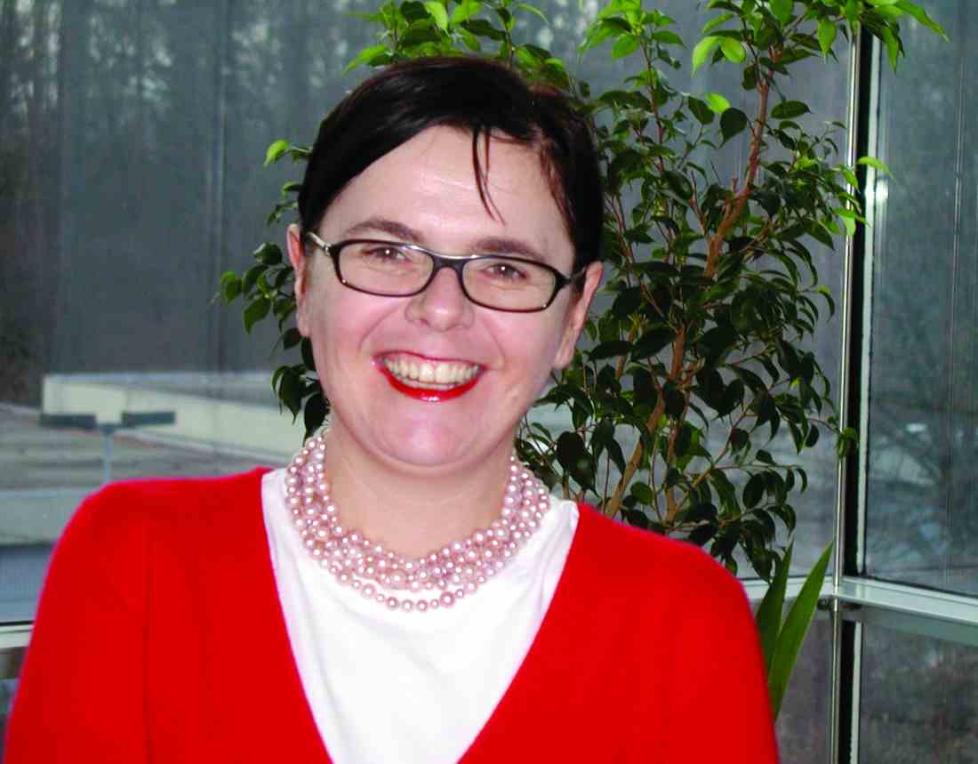
Dr. Brodmann said that investigators need to look beyond the usual measures, such as late lumen loss, which prove only that the drug is working in the vessel – and toward outcomes that better reflect a patient’s real-world quality of life after the intervention.
“The first time you have a DCB or other technology, you’re really proving efficacy just in terms of treating the stenosis or the occlusion,” she said. “But for the larger RCTs or the registries, you have to not just prove efficacy, you have to go into other endpoints, and that’s something we have to change when we look at these devices,” she said.
“For example, take a patient with intermittent claudication. If you keep that patient mobile and he’s able to walk and do regular daily activity, that’s one of the things we should look at in the future when we design trials and registries. Because if this patient, after the intervention, is able to resume his daily exercise routine and activities without feeling limited, he is also going to be reducing his cardiovascular morbidity and mortality,” Dr. Brodmann said.
Similarly, future studies should take a closer look at re-intervention as an outcome measure. “One of the most important things we can do for our patients is not have to bring them back into the cath lab,” Dr. Brodmann said. “It may not carry the same risk as surgery, but each endovascular procedure you perform still has risks – bleeding at the puncture site, allergic reactions to contrast media. Reducing the patient’s likelihood of going back is another quality-of-life improvement.”
Right now, “quality of life measures are insufficiently captured” in randomized controlled trials (RTCs). “There’s only one DCB trial that’s looked at it. But its increasingly being discussed in expert circles that it’s more and more necessary to focus on quality of life, and to expand our endpoints. We may find we do see something good for our patients beyond the endpoints we have right now,” Dr. Brodmann said.
To understand the full efficacy picture with DCBs, Dr. Brodmann said, it’s important to look both at registry and RCT evidence. “The large RCTs we have establish proof of efficacy but then I would highlight the manufacturer registries, because these registries represent the real-world scenarios you see when you treat patients with a DCB.”
Session 99: New Developments in the Treatment of Diseases of the Lower Extremities
Grand Ballroom East, 3rd Floor
Differences and Similarities in all the DCB Registries and RCTs: All DCBs and All Studies Are Not Equal
Saturday 7:58 .m. – 8:03 a.m.
Friday/Saturday Debates
FRIDAY
Session 75: More Carotid Disease And Treatment Related Topics And Controversies; Transcervical Cas (Tcar) And New Mesh Covered Carotid Stents
1:18 p.m. – 1:23 p.m.
Debate: Early CEA After Symptom Onset Is Beneficial To Patients: The Earlier The Better After Certain Requirements Are Met
Presenter: Ross Naylor, MD, FRCS
1:24 p.m. – 1:29 p.m.
Debate: Early CEA after Symptoms (TIA or Small Stroke):
Timing is Everything: Within 48 Hours is Bad:
Within 3-14 Days is Good: Why
Presenter: Ian Loftus, MD
1:30 p.m. – 1:35 p.m.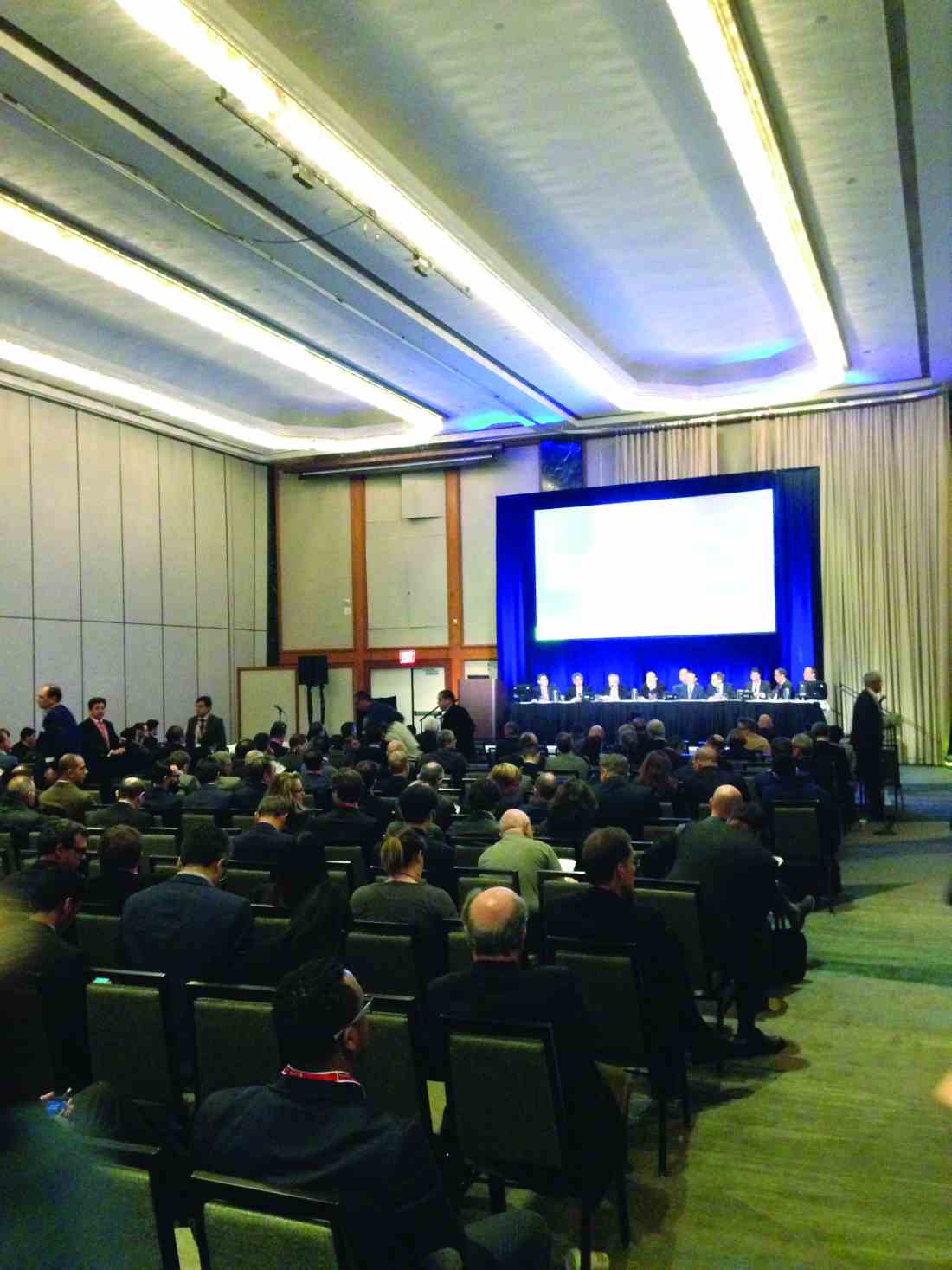
Presenter: Laura Capoccia, MD, PhD
1:36 p.m. – 1:41 p.m.
Debate: Another Balanced View: When is Early CEA after Symptom Onset in Patients with Carotid Stenosis Safe and Beneficial and When is It Not
Presenter: Martin Bjorck, MD, PhD
Session 77: New Carotid Concepts and Updates
4:12 p.m. – 4:17 p.m.
Debate: CAS Has No Increased Cost Consequences Compared to CEA
Presenters: Brajesh K. Lal, MD / Thomas G. Brott, MD
4:18 p.m. – 4:23 p.m.
Debate: Not So: CEA Costs Less Than CAS: Why the Discrepancy
Presenter: Kosmas I. Paraskevas, MD
Session 79: New Developments in the Treatment of Aneurysms and Other Diseases Involving the Popliteal Artery
6:52 a.m. – 6:57 a.m.
Debate: Endovascular Repair of Popliteal Aneurysms is Less Risky Than Open Repair and Should Be the Procedure of Choice: What Percent of Patients Should Be Treated Endo
Presenter: Irwin V. Mohan, MBBS, MD, FRCS, FEBVS, FRACS
6:58 a.m. – 7:03 a.m.
Debate: Not So: Open Repair is Better for Most Popliteal Aneurysm Patients: Endo Repair Is Sometimes a Failed Experiment
Presenter: Martin Bjorck, MD, PhD
Session 80: Infected Arteries and Arterial Grafts and Their Treatment: Infected EVARs; Mycotic AAAs; Infected Aortic/Arterial Prosthetic Grafts; Treatment Of Aorto-Esophageal Fistula
7:40 a.m. – 7:45 a.m.
Debate: Update on Treatment of Infected Aortic Endografts: Open Surgical Graft Excision is Always Indicated
Presenter: Kamphol Laohapensang, MD
7:46 a.m. – 7:51 a.m.
Debate: Not So: Semi-Conservative Treatment Without Graft Excision and with Drainage and Antibiotic Irrigation of AAA Sac Can be Effective Treatment: Longer-Term Results Prove It
Presenter: Martin Malina, MD, PhD
Session 87: Venous Cross-Sectional Imaging Techniques, Pelvic Venous Disorders
8:35 a.m. – 8:40 a.m.
Debate: Renal Vein Transposition (with Patch) is the Ideal Treatment for Nutcracker Syndrome, Not Stenting
Presenter: Olivier Hartung, MD
8:41 a.m. – 8:46 a.m.
Debate: Gonadal Vein Transposition is the Ideal Treatment for Nutcracker Syndrome
Presenter: Cynthia K. Shortell, MD
8:47 a.m. – 8:52 a.m.
Debate: Stenting is the Ideal Treatment or Nutcracker Syndrome
Presenter: Thomas S. Maldonado, MD
8:53 a.m. – 8:58 a.m.
Debate: Hybrid Endo-Open Surgery is the Ideal Treatment for Nutcracker Syndrome
Manju Kalra, MBBS
Session 88: Femoro-Iliocaval Interventional Strategies to Reduce Venous Hypertension, Hot Ideas for Recanalizing Chronic Total Occlusions
9:11 a.m. – 9:16 a.m.
Debate: Open Excisional Surgery for Post-Thrombotic Common Femoral Vein Obstruction (Endophlebectomy) is of Limited Value in the Endovascular Era
Presenter: Jose I. Almeida, MD, FACS, RPVI, RVT
9:17 a.m. – 9:22 a.m.
Debate: Open Excisional Surgery for Post-Thrombotic Common Femoral Vein Obstruction (Endophlebectomy) is Standard of Care
Presenter: Cees H.A. Wittens, MD, PhD
Session Tackles Vascular Malformations
A session on Friday morning entitled “Introduction to Vascular Malformations” will consider the classification, imaging, and surgical approaches to the congenital anomalies that occur in veins, lymph nodes, both, or in arteries and veins. The session will be co-moderated by Dr. Krassi Ivancev, University Hospital Hamburg-Eppendorf, Hamburg, Germany, and Dr. Furuzan Numan, Istanbul University Cerrahpasa Medical Faculty, Istanbul, Turkey.

Vascular malformations are present at birth and become apparent sometime later in life. Surgery often does not completely remove a malformation, which can lead to recurrence.
“Vascular malformations constitute the most difficult challenge in the world of vascular medicine and as a group are the most challenging of all vascular disease entities. Compounding their inherent difficulty in clinical management is the extreme rarity of these complex vascular lesions. Most malformations are in surgically difficult anatomies and many are impossible to resect, save by amputation,” said Dr. Ivancev.
Since vascular anomalies represent a spectrum of disorders, incorrect identification and misdiagnosis can occur, which can lead clinicians in the wrong direction when treating patients. As will be discussed by Dr. Leo J. Schultze Kool of Radboud University Nijmegen in the Netherlands, the International Society for the Study of Vascular Anomalies (ISSVA) classification system stratifies vascular lesions into vascular malformations and proliferative vascular lesions.
Dr. Wayne Yakes of the Yakes Vascular Malformation Center, will discuss an arteriovenous malformation (AVM) classification system he pioneered that defines all the angioarchitectures and their associated treatment strategies. Dr. Yakes will also talk about the therapeutic implications of the AVM classification when confronting challenging cases.
Diagnostic classification of vascular anomalies based on their visual appearance has been an area of innovation and invention in recent years. Imaging of congenital vascular malformations using magnetic resonance and using this information as a guide to treatment will be addressed by Dr. R. Sean Pakbaz of Scripps Healthcare.
Embolization techniques began to be used about 30 years ago, and have proven to be effective. Dr. James Donaldson of Northwestern University Feinberg School of Medicine will discuss endovascular access and embolization in neonates and children.
Session 92: Introduction to Vascular Malformations
Friday 6:45 a.m. – 7:30 a.m.
Gramercy Suites East and West, 2nd Floor
A session on Friday morning entitled “Introduction to Vascular Malformations” will consider the classification, imaging, and surgical approaches to the congenital anomalies that occur in veins, lymph nodes, both, or in arteries and veins. The session will be co-moderated by Dr. Krassi Ivancev, University Hospital Hamburg-Eppendorf, Hamburg, Germany, and Dr. Furuzan Numan, Istanbul University Cerrahpasa Medical Faculty, Istanbul, Turkey.
Vascular malformations are present at birth and become apparent sometime later in life. Surgery often does not completely remove a malformation, which can lead to recurrence.
“Vascular malformations constitute the most difficult challenge in the world of vascular medicine and as a group are the most challenging of all vascular disease entities. Compounding their inherent difficulty in clinical management is the extreme rarity of these complex vascular lesions. Most malformations are in surgically difficult anatomies and many are impossible to resect, save by amputation,” said Dr. Ivancev.
Since vascular anomalies represent a spectrum of disorders, incorrect identification and misdiagnosis can occur, which can lead clinicians in the wrong direction when treating patients. As will be discussed by Dr. Leo J. Schultze Kool of Radboud University Nijmegen in the Netherlands, the International Society for the Study of Vascular Anomalies (ISSVA) classification system stratifies vascular lesions into vascular malformations and proliferative vascular lesions.
Dr. Wayne Yakes of the Yakes Vascular Malformation Center, will discuss an arteriovenous malformation (AVM) classification system he pioneered that defines all the angioarchitectures and their associated treatment strategies. Dr. Yakes will also talk about the therapeutic implications of the AVM classification when confronting challenging cases.
Diagnostic classification of vascular anomalies based on their visual appearance has been an area of innovation and invention in recent years. Imaging of congenital vascular malformations using magnetic resonance and using this information as a guide to treatment will be addressed by Dr. R. Sean Pakbaz of Scripps Healthcare.
Embolization techniques began to be used about 30 years ago, and have proven to be effective. Dr. James Donaldson of Northwestern University Feinberg School of Medicine will discuss endovascular access and embolization in neonates and children.
Session 92: Introduction to Vascular Malformations
Friday 6:45 a.m. – 7:30 a.m.
Gramercy Suites East and West, 2nd Floor
Caval Interruption Strategies in the Spotlight
Caval interruption, typically by the installation of a filter, traps dislodged blood clots in the inferior vena cava, before they can reach the heart and lungs. The strategy will be the topic of the second half of the Friday afternoon session entitled “Endovascular and Open Solutions for Inferior Vena Cava Tumors and Occlusions, Vena Cava Filtration Strategies, Pitfalls, and Complications and More About Iliac Vein Stenting.”
The first half of the session will focus on the general topic of interventions of the inferior vena cava. This will include everything from percutaneous recanalization for occluded IVC filters to open and robotic vena cava reconstruction for the removal of benign and malignant tumors. The second half will cover the current controversies involving inferior vena cava filter indications for placement, retrieval, and the management of their complications.
“Session attendees will be able to understand current methods and indications for IVC filter placement, follow-up and retrieval. In addition, they will get an update on current research to improve the care of our patients requiring IVC interruption. The session will aid practitioners to better understand the requirements for open and endovascular management of vena cava tumors and obstruction,” said Dr. Gillespie.
Dr. John Rectenwald, the co-moderator, will consider the indications for IVC filters and discuss whether these are being observed in clinical practice. He will be followed by Dr. David Rosenthal of the Medical College of Georgia, who will provide an update on the Sentry bioconvertible non-retrieval IVS filter. The Sentry device is designed to provide protection against pulmonary embolism at the time of highest risk of embolism, followed by bioconversion to leave an unobstructed IVC lumen. The next speaker will be Dr. Gillespie, who will discuss the retrieval of IVC filters and the influence on retrieval rate by the filter design, institutional practice, and the individual surgeon. Dr. Steven Abramowitz of Georgetown University School of Medicine will talk about ulcer healing and quality of life following endovascular iliocaval reconstructive stenting in patients who have both IVC and iliac occlusive disease.
Dr. Rectenwald will discuss the Prevention du Risque d’Embolie Pulmonaire par Interruption Cav (PREPIC) trial of over 400 patients randomized to receive or not receive a filter along with standard anticoagulation treatment. Recommendations regarding IVC filters have been based on the PREPIC trial and follow-up data. The follow-up data showed no survival benefit. Whether the evidence supports the use of IVC filters will be discussed. Continuing this theme, Dr. Gillespie will update the physician-initiated, Investigational Device Exemption vena cava PRESERVE study, a large-scale, multi-specialty, prospective study launched in October 2016, which is assessing the use of IVC filters in the United States. The study involves all commercially available inferior vena cava filters placed in subjects for the prevention of death from fatal or symptomatic pulmonary embolism.
IVC filters are subject to complications. How to deal with these is a major challenge. Complications and how to avoid them will be the topic of a presentation by Dr. Clifford Sales of Mount Sinai School of Medicine, and how best to deal with filters that are difficult to retrieve will be discussed by Dr. Brian DeRubertis of UCLA Division of Vascular Surgery, Los Angeles, Calif. What to do when filters actually fracture and generate embolic fragments will be the topic of a presentation by Dr. Constantino Peña, University of South Florida. Finally, Dr. Nicholas Gargiulo, III, of The Clinch Valley Medical Center will consider prophylactic caval filtration in term of bariatric surgery patients who are the best candidates.
“There are special challenges involved in the open and endovascular management of inferior vena cava problems. These represent opportunities for practitioners to grow their practice. These patients are some of the most complicated patients we care for. Their management requires advanced techniques and dedicated clinicians to their care and management,” said Dr. Gillespie.
Session 91:
Endovascular and Open Solutions for Inferior Vena Cava Tumors and Occusions, Vena Cava Filtration Strategies, Pitfalls, and Complications and More About Iliac Vein Stenting
Caval Interruption
Friday 4:00 p.m. to 4:53 p.m.
Trianon Ballroom, 3rd Floor
Updates Presented on Robotic Vena Cava Surgery
Developments in robotic vena cava surgery will be presented Friday afternoon during the session entitled “Endovascular and Open Solutions for Inferior Vena Cava Tumors and Occusions, Vena Cava Filtration Strategies, Pitfalls, and Complications and More About Iliac Vein Stenting.”
“The selective use of robotic surgery on the vena cava is a tremendous step forward because of its minimal invasiveness and also because of the magnification that the robot gives and the technical advantages the robot gives over open surgery or laparoscopic surgery,” said Dr. Money.
Traditionally, inferior vena cava thrombectomy is done using a large open incision, since it is difficult otherwise to gain access to the vein. The surgery is done most often to address involvement of the inferior vena cava by an adjacent tumor. In addition, surgery may be needed to remove filters inserted to prevent migration of pulmonary emboli when problems develop with the filter.
The open incision approach carries a considerable risk of morbidity with the approach. A minimally invasive treatment option has been needed. Minimally invasive laparoscopy is an option. But drawbacks include the technically challenging nature of the procedure which makes for a steep learning curve.
Within the past decade, the use of robotic surgery has been utilized for interior vena cava thrombectomy. The approach involves multiple but smaller incisions to allow access of robotic tools. The robotic approach provides better visualization with magnification, three-dimensional vision, tremor filtration, and seven degrees of freedom, among other advantages.
“The overall take-home message from my presentation is that robotic vena caval surgery can be done and it should be used. Overall, robotic surgery for the vena cava is a field that is developing. It requires collaboration with other specialties and a realization that minimally invasive procedures are not just endovascular procedures but may be laparoscopic or robotic in nature,” said Dr. Money.
Dr. Money will describe the outcomes of a prospective study undertaken at the Scottsdale Mayo Clinic that evaluated the efficacy of a vena cava surgical system for radical nephrectomy and inferior vena cava tumor thrombectomy and, in a few patients, inferior vena cava filter removal. More recently, the robotic surgery was used to treat compression of the left renal vein between the superior mesenteric artery and aorta.
A focus of the presentation will be the key importance of the collaboration between vascular and robotic surgeons. “As time has developed I have become more aware and more adept at what the robot can do. However, it is not a routine part of my practice. By collaborating with others who use it more frequently, we have developed a practice where vena caval surgery can be done in a minimally invasive fashion with patients going home following a shorter length of stay, with less significant blood loss, and overall an easier recovery,” said Dr. Money.
Session 91:Endovascular and Open Solutions for Inferior Vena Cava Tumors and Occusions, Vena Cava Filtration Strategies, Pitfalls, and Complications and More About Iliac Vein Stenting
Robotic Vena Cava Surgery
Friday, 3:48 p.m. – 3:53 p.m.
Trianon Ballroom, 3rd Floor
Don’t Miss Out On. . .
The Challenging World of the Diagnosis and Treatment of Vascular Malformations: An Orphan Disease that Has Now Come of Age
(Sessions 92-98; Friday, 6:45 a.m. to 1:30 p.m.)
Location: Gramercy Suites East and West, 2nd Floor
Vascular malformations constitute one of the most challenging clinical entities encountered in Vascular Medicine today. They can occur in every anatomy in the body insinuating themselves throughout an organ or tissue. Being totally comprised of vascular structures, any surgical attempt at resection is fraught with potential hemorrhagic complications. Compounding their inherent difficulty in management , they are also very rare entities. The clinical presentations are extremely protean and can range from an asymptomatic birthmark, to fulminant, life-threatening CHF. Attributing any of these extremely varied symptoms that a patient may present with to a vascular malformation can be challenging to the most experienced clinician. Patients typically bounce from clinician to clinician experiencing disappointing outcomes, complications, and recurrence or worsening of their presenting symptoms. Due to their extreme rarity (>1% of the population), it is difficult for clinicians to gain any experience at all in their diagnosis and optimal management and make definitive statements.
The purpose of these Vascular Malformation Sessions is to offer the attendee the current state-of-the-art multi-disciplinary endovascular and surgical approaches to accurately diagnose and optimally treat all types of vascular malformations (AVMs, AVFs, venous malformations, lymphatic malformations, capillary-venous malformations, and mixed lesions) in all the various problematic anatomies in which they occur. Being that there are controversies regarding the various treatment strategies regarding vascular malformations, the attendees will be exposed to those multiple approaches and philosophies regarding the science, the multiple management strategies, the results, the long-term outcomes, and the complications inherent in the various palliative and curative treatments proffered by international experts.
Improving Outcomes in Hemodialysis Access
(Sessions 106-110; Saturday; 7:55 a.m. – 4:00 p.m.) Registration Begins At 6:00 a.m.
Location: Grand Ballroom West, 3rd Floor
The Hemodialysis Access sessions on Saturday (Sessions 106 – 110) will include presentations on planning, optimizing outcomes, political, economic and legal issues, new technologies and an update on clinical issues related to hemodialysis access. Experts will address specific topics including vein preservation and planning for access, use of ultrasound to facilitate cannulation, use of simulators, the role of drug eluting balloons and stents, management of complications including steal syndrome, infections and access hemorrhage, coding for access procedures, pharmacologic and mechanical approaches to improve fistula outcomes, treatment of fistula aneurysms, use of the HeRO graft and many more important topics. The keynote speaker will be Dr. Michael Brescia who will give his unique historical perspective on the development of the AV fistula. This session should be of interest to surgeons, nephrologists, interventionalists, nurses, dialysis technicians and others interested in the care of patients with end stage renal disease.
The Robots Are Coming to Vascular Surgery
Emerging technologies in robotics systems will be the subject of a key session at the 2016 Veithsymposium, entitled “Vascular Robotics and Guidance Systems” and co-chaired by Dr. Kenneth Ouriel, founder of Syntactx, and Dr. Anton N. Sidawy of George Washington University.
According to Dr. Sidawy, the major focus of the session will be the Magellan System, which is a steerable catheter system controlled by a remote physician console. The system has two major components: a robotic arm and the aforementioned remote console. This design is meant to keep the operator away from the table, thereby decreasing exposure to the radiation source.
The promise of such technology is becoming increasingly acknowledged in the field of vascular surgery. The Magellan can be used in a number of endovascular procedures, including – but not necessarily limited to – embolization, endovascular aneurysm repair (EVAR), angioplasty, stenting, crossing total occlusions, and venous procedures. Such applications will be highlighted in-depth during this session, with presentations by some of the most prominent names in the field of vascular robotic surgery.
The session will conclude with a panel discussion involving all the speakers and which will cover the current state and future of robotics in vascular surgery.
Thursday, November 17, 2016
Session 53: VASCULAR ROBOTICS AND GUIDANCE SYSTEMS
Moderators: Kenneth Ouriel, MD, MBA / Anton N. Sidawy, MD, MPH
3:52 PM - 4:52 PM
Lower Extremity Occlusive Disease and Alternative Graft Options
Vascular surgeons spend much of their clinical time taking care of patients with lower extremity occlusive disease, so the “New Devices and Techniques for Treating Lower Extremity Occlusive Disease; Prosthetic Grafts and Heparin Bonding” session on Thursday afternoon will be particularly useful for the practicing surgeon, according to Dr. Russell Samson who will co-moderate the session along with Dr. Ali F. AbuRahma of West Virginia University.
Two recent advances with respect to such alternative graft options, which will be addressed during the session, are heparin-bonded ePTFE and spiral laminar flow grafts.
“Whether these two modalities improve patency rates compared to standard ePTFE grafts remains controversial,” Dr. Samson said.
Heparin-bonded ePTFE will be the topic of a debate presented by Dr. Richard Neville of George Washington University Hospital, and Dr. Jonathan Beard of Sheffield Vascular Institute, in the United Kingdom. These two experts will conduct a debate. Dr. Neville will discuss long-term patency data suggesting that heparin bonding is valuable in PTFE Fempop Bypass Grafts, and Dr. Beard will argue that it does not improve results.
Additionally, Dr. Yann Gouëffic of the University of Nantes, France, will address the cost-efficiency of heparin bonding.
“In an age where surgeons are becoming more cost conscious, new graft configurations, which are often more costly, need to offer significant improvement in long-term outcomes,” Dr. Samson said.
As for spiral laminar flow grafts, Dr. Hosam F. El Sayed of Ohio State University will provide insight into a specific format of PTFE that creates a spiral laminar flow that more accurately represents flow within the arterial system, Dr. Samson noted.
Therefore, Dr. Enrico Ascher of Lutheran Medical Center, who is highly regarded for his experience in performing distal tibial bypass, will provide insight into how to deal with “pipe-like” calcified arteries that some surgeons may consider inoperable, Dr. Samson added.
Further, since many surgeons may prefer an endovascular procedure as a first-line alternative to bypass, this session will also include a talk by Dr. Hiroyoshi Yokoi of Fukuoka Sanno Hospital in Japan on advanced guidewire techniques to open these distal vessels, and another by noted inventor, Dr. Timothy A.M. Chuter of the University of California, San Francisco, who will discuss advantages and early clinical experience with a novel balloon that does not straighten when it inflates.
Another talk during the session, which will be given by Dr. Joseph L. Mills of Baylor College of Medicine, will focus on the impact of foot infections on leg bypass outcomes in CLI patients and what can be done to offset that impact.
“Dr. Mills will remind us that no matter what treatment we choose, the final result can be spoiled by an infection, so managing foot infections in patients requiring surgical bypass becomes a preeminent consideration,” Dr. Samson said.
“The practicing vascular surgeon deals with challenging infrainguinal atherosclerotic conditions on a daily basis. Therefore, information gained from attending this program should enhance their ability to offer long-lasting therapies,” he added.
Session 59
“New Devices and Techniques for Treating Lower Extremity Occlusive Disease; Prosthetic Grafts and Heparin Bonding”
Thursday, 1:00 p.m. – 1:54 p.m.
Grand Ballroom West, 3rd Floor