User login
Are serum uric acid levels always elevated in acute gout?
NO. Many patients with acute gout (11%-49%) have normal serum uric acid (SUA) levels (strength of recommendation [SOR]: A, prospective cohort studies). Patients taking allopurinol are significantly more likely to have normal uric acid levels during acute gout attacks (SOR: B, extrapolated from prospective cohorts).
Evidence summary
Six studies have evaluated SUA levels in patients with acute gout. Despite variations in diagnostic approach (clinical criteria vs synovial crystal analysis) and definitions of normal SUA (based on laboratory methods and sex), all 6 studies found normal levels in 11% to 49% of patients with acute gout (TABLE 1).
TABLE 1
Serum uric acid and acute gout: The evidence
| Type of cohort (n) | LOE* | Setting | Method of diagnosis | % with normal serum uric acid |
|---|---|---|---|---|
| Prospective1 (28) | 1b | Veterans Administration rheumatology clinic | Crystal positivity | 11% |
| Prospective2 (38) | 1b | Multiple settings (eg, inpatient, clinic, ED) | Clinical criteria or crystal positivity | 43% |
| Retrospective3 (226) | 2b | Hospitalized patients | Clinical criteria or crystal positivity | 12% |
| Retrospective4 (339) | 2b | Multiple settings | Crystal positivity | 32% |
| Retrospective5 (41) | 2b | Rheumatology clinic | Clinical criteria | 49% |
| Retrospective6 (69) | 2b | Multiple settings | Clinical criteria | 33%† |
| ED, emergency department; LOE, level of evidence. *1b, prospective cohort study with good follow-up (>80%); 2b, retrospective cohort study or prospective study with poor follow-up. †Not necessarily during acute gout. | ||||
Elevated SUA can be an indicator of gout—or not
A prospective cohort study of 82 patients at a Veterans Administration rheumatology clinic found elevated SUA to be the most sensitive indicator among various clinical criteria for diagnosing acute gout. However, 3 (11%) of the 28 patients who had crystal-proven gout also had a normal SUA.1
A second prospective cohort study that evaluated 38 patients during 42 episodes of acute gout in various clinical settings reported a normal SUA in 43% of patients diagnosed on clinical grounds or by joint aspiration.2
Some patients become hyperuricemic after diagnosis
The largest retrospective cohort study evaluated 226 Korean inpatients with acute gout diagnosed either by synovial crystals or American College of Rheumatology (ACR) criteria (TABLE 2). It found that 12% (27) had a normal SUA at diagnosis. Interestingly, 81% became hyperuricemic some time after diagnosis.3
TABLE 2
American College of Rheumatology criteria for classifying acute gouty arthritis
|
| Source: Wallace SL et al. Arthritis Rheum. 1977.7 |
What is a normal SUA value?
Another study reviewed SUA levels in a cohort derived from 2 large prospective RCTs of etoricoxib in patients diagnosed with acute gout by crystal analysis. The proportion of patients with a normal SUA varied substantially according to the definition of a normal value: 32% were normal using a value of 0.48 mmol/L; 11% had normal SUA levels when 0.36 mmol/L was used as the cutoff.4
A secondary analysis evaluated the effect of allopurinol on SUA. The proportion of patients on allopurinol with a normal SUA level compared with patients not taking allopurinol was 49% vs 29% using the higher normal cutoff value, and 29% vs 11% using the lower normal value (P<.001).4
Two studies find many gout patients with a normal SUA
A Japanese retrospective cohort study using ACR criteria found that nearly half of patients diagnosed with acute gout had a normal SUA level.5 A 1967 retrospective examination of Framingham Heart Study data found that one-third of patients clinically diagnosed with gout had a normal level. Some of the patients hadn’t been diagnosed at the time their SUA was measured, however.6
Recommendations
The ACR’s 1977 criteria for diagnosing gout include hyperuricemia as one potential indicator.7 The European League Against Rheumatism advises that normal SUA levels may accompany crystal-proven gout because uric acid either acts as a negative acute-phase reactant or increases in renal excretion during acute episodes. They conclude that SUA has “limited diagnostic value,” especially during acute gout.8
1. Malik A, Schumacher HR, Dinnella JE, et al. Clinical diagnostic criteria for gout: comparison with the gold standard of synovial fluid crystal analysis. J Clin Rheumatol. 2009;15:22-24.
2. Logan JA, Morrison E, McGill PE. Serum uric acid in acute gout. Ann Rheum Dis. 1997;56:696-697.
3. Park YB, Park YS, Lee SC, et al. Clinical analysis of gouty patients with normouricaemia at diagnosis. Ann Rheum Dis. 2003;62:90-92.
4. Schlesinger N, Norquist JM, Watson DJ. Serum urate during acute gout. J Rheumatol. 2009;36:1287-1289.
5. Urano W, Yamanaka H, Tsutani H, et al. The inflammatory process in the mechanism of decreased serum uric acid concentrations during acute gouty arthritis. J Rheumatol. 2002;29:1950-1953.
6. Hall AP, Barry PE, Dawber TR, et al. Epidemiology of gout and hyperuricemia. A long-term population study. Am J Med. 1967;42:27-37.
7. Wallace SL, Robinson H, Masi AT, et al. Preliminary criteria for the classification of the acute arthritis of primary gout. Arthritis Rheum. 1977;20:895-900.
8. Zhang W, Doherty M, Pascual E, et al. EULAR evidence based recommendations for gout. Part I: diagnosis. Report of a task force of the Standing Committee for International Clinical Studies Including Therapeutics (ESCISIT). Ann Rheum Dis. 2006;65:1301-1311.
NO. Many patients with acute gout (11%-49%) have normal serum uric acid (SUA) levels (strength of recommendation [SOR]: A, prospective cohort studies). Patients taking allopurinol are significantly more likely to have normal uric acid levels during acute gout attacks (SOR: B, extrapolated from prospective cohorts).
Evidence summary
Six studies have evaluated SUA levels in patients with acute gout. Despite variations in diagnostic approach (clinical criteria vs synovial crystal analysis) and definitions of normal SUA (based on laboratory methods and sex), all 6 studies found normal levels in 11% to 49% of patients with acute gout (TABLE 1).
TABLE 1
Serum uric acid and acute gout: The evidence
| Type of cohort (n) | LOE* | Setting | Method of diagnosis | % with normal serum uric acid |
|---|---|---|---|---|
| Prospective1 (28) | 1b | Veterans Administration rheumatology clinic | Crystal positivity | 11% |
| Prospective2 (38) | 1b | Multiple settings (eg, inpatient, clinic, ED) | Clinical criteria or crystal positivity | 43% |
| Retrospective3 (226) | 2b | Hospitalized patients | Clinical criteria or crystal positivity | 12% |
| Retrospective4 (339) | 2b | Multiple settings | Crystal positivity | 32% |
| Retrospective5 (41) | 2b | Rheumatology clinic | Clinical criteria | 49% |
| Retrospective6 (69) | 2b | Multiple settings | Clinical criteria | 33%† |
| ED, emergency department; LOE, level of evidence. *1b, prospective cohort study with good follow-up (>80%); 2b, retrospective cohort study or prospective study with poor follow-up. †Not necessarily during acute gout. | ||||
Elevated SUA can be an indicator of gout—or not
A prospective cohort study of 82 patients at a Veterans Administration rheumatology clinic found elevated SUA to be the most sensitive indicator among various clinical criteria for diagnosing acute gout. However, 3 (11%) of the 28 patients who had crystal-proven gout also had a normal SUA.1
A second prospective cohort study that evaluated 38 patients during 42 episodes of acute gout in various clinical settings reported a normal SUA in 43% of patients diagnosed on clinical grounds or by joint aspiration.2
Some patients become hyperuricemic after diagnosis
The largest retrospective cohort study evaluated 226 Korean inpatients with acute gout diagnosed either by synovial crystals or American College of Rheumatology (ACR) criteria (TABLE 2). It found that 12% (27) had a normal SUA at diagnosis. Interestingly, 81% became hyperuricemic some time after diagnosis.3
TABLE 2
American College of Rheumatology criteria for classifying acute gouty arthritis
|
| Source: Wallace SL et al. Arthritis Rheum. 1977.7 |
What is a normal SUA value?
Another study reviewed SUA levels in a cohort derived from 2 large prospective RCTs of etoricoxib in patients diagnosed with acute gout by crystal analysis. The proportion of patients with a normal SUA varied substantially according to the definition of a normal value: 32% were normal using a value of 0.48 mmol/L; 11% had normal SUA levels when 0.36 mmol/L was used as the cutoff.4
A secondary analysis evaluated the effect of allopurinol on SUA. The proportion of patients on allopurinol with a normal SUA level compared with patients not taking allopurinol was 49% vs 29% using the higher normal cutoff value, and 29% vs 11% using the lower normal value (P<.001).4
Two studies find many gout patients with a normal SUA
A Japanese retrospective cohort study using ACR criteria found that nearly half of patients diagnosed with acute gout had a normal SUA level.5 A 1967 retrospective examination of Framingham Heart Study data found that one-third of patients clinically diagnosed with gout had a normal level. Some of the patients hadn’t been diagnosed at the time their SUA was measured, however.6
Recommendations
The ACR’s 1977 criteria for diagnosing gout include hyperuricemia as one potential indicator.7 The European League Against Rheumatism advises that normal SUA levels may accompany crystal-proven gout because uric acid either acts as a negative acute-phase reactant or increases in renal excretion during acute episodes. They conclude that SUA has “limited diagnostic value,” especially during acute gout.8
1. Malik A, Schumacher HR, Dinnella JE, et al. Clinical diagnostic criteria for gout: comparison with the gold standard of synovial fluid crystal analysis. J Clin Rheumatol. 2009;15:22-24.
2. Logan JA, Morrison E, McGill PE. Serum uric acid in acute gout. Ann Rheum Dis. 1997;56:696-697.
3. Park YB, Park YS, Lee SC, et al. Clinical analysis of gouty patients with normouricaemia at diagnosis. Ann Rheum Dis. 2003;62:90-92.
4. Schlesinger N, Norquist JM, Watson DJ. Serum urate during acute gout. J Rheumatol. 2009;36:1287-1289.
5. Urano W, Yamanaka H, Tsutani H, et al. The inflammatory process in the mechanism of decreased serum uric acid concentrations during acute gouty arthritis. J Rheumatol. 2002;29:1950-1953.
6. Hall AP, Barry PE, Dawber TR, et al. Epidemiology of gout and hyperuricemia. A long-term population study. Am J Med. 1967;42:27-37.
7. Wallace SL, Robinson H, Masi AT, et al. Preliminary criteria for the classification of the acute arthritis of primary gout. Arthritis Rheum. 1977;20:895-900.
8. Zhang W, Doherty M, Pascual E, et al. EULAR evidence based recommendations for gout. Part I: diagnosis. Report of a task force of the Standing Committee for International Clinical Studies Including Therapeutics (ESCISIT). Ann Rheum Dis. 2006;65:1301-1311.
Evidence-based answers from the Family Physicians Inquiries Network
What is the best way to treat Morton’s neuroma?
NO SINGLE TREATMENT HAS BEEN IDENTIFIED in the literature. That said, a protocol of stepped care that showed good results in an uncontrolled trial seems reasonable: patient education and foot-wear or insole changes, followed by corticosteroid injections and, finally, surgery (strength of recommendation [SOR]: C, case series).
Injecting sclerosing alcohol depends on the provider’s access to and comfort with ultrasound, but the evidence is insufficient to recommend it routinely (SOR: C, case series).
Evidence summary
Options for treating Morton’s neuroma include changing shoe type, using insoles or metatarsal pads, taking nonsteroidal anti-inflammatory drugs (NSAIDs), giving corticosteroid or sclerosing alcohol injections, and surgically excising or transposing the offending nerve.1-3
Different conservative measures produce similar results
A small randomized prospective study of 23 patients compared reduction in neuroma pain using supinatory or pronatory insoles.4 No explicit inclusion or exclusion criteria other than clinical diagnosis were mentioned. Neither participants nor evaluators were blind to intervention allocations.
Two patients (13%) dropped out at 1 month. At 12 months, pain reduction in the supination and pronation insole groups was 50% and 45%, respectively (not significant).
Injections improve symptoms with minimal adverse effects
A prospective randomized study of 82 patients compared steroid injections alone with shoe modifications.5 Primary outcomes were patient satisfaction (presence or absence of pain), amount of pain, and return of pain.
Steroid injections yielded better patient satisfaction compared with shoe modifications alone at 1 and 6 months. Twenty-three percent of shoe-modification patients achieved complete satisfaction at 1 month, compared with 50% of injection patients (P<.01; number needed to treat [NNT]=3.7). At 6 months, the results were 28.6% satisfaction with shoe modification and 73.5% satisfaction with injection (P<.001; NNT=2.3).
The difference disappeared at 1 year (63% satisfaction with shoe modification compared with 82% satisfaction with injection; P>.05), although patients were allowed to cross over at 6 months. No complications occurred. The study was limited by a high rate of crossover from the shoe modification to the injection group at 6 months, elimination of dropouts from the final analysis, and lack of intent-to-treat analysis.
Another technique uses the sclerosing effects of alcohol6 delivered by multiple ultrasound-guided injections over time.7,8 Improvement of symptoms with no long-term adverse events were reported in several case series, although in each study a small number of patients reported localized pain at the site of injection.6-10 The TABLE summarizes injection studies.5-11
TABLE
How injection therapies for Morton’s neuroma compare
| Study | Injection materials | Type of study | Number of cases | Average follow-up, (mo) | Average number of injections | Results |
|---|---|---|---|---|---|---|
| Greenfield 19849 | Steroid | Retrospective case series | 67 | 24 | 3 | 80% complete relief |
| Saygi 20055 | Steroid | Prospective quasirandomized injection vs footwear modification | 82 | 12 | 2-3 | 82% vs 63% complete or partial pain relief in 2 groups, respectively |
| Markovic 200811 | Steroid* | Prospective case series | 39 | 9 | 1 | 38% complete satisfaction and 28% satisfaction with minor reservations |
| Dockery 19996 | Alcohol | Prospective case series | 100 | 13 | 5.5 | 89% resolution or improved symptoms |
| Fanucci 20048 | Alcohol* | Prospective case series | 40 | 10 | 4 | 90% resolution or improved symptoms |
| Hughes 20077 | Alcohol* | Prospective case series | 101 | 21.1 | 4.1 | 94% resolution or improved symptoms |
| Mozena 200710 | Alcohol | Retrospective case series | 42 | 11 | 3-7 | 61% resolution or improved symptoms. Patients with ≥5 injections (74%) were more likely to respond (P=.0072) |
| *Ultrasound guided. | ||||||
Surgery: Consider cost and risk of complications
Most surgical studies enrolled patients who had initially failed conservative treatments. Costs and risks of complications must be weighed, including infection, scar sensitivity, residual pain, sensory deficits, and other wound-related morbidities.
Technique varies considerably among surgeons.12 A Cochrane systematic review of 3 randomized controlled trials comprising 121 patients concluded that, at most, very limited evidence exists that transposition of the transected plantar digital nerve may yield better long-term results than standard resection.3
In a retrospective case series, 82 patients with primary Morton’s neuroma were treated by a single surgeon and a single technique (dorsal incision and nerve transection).13 All patients had failed conservative management with orthotics, shoe modification, and NSAIDs (lidocaine injections were used only to pinpoint neuroma location). Sixty-six (81%) returned for follow-up.
Average follow-up evaluation was 5.8 years. Of the patients who were followed, 85% rated overall satisfaction as excellent or good.
Stepped care gets results
A prospective case series enrolled 115 subjects out of 340 consecutive patients who presented to a private orthopedic clinic with a diagnosis of Morton’s neuroma.1 The authors assessed a 3-stage protocol of stepped care, progressing to the next stage if improvement was inadequate after 3 months: Stage I comprised patient education, footwear modifications, and a metatarsal pad placed proximal to the involved nerve; stage II, injection of steroids with local anesthetic or local anesthetic alone; and stage III, surgery.
Of 57 patients treated only with footwear modifications, 47 (41%) improved and required no further treatment. Twenty-seven (47%) of the 58 patients who received injections improved and required no further treatment. Of the 24 patients who advanced to stage III, 96% improved with surgery.
Recommendations
No consensus exists regarding definitive treatment of Morton’s neuroma. The American College of Occupational and Environmental Medicine recommends excision of the neuroma if nonsurgical treatment fails.14
A 2003 Cochrane systematic review of 107 studies found insufficient evidence to assess efficacy of surgical and nonsurgical interventions.3 Stepped care has been adopted by many third-party payers who require conservative care before reimbursing for injections, and treatment with injections before reimbursing for surgery.
1. Bennett GL, Graham CE, Mauldin DM. Morton’s interdigital neuroma: a comprehensive treatment protocol. Foot Ankle Int. 1995;16:760-763.
2. Wu KK. Morton’s neuroma and metatarsalgia. Curr Opin Rheumatol. 2000;12:131-142.
3. Thomson CE, Gibson JN, Martin D. Interventions for the treatment of Morton’s neuroma. Cochrane Database Syst Rev. 2004;(3):CD003118.-
4. Kilmartin TE, Wallace WA. Effect of pronation and supination orthosis on Morton’s neuroma and lower extremity function. Foot Ankle Int. 1994;15:256-262.
5. Saygi B, Yildirim Y, Saygi EK, et al. Morton’s neuroma: comparative results of two conservative methods. Foot Ankle Int. 2005;26:556-559.
6. Dockery GL. The treatment of intermetatarsal neuromas with 4% alcohol sclerosing injections. J Foot Ankle Surg. 1999;38:403-408.
7. Hughes RJ, Ali K, Jones H, et al. Treatment of Morton’s neuroma with alcohol injection under sonographic guidance: follow-up of 101 cases. Am J Roentgenol. 2007;188:1535-1539.
8. Fanucci E, Masala S, Fabiano S, et al. Treatment of intermetatarsal Morton’s neuroma with alcohol injection under US guide: 10-month follow-up. Eur Radiol. 2004;14:514-518.
9. Greenfield J, Rea J, Jr, Ilfeld FW. Morton’s interdigital neuroma: indications for treatment by local injections versus surgery. Clin Orthop Relat Res. 1984;185:142-144.
10. Mozena JD, Clifford JT. Efficacy of chemical neurolysis for the treatment of interdigital nerve compression of the foot: a retrospective study. J Am Podiatr Med Assoc. 2007;97:203-206.
11. Markovic M, Crichton K, Read JW, et al. Effectiveness of ultrasound-guided corticosteroid injection in the treatment of Morton’s neuroma. Foot Ankle Int. 2008;29:483-487.
12. Hassouna H, Singh D. Morton’s metatarsalgia: pathogenesis, aetiology and current management. Acta Orthop Belg. 2005;71:646-655.
13. Coughlin MJ, Pinsonneault T. Operative treatment of interdigital neuroma: a long-term follow-up study. J Bone Joint Surg Am. 2001;83-A:1321-1328.
14. Clinical Practice Guideline Forefoot Disorders Panel, Thomas JL, Blitch EL, IV, Chaney DM, et al. Diagnosis and treatment of forefoot disorders. Section 3. Morton’s intermetatarsal neuroma. J Foot Ankle Surg. 2009;48:251-256.Available at: . Accessed July 5, 2010.
NO SINGLE TREATMENT HAS BEEN IDENTIFIED in the literature. That said, a protocol of stepped care that showed good results in an uncontrolled trial seems reasonable: patient education and foot-wear or insole changes, followed by corticosteroid injections and, finally, surgery (strength of recommendation [SOR]: C, case series).
Injecting sclerosing alcohol depends on the provider’s access to and comfort with ultrasound, but the evidence is insufficient to recommend it routinely (SOR: C, case series).
Evidence summary
Options for treating Morton’s neuroma include changing shoe type, using insoles or metatarsal pads, taking nonsteroidal anti-inflammatory drugs (NSAIDs), giving corticosteroid or sclerosing alcohol injections, and surgically excising or transposing the offending nerve.1-3
Different conservative measures produce similar results
A small randomized prospective study of 23 patients compared reduction in neuroma pain using supinatory or pronatory insoles.4 No explicit inclusion or exclusion criteria other than clinical diagnosis were mentioned. Neither participants nor evaluators were blind to intervention allocations.
Two patients (13%) dropped out at 1 month. At 12 months, pain reduction in the supination and pronation insole groups was 50% and 45%, respectively (not significant).
Injections improve symptoms with minimal adverse effects
A prospective randomized study of 82 patients compared steroid injections alone with shoe modifications.5 Primary outcomes were patient satisfaction (presence or absence of pain), amount of pain, and return of pain.
Steroid injections yielded better patient satisfaction compared with shoe modifications alone at 1 and 6 months. Twenty-three percent of shoe-modification patients achieved complete satisfaction at 1 month, compared with 50% of injection patients (P<.01; number needed to treat [NNT]=3.7). At 6 months, the results were 28.6% satisfaction with shoe modification and 73.5% satisfaction with injection (P<.001; NNT=2.3).
The difference disappeared at 1 year (63% satisfaction with shoe modification compared with 82% satisfaction with injection; P>.05), although patients were allowed to cross over at 6 months. No complications occurred. The study was limited by a high rate of crossover from the shoe modification to the injection group at 6 months, elimination of dropouts from the final analysis, and lack of intent-to-treat analysis.
Another technique uses the sclerosing effects of alcohol6 delivered by multiple ultrasound-guided injections over time.7,8 Improvement of symptoms with no long-term adverse events were reported in several case series, although in each study a small number of patients reported localized pain at the site of injection.6-10 The TABLE summarizes injection studies.5-11
TABLE
How injection therapies for Morton’s neuroma compare
| Study | Injection materials | Type of study | Number of cases | Average follow-up, (mo) | Average number of injections | Results |
|---|---|---|---|---|---|---|
| Greenfield 19849 | Steroid | Retrospective case series | 67 | 24 | 3 | 80% complete relief |
| Saygi 20055 | Steroid | Prospective quasirandomized injection vs footwear modification | 82 | 12 | 2-3 | 82% vs 63% complete or partial pain relief in 2 groups, respectively |
| Markovic 200811 | Steroid* | Prospective case series | 39 | 9 | 1 | 38% complete satisfaction and 28% satisfaction with minor reservations |
| Dockery 19996 | Alcohol | Prospective case series | 100 | 13 | 5.5 | 89% resolution or improved symptoms |
| Fanucci 20048 | Alcohol* | Prospective case series | 40 | 10 | 4 | 90% resolution or improved symptoms |
| Hughes 20077 | Alcohol* | Prospective case series | 101 | 21.1 | 4.1 | 94% resolution or improved symptoms |
| Mozena 200710 | Alcohol | Retrospective case series | 42 | 11 | 3-7 | 61% resolution or improved symptoms. Patients with ≥5 injections (74%) were more likely to respond (P=.0072) |
| *Ultrasound guided. | ||||||
Surgery: Consider cost and risk of complications
Most surgical studies enrolled patients who had initially failed conservative treatments. Costs and risks of complications must be weighed, including infection, scar sensitivity, residual pain, sensory deficits, and other wound-related morbidities.
Technique varies considerably among surgeons.12 A Cochrane systematic review of 3 randomized controlled trials comprising 121 patients concluded that, at most, very limited evidence exists that transposition of the transected plantar digital nerve may yield better long-term results than standard resection.3
In a retrospective case series, 82 patients with primary Morton’s neuroma were treated by a single surgeon and a single technique (dorsal incision and nerve transection).13 All patients had failed conservative management with orthotics, shoe modification, and NSAIDs (lidocaine injections were used only to pinpoint neuroma location). Sixty-six (81%) returned for follow-up.
Average follow-up evaluation was 5.8 years. Of the patients who were followed, 85% rated overall satisfaction as excellent or good.
Stepped care gets results
A prospective case series enrolled 115 subjects out of 340 consecutive patients who presented to a private orthopedic clinic with a diagnosis of Morton’s neuroma.1 The authors assessed a 3-stage protocol of stepped care, progressing to the next stage if improvement was inadequate after 3 months: Stage I comprised patient education, footwear modifications, and a metatarsal pad placed proximal to the involved nerve; stage II, injection of steroids with local anesthetic or local anesthetic alone; and stage III, surgery.
Of 57 patients treated only with footwear modifications, 47 (41%) improved and required no further treatment. Twenty-seven (47%) of the 58 patients who received injections improved and required no further treatment. Of the 24 patients who advanced to stage III, 96% improved with surgery.
Recommendations
No consensus exists regarding definitive treatment of Morton’s neuroma. The American College of Occupational and Environmental Medicine recommends excision of the neuroma if nonsurgical treatment fails.14
A 2003 Cochrane systematic review of 107 studies found insufficient evidence to assess efficacy of surgical and nonsurgical interventions.3 Stepped care has been adopted by many third-party payers who require conservative care before reimbursing for injections, and treatment with injections before reimbursing for surgery.
1. Bennett GL, Graham CE, Mauldin DM. Morton’s interdigital neuroma: a comprehensive treatment protocol. Foot Ankle Int. 1995;16:760-763.
2. Wu KK. Morton’s neuroma and metatarsalgia. Curr Opin Rheumatol. 2000;12:131-142.
3. Thomson CE, Gibson JN, Martin D. Interventions for the treatment of Morton’s neuroma. Cochrane Database Syst Rev. 2004;(3):CD003118.-
4. Kilmartin TE, Wallace WA. Effect of pronation and supination orthosis on Morton’s neuroma and lower extremity function. Foot Ankle Int. 1994;15:256-262.
5. Saygi B, Yildirim Y, Saygi EK, et al. Morton’s neuroma: comparative results of two conservative methods. Foot Ankle Int. 2005;26:556-559.
6. Dockery GL. The treatment of intermetatarsal neuromas with 4% alcohol sclerosing injections. J Foot Ankle Surg. 1999;38:403-408.
7. Hughes RJ, Ali K, Jones H, et al. Treatment of Morton’s neuroma with alcohol injection under sonographic guidance: follow-up of 101 cases. Am J Roentgenol. 2007;188:1535-1539.
8. Fanucci E, Masala S, Fabiano S, et al. Treatment of intermetatarsal Morton’s neuroma with alcohol injection under US guide: 10-month follow-up. Eur Radiol. 2004;14:514-518.
9. Greenfield J, Rea J, Jr, Ilfeld FW. Morton’s interdigital neuroma: indications for treatment by local injections versus surgery. Clin Orthop Relat Res. 1984;185:142-144.
10. Mozena JD, Clifford JT. Efficacy of chemical neurolysis for the treatment of interdigital nerve compression of the foot: a retrospective study. J Am Podiatr Med Assoc. 2007;97:203-206.
11. Markovic M, Crichton K, Read JW, et al. Effectiveness of ultrasound-guided corticosteroid injection in the treatment of Morton’s neuroma. Foot Ankle Int. 2008;29:483-487.
12. Hassouna H, Singh D. Morton’s metatarsalgia: pathogenesis, aetiology and current management. Acta Orthop Belg. 2005;71:646-655.
13. Coughlin MJ, Pinsonneault T. Operative treatment of interdigital neuroma: a long-term follow-up study. J Bone Joint Surg Am. 2001;83-A:1321-1328.
14. Clinical Practice Guideline Forefoot Disorders Panel, Thomas JL, Blitch EL, IV, Chaney DM, et al. Diagnosis and treatment of forefoot disorders. Section 3. Morton’s intermetatarsal neuroma. J Foot Ankle Surg. 2009;48:251-256.Available at: . Accessed July 5, 2010.
Evidence-based answers from the Family Physicians Inquiries Network
What’s the best way to treat Achilles tendonopathy?
Rest and ice are considered first-line therapy for acute Achilles tendonopathy (strength of recommendation [SOR]: C, expert opinion), as is nonsteroidal anti-inflammatory drugs (NSAIDs) (SOR: B, systematic review).
Chronic noninsertional Achilles tendonopathy should be treated with eccentric calf-muscle training (ECMT) (SOR: B, 3 randomized controlled trials [RCTs]). Continuing an activity such as running, while monitoring pain, is as effective as resting while enrolled in an eccentric strengthening program (SOR: B, 1 RCT). If conservative management fails, surgery may help (SOR: C, case series).
Treat tendonopathy one step at a time
Charles W. Webb, DO
Oregon Health and Science University, Portland, Ore
I’ve found stepwise treatment of Achilles tendonopathy to be the most useful approach for my patients. Initially, I recommend NSAIDs, stretching, and decreasing the offending activity. If the patient hasn’t improved in 2 weeks, I refer him for physical therapy with a focus on flexibility and eccentric strengthening. I recommend applying ice for 20 minutes after each therapy or exercise session and 2 or 3 more times a day.
If the patient doesn’t improve in another 2 to 3 weeks, I have him (or her) wear a cam walking boot (fracture boot) constantly, except when bathing, for 2 to 6 weeks. The boot maintains the ankle in a neutral position, facilitating the rest required for healing. I’ve found that most patients can avoid surgery by wearing the boot until they are free of pain for a week (but no longer than 6 weeks) and then undergoing aggressive physical therapy to strengthen the ankle.
Evidence summary
While patients may refer to this condition as “Achilles tendonitis,” the phrase is actually a misnomer. That’s because in tendonitis, the inflammation is limited to the surrounding structures, but doesn’t involve the tendon itself.
Acute symptoms respond to NSAIDs
A Cochrane review of treatments for acute Achilles tendonopathy included 9 randomized trials with a total of 697 patients. Two RCTs showed evidence that NSAIDs alleviate acute symptoms. In 1 trial of 212 patients who received an oral NSAID or placebo for 14 days, significantly fewer patients in the placebo group achieved a good or excellent symptom relief (relative risk [RR]=0.42; 95% confidence interval [CI], 0.21-0.86). A second RCT of 243 patients compared treatment with a topical NSAID to placebo for 21 days; significantly fewer participants who took placebo returned to their pre-injury level of activity (RR=0.78; 95% CI, 0.63-0.95). Low-dose heparin injection, heel pads, topical laser therapy, and peritendinous steroid injection produced no significant decrease in pain compared with placebo.1
Continue to exercise?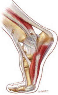
Patients with chronic noninsertional Achilles tendonopathy may be able to continue activity during eccentric calf-muscle training without exacerbating the tendonopathy, according to an RCT of 38 patients that compared rest with eccentric training and continued activity with eccentric training.
SHOWN HERE: MEDIAL FOOT AND ANKLE WITHOUT INJURY.
Eccentric calf-muscle training helps chronic tendonopathy
ECMT is an effective intervention for chronic noninsertional Achilles tendonopathy. An observational study of ECMT in 78 patients (101 tendons total) with chronic noninsertional Achilles tendonopathy found that 89% were back to their preinjury activity level after a 12-week regimen of ECMT, as indicated by significantly lower scores on a 100-point visual analog scale (67 at baseline vs 10.2 at 12 weeks; P<0.001).2
An RCT compared ECMT or repetitive shock wave therapy to a “wait-and-see” control group (25 patients in each group). After 4 months, both the ECMT and shock wave therapy groups reported a significant decrease in pain compared with controls (P< 0.001; mean difference in 10-point pain score, 2.4; 95% CI, 1.3-3.5). Sixty percent of participants in the ECMT group reported complete recovery or significant improvement (P<0.001; number needed to treat [NNT]=3; mean difference in 6-point Likert scale=–1.6; 95% CI, –2.4 to 0.8).3
During an ECMT program, patients may be able to continue activity without exacerbating Achilles tendonopathy. An RCT of 38 patients compared rest with eccentric training and continued activity and eccentric training for 12 weeks to 6 months using a pain-monitored model. Both groups showed similar improvement in pain and function when measured with the Swedish version of the Achilles assessment questionnaire (VISA-A-S 100-point scale: 0=worst function, 100=completely recovered). The baseline mean VISA-A-S scores were 57 for both groups; the mean scores after 12 months were 85 for the exercise group and 91 for the rest group.4
Another option: The AirHeel brace
An AirHeel brace may be a viable alternative to ECMT, especially if the patient can’t tolerate eccentric muscle training because of pain. The brace was designed for Achilles tendonopathy and posterior heel pain. It applies intermittent compression to minimize swelling and promote circulation.
An RCT evaluated 100 patients with chronic noninsertional Achilles tendonopathy who were treated with ECMT, an AirHeel Brace, or a combination of the 2 for 12 weeks. At 1-year follow-up, all 3 groups showed a significant reduction in pain measured by a 10-point visual analog scale (ECMT 30%, brace 27%, combination 53%; P<0.0001). All the groups also showed significantly improved function as measured by an American Orthopaedic Foot and Ankle Society Score (10%, 10%, and 30%, respectively; P<0.001). No significant differences in outcomes were found across the treatment groups, and 90% of participants reached their preinjury activity level after 1 year.4
Shock wave therapy and topical glyceryl trinitrate
The role of repetitive shock wave therapy and topical glyceryl trinitrate in the treatment of chronic noninsertional Achilles tendonopathy is less clear. In a double-blinded RCT of 49 patients, shock wave therapy did not produce better outcomes than placebo.5
However, a prospective cohort study of 68 patients found that patients who received shock wave therapy had significantly lower mean visual analog scale scores for pain compared with controls after 1 month (P<0.001), 3 months (P<0.001), and 12 months (P<0.001) of treatment.6 In a double-blinded RCT of 65 patients, more patients treated with topical glyceryl trinitrate therapy were asymptomatic during activities of daily living at 6 months compared with placebo (P<0.001).7
Both shock wave therapy and topical glyceryl trinitrate may have a significant role to play in treating chronic noninsertional Achilles tendonopathy, but more studies are needed to support their use.
Surgery appears to work better for athletes
Surgery is an option for patients with chronic noninsertional Achilles tendonopathy who have failed conservative measures and a 3- to 6-month rehabilitation program. In a systematic review of 26 studies of patients managed surgically, the mean success rate (full return to preinjury activity level) was 77%. However, a negative correlation was observed between the reported success rate and the overall methods score, a rating of the quality of studies (r=0.53; P<0.01). Only 5 of the studies reviewed were prospective cohort studies; the remaining 21 were retrospective cohort studies and case studies.8
Athletes responded significantly better to surgery than nonathletic patients, returning to full activity in 4.5 months compared with 7.1 months for nonathletes (P<0.03). Fewer athletes had surgical complications (9% compared with 19% of nonathletes).9 More studies are needed to clarify the role of surgery in managing Achilles tendonopathy.
Recommendations
The American College of Foot and Ankle Surgeons recommends initial management by reducing pressure around the affected area combined with heel lifts, orthotics, NSAID therapy, and physical therapy. Immobilization may be used on a case-by-case basis. Local steroid injections aren’t recommended. Patients with resistant tendonopathy should be referred to a foot and ankle surgeon.10
1. McLaughlan G, Handoll H. Interventions for treating acute and chronic Achilles tendonitis. Cochrane Database Syst Rev. 2001;(2):CD000232.-
2. Fahlstrom M, Jonsson P, Lorentzon R, et al. Chronic Achilles tendon pain treated with eccentric calf-muscle training. Knee Surg Sports Traumatol Arthrosc. 2003;11:327-333.
3. Rompe J, Nafe B, Furia J, et al. Eccentric loading, shock-wave treatment, or a wait-and-see policy for tendinopathy of the main body of tendo Achillis. Am J Sports Med. 2007;35:374-383.
4. Silbernagel K, Thomee R, Eriksson B, et al. Continued sports activity, using a pain-monitoring model, during rehabilitation in patients with Achilles tendinopathy. Am J Sports Med. 2007;35:897-906.
5. Costa M, Shepstone L, Donnell S, et al. Shock wave therapy for chronic Achilles tendon pain. Clin Orthop. 2005;440:199-204.
6. Furia J. High-energy extracorporeal shock wave therapy as a treatment for insertional Achilles tendinopathy. Am J Sports Med. 2006;34:733-740.
7. Paoloni J, Appleyard R, Nelson J, et al. Topical glyceryl trinitrate treatment of chronic noninsertional Achilles tendinopathy. J Bone Joint Surg Am. 2004;86:916-922.
8. Tallon C, Coleman B, Khan K, et al. Outcome of surgery for chronic Achilles tendinopathy. Am J Sports Med. 2001;29:315-320.
9. Maffuli N, Testa V, Capasso G, et al. Surgery for chronic Achilles tendinopathy yields worse results in nonathletic patients. Clin J Sport Med. 2006;16:123-128.
10. Thomas J, Christensen J, Kravitz S, et al. The diagnosis and treatment of heel pain. J Foot Ankle Surg. 2001;40:329-337.
Rest and ice are considered first-line therapy for acute Achilles tendonopathy (strength of recommendation [SOR]: C, expert opinion), as is nonsteroidal anti-inflammatory drugs (NSAIDs) (SOR: B, systematic review).
Chronic noninsertional Achilles tendonopathy should be treated with eccentric calf-muscle training (ECMT) (SOR: B, 3 randomized controlled trials [RCTs]). Continuing an activity such as running, while monitoring pain, is as effective as resting while enrolled in an eccentric strengthening program (SOR: B, 1 RCT). If conservative management fails, surgery may help (SOR: C, case series).
Treat tendonopathy one step at a time
Charles W. Webb, DO
Oregon Health and Science University, Portland, Ore
I’ve found stepwise treatment of Achilles tendonopathy to be the most useful approach for my patients. Initially, I recommend NSAIDs, stretching, and decreasing the offending activity. If the patient hasn’t improved in 2 weeks, I refer him for physical therapy with a focus on flexibility and eccentric strengthening. I recommend applying ice for 20 minutes after each therapy or exercise session and 2 or 3 more times a day.
If the patient doesn’t improve in another 2 to 3 weeks, I have him (or her) wear a cam walking boot (fracture boot) constantly, except when bathing, for 2 to 6 weeks. The boot maintains the ankle in a neutral position, facilitating the rest required for healing. I’ve found that most patients can avoid surgery by wearing the boot until they are free of pain for a week (but no longer than 6 weeks) and then undergoing aggressive physical therapy to strengthen the ankle.
Evidence summary
While patients may refer to this condition as “Achilles tendonitis,” the phrase is actually a misnomer. That’s because in tendonitis, the inflammation is limited to the surrounding structures, but doesn’t involve the tendon itself.
Acute symptoms respond to NSAIDs
A Cochrane review of treatments for acute Achilles tendonopathy included 9 randomized trials with a total of 697 patients. Two RCTs showed evidence that NSAIDs alleviate acute symptoms. In 1 trial of 212 patients who received an oral NSAID or placebo for 14 days, significantly fewer patients in the placebo group achieved a good or excellent symptom relief (relative risk [RR]=0.42; 95% confidence interval [CI], 0.21-0.86). A second RCT of 243 patients compared treatment with a topical NSAID to placebo for 21 days; significantly fewer participants who took placebo returned to their pre-injury level of activity (RR=0.78; 95% CI, 0.63-0.95). Low-dose heparin injection, heel pads, topical laser therapy, and peritendinous steroid injection produced no significant decrease in pain compared with placebo.1
Continue to exercise?
Patients with chronic noninsertional Achilles tendonopathy may be able to continue activity during eccentric calf-muscle training without exacerbating the tendonopathy, according to an RCT of 38 patients that compared rest with eccentric training and continued activity with eccentric training.
SHOWN HERE: MEDIAL FOOT AND ANKLE WITHOUT INJURY.
Eccentric calf-muscle training helps chronic tendonopathy
ECMT is an effective intervention for chronic noninsertional Achilles tendonopathy. An observational study of ECMT in 78 patients (101 tendons total) with chronic noninsertional Achilles tendonopathy found that 89% were back to their preinjury activity level after a 12-week regimen of ECMT, as indicated by significantly lower scores on a 100-point visual analog scale (67 at baseline vs 10.2 at 12 weeks; P<0.001).2
An RCT compared ECMT or repetitive shock wave therapy to a “wait-and-see” control group (25 patients in each group). After 4 months, both the ECMT and shock wave therapy groups reported a significant decrease in pain compared with controls (P< 0.001; mean difference in 10-point pain score, 2.4; 95% CI, 1.3-3.5). Sixty percent of participants in the ECMT group reported complete recovery or significant improvement (P<0.001; number needed to treat [NNT]=3; mean difference in 6-point Likert scale=–1.6; 95% CI, –2.4 to 0.8).3
During an ECMT program, patients may be able to continue activity without exacerbating Achilles tendonopathy. An RCT of 38 patients compared rest with eccentric training and continued activity and eccentric training for 12 weeks to 6 months using a pain-monitored model. Both groups showed similar improvement in pain and function when measured with the Swedish version of the Achilles assessment questionnaire (VISA-A-S 100-point scale: 0=worst function, 100=completely recovered). The baseline mean VISA-A-S scores were 57 for both groups; the mean scores after 12 months were 85 for the exercise group and 91 for the rest group.4
Another option: The AirHeel brace
An AirHeel brace may be a viable alternative to ECMT, especially if the patient can’t tolerate eccentric muscle training because of pain. The brace was designed for Achilles tendonopathy and posterior heel pain. It applies intermittent compression to minimize swelling and promote circulation.
An RCT evaluated 100 patients with chronic noninsertional Achilles tendonopathy who were treated with ECMT, an AirHeel Brace, or a combination of the 2 for 12 weeks. At 1-year follow-up, all 3 groups showed a significant reduction in pain measured by a 10-point visual analog scale (ECMT 30%, brace 27%, combination 53%; P<0.0001). All the groups also showed significantly improved function as measured by an American Orthopaedic Foot and Ankle Society Score (10%, 10%, and 30%, respectively; P<0.001). No significant differences in outcomes were found across the treatment groups, and 90% of participants reached their preinjury activity level after 1 year.4
Shock wave therapy and topical glyceryl trinitrate
The role of repetitive shock wave therapy and topical glyceryl trinitrate in the treatment of chronic noninsertional Achilles tendonopathy is less clear. In a double-blinded RCT of 49 patients, shock wave therapy did not produce better outcomes than placebo.5
However, a prospective cohort study of 68 patients found that patients who received shock wave therapy had significantly lower mean visual analog scale scores for pain compared with controls after 1 month (P<0.001), 3 months (P<0.001), and 12 months (P<0.001) of treatment.6 In a double-blinded RCT of 65 patients, more patients treated with topical glyceryl trinitrate therapy were asymptomatic during activities of daily living at 6 months compared with placebo (P<0.001).7
Both shock wave therapy and topical glyceryl trinitrate may have a significant role to play in treating chronic noninsertional Achilles tendonopathy, but more studies are needed to support their use.
Surgery appears to work better for athletes
Surgery is an option for patients with chronic noninsertional Achilles tendonopathy who have failed conservative measures and a 3- to 6-month rehabilitation program. In a systematic review of 26 studies of patients managed surgically, the mean success rate (full return to preinjury activity level) was 77%. However, a negative correlation was observed between the reported success rate and the overall methods score, a rating of the quality of studies (r=0.53; P<0.01). Only 5 of the studies reviewed were prospective cohort studies; the remaining 21 were retrospective cohort studies and case studies.8
Athletes responded significantly better to surgery than nonathletic patients, returning to full activity in 4.5 months compared with 7.1 months for nonathletes (P<0.03). Fewer athletes had surgical complications (9% compared with 19% of nonathletes).9 More studies are needed to clarify the role of surgery in managing Achilles tendonopathy.
Recommendations
The American College of Foot and Ankle Surgeons recommends initial management by reducing pressure around the affected area combined with heel lifts, orthotics, NSAID therapy, and physical therapy. Immobilization may be used on a case-by-case basis. Local steroid injections aren’t recommended. Patients with resistant tendonopathy should be referred to a foot and ankle surgeon.10
1. McLaughlan G, Handoll H. Interventions for treating acute and chronic Achilles tendonitis. Cochrane Database Syst Rev. 2001;(2):CD000232.-
2. Fahlstrom M, Jonsson P, Lorentzon R, et al. Chronic Achilles tendon pain treated with eccentric calf-muscle training. Knee Surg Sports Traumatol Arthrosc. 2003;11:327-333.
3. Rompe J, Nafe B, Furia J, et al. Eccentric loading, shock-wave treatment, or a wait-and-see policy for tendinopathy of the main body of tendo Achillis. Am J Sports Med. 2007;35:374-383.
4. Silbernagel K, Thomee R, Eriksson B, et al. Continued sports activity, using a pain-monitoring model, during rehabilitation in patients with Achilles tendinopathy. Am J Sports Med. 2007;35:897-906.
5. Costa M, Shepstone L, Donnell S, et al. Shock wave therapy for chronic Achilles tendon pain. Clin Orthop. 2005;440:199-204.
6. Furia J. High-energy extracorporeal shock wave therapy as a treatment for insertional Achilles tendinopathy. Am J Sports Med. 2006;34:733-740.
7. Paoloni J, Appleyard R, Nelson J, et al. Topical glyceryl trinitrate treatment of chronic noninsertional Achilles tendinopathy. J Bone Joint Surg Am. 2004;86:916-922.
8. Tallon C, Coleman B, Khan K, et al. Outcome of surgery for chronic Achilles tendinopathy. Am J Sports Med. 2001;29:315-320.
9. Maffuli N, Testa V, Capasso G, et al. Surgery for chronic Achilles tendinopathy yields worse results in nonathletic patients. Clin J Sport Med. 2006;16:123-128.
10. Thomas J, Christensen J, Kravitz S, et al. The diagnosis and treatment of heel pain. J Foot Ankle Surg. 2001;40:329-337.
Evidence-based answers from the Family Physicians Inquiries Network
Do glucosamine and chondroitin worsen blood sugar control in diabetes?
Despite theoretical risks based on animal models given high intravenous doses, glucosamine/chondroitin (1500 mg/1200 mg daily) does not adversely affect short-term glycemic control for patients whose diabetes is well-controlled, or for those without diabetes or glucose intolerance (SOR: A, consistent, good-quality patient-oriented evidence). Some preliminary evidence suggests that glucosamine may worsen glucose intolerance for patients with untreated or undiagnosed glucose intolerance or diabetes (SOR: C, extrapolation from disease-oriented evidence).
Long-term effects are unknown; however, no compelling theoretical or incidental data suggest that long-term results should be different (SOR: C, expert opinion). Further studies are required to clarify the effects of glucosamine on patients with poorly controlled diabetes or glucose intolerance.
These products seem to be a safe alternative to NSAIDs
Lisa Brandes, MD
University of Wyoming, Cheyenne
Glucosamine/chondroitin is a popular over-the-counter supplement used by many patients; it appears to be without any serious adverse affects or drug interactions. It does not seem to have much effect on blood sugar for patients with diabetes. It may relieve symptoms for some patients with pain due to osteoarthritis. As such, glucosamine/chondroitin seems to be a safe alternative to nonsteroidal antiinflammatory drugs (NSAIDs) for patients with osteoarthritis.
I would monitor blood sugars more frequently for patients with diabetes given the low numbers in the studies cited above. I would avoid glucosamine/chondroitin during pregnancy and lactation for the younger symptomatic female patient. The cost of this product varies widely, and this can be a factor for patients since they are paying out of pocket.
Evidence summary
Diabetes mellitus and osteoarthritis commonly overlap in patients, many of whom have looked to the nutritional supplement combination of glucosamine and chondroitin sulfates for pain relief. The effectiveness of these supplements in improving patient-oriented outcomes for osteoarthritis is still being evaluated. However, regardless of their effectiveness they remain popular supplements, representing up to a third of the specialized supplement market in the US.1
The mechanism by which glucosamine is hypothesized to affect blood glucose involves the roles of glucosamine and the hexosamine biosynthesis pathway in the regulation of glucose transport. Overexpression of enzymes involved in this pathway have led to high levels of glucose and insulin resistance in animals given huge doses of intravenous glucosamine (100–200 times higher than oral therapeutic doses in humans).2 Studies have specifically investigated the effects of intravenous glucosamine infusion in healthy humans, and it did not show any effect on insulin sensitivity or plasma glucose.
A Cochrane systematic review of 20 randomized controlled trials (RCTs) including 2570 patients in order to evaluate the effectiveness and toxicity of glucosamine in osteoarthritis found that glucosamine was as safe as placebo in terms of adverse reactions. However, they did not comment specifically on diabetic patients or hyperglycemia per se.3
An RCT published in 2004 tested whether glucose intolerance occurs when healthy adults consume normal, recommended dosages of glucosamine sulfate. Nineteen healthy adults were randomized to receive either 1500 mg of glucosamine sulfate or placebo orally each day for 12 weeks. Three-hour oral glucose tolerance tests were performed using 75 g of dextrose. These occurred before the study, at 6 weeks, and at 12 weeks. There were no significant differences between fasting levels of serum insulin or blood glucose.4 Glucosamine sulfate supplementation did not alter serum insulin or plasma glucose during the tests. Limitations to the study include the small number of subjects, the short duration period, and the fact that the tests were performed before the patient’s daily glucosamine dosing.
A recent study examined insulin and glucose levels with and without the simultaneous ingestion of 1500 mg of glucosamine. Sixteen fasting volunteers with osteoarthritis but without known diabetes or glucose intolerance received 7 g of glucose with or without ingestion of 1500 mg glucosamine sulfate. The authors unexpectedly uncovered undiagnosed diabetes or impaired glucose tolerance in 3 subjects. These 3 subjects showed a statistically significant (P=.04) 31% increase in the area under the curve of glucose levels following the test. There was no effect of glucosamine sulfate ingestion on patients with normal baseline glucose testing or on insulin levels. Their results might be important since they are the first to suggest that glucosamine ingestion may affect glucose levels in individuals who have untreated diabetes or glucose intolerance.5
One double-blinded RCT evaluated whether oral glucosamine supplementation altered glycosylated hemoglobin (HbA1c) concentrations for patients with well-controlled diabetes mellitus. Thirty-eight patients were randomized to receive either treatment with glucosamine/chondroitin at the recommended doses or placebo. After 3 months of treatment HbA1c levels did not change and were not significantly different between groups (P=.2).6
Another study addressed whether glucosamine taken at recommended doses for the treatment of osteoarthritis had any detrimental effect on glucose metabolism. Fourteen patients participated and had a baseline 4-hour meal tolerance test and a frequently sampled intravenous glucose tolerance test, before and after 4 weeks of glucosamine sulfate treatment (500 mg orally 3 times daily). After 4 weeks they found no change in fasting plasma glucose, insulin, glucose tolerance, or difference in insulin sensitivity in the group of subjects.7 Again, the study was limited by a small subject number and short duration of study.
Recommendations from others
The PDR for Nonprescription Drugs and Dietary Supplements states that “glucosamine is likely safe for patients with diabetes that is well controlled with diet only or with one or two oral antidiabetic agents (HbA1c less than 6.5%). For patients with higher HbA1c concentrations or for those requiring insulin, closely monitor blood glucose concentrations.”8
The American Pain Society encourages adults with osteoarthritis to take 1500 mg of glucosamine daily as a dietary supplement but does not specifically recommend it as pharmacologic management for pain.9 The American College of Rheumatology Subcommittee on Osteoarthritis has no recommendations regarding the use of glucosamine or chondroitin in the treatment of knee osteoarthritis.10
1. Clough T. Specialty ingredients market overview. Chemical Market Reporter, July 2003. Available at: www.health-strategy.com/contentmgr/showdetails.php/id/172. Accessed on October 31, 2006.
2. Anderson JW, Nicolosi RJ, Borzelleca JF. Glucosamine effects in humans: a review of effects on glucose metabolism, side effects, safety considerations and efficacy. Food Chem Toxicol 2005;43:187-201.
3. Towheed TE, Maxwell L, Anastassiades TP, et al. Glucosamine therapy for treating osteoarthritis. Cochrane Database Syst Rev 2005;(2):CD002946.-
4. Tannis AJ, Barban J, Conquer JA. Effect of glucosamine supplementation on fasting and non-fasting plasma glucose and serum insulin concentrations in healthy individuals. Osteoarthritis Cartilage 2004;12:506-511.
5. Biggee BA, Blinn CM, Nuite M, Silbert JE, McAlindon TE. Effects of oral glucosamine sulphate on serum glucose and insulin during an oral glucose tolerance test of subjects with osteoarthritis. Ann Rheum Dis 2006 Jul 3; [Epub ahead of print].
6. Scroggie DA, Albright A, Harris MD. The effect of glucosamine-chondroitin supplementation on glycosylated hemoglobin levels in patients with type 2 diabetes mellitus. Arch Int Med 2003;163:1587-1590.
7. Yu JG, Boies SM, Olefsky JM. The effect of oral glucosamine sulfate on insulin sensitivity in human subjects. Diabetes Care 2003;26:1941-1942.
8. PDR for Nonprescription Drugs, Dietary Supplements, and Herbs: The Definitive Guide to OTC Medications Montvale, NJ: Thomson PDR, 2006.
9. Pain in osteoarthritis, rheumatoid arthritis, and juvenile chronic arthritis. National Guideline Clearinghouse. July 9, 2003. Updated June 16, 2005. Available at: www.guideline.gov/summary/summary.aspx?doc_id=3691. Accessed on October 31, 2006.
10. American College of Rheumatology Subcommittee on Osteoarthritis Guidelines. Recommendations for the medical management of osteoarthritis of the hip and knee: 2000 update. Arthritis Rheum 2000;43:1905-1915.
Despite theoretical risks based on animal models given high intravenous doses, glucosamine/chondroitin (1500 mg/1200 mg daily) does not adversely affect short-term glycemic control for patients whose diabetes is well-controlled, or for those without diabetes or glucose intolerance (SOR: A, consistent, good-quality patient-oriented evidence). Some preliminary evidence suggests that glucosamine may worsen glucose intolerance for patients with untreated or undiagnosed glucose intolerance or diabetes (SOR: C, extrapolation from disease-oriented evidence).
Long-term effects are unknown; however, no compelling theoretical or incidental data suggest that long-term results should be different (SOR: C, expert opinion). Further studies are required to clarify the effects of glucosamine on patients with poorly controlled diabetes or glucose intolerance.
These products seem to be a safe alternative to NSAIDs
Lisa Brandes, MD
University of Wyoming, Cheyenne
Glucosamine/chondroitin is a popular over-the-counter supplement used by many patients; it appears to be without any serious adverse affects or drug interactions. It does not seem to have much effect on blood sugar for patients with diabetes. It may relieve symptoms for some patients with pain due to osteoarthritis. As such, glucosamine/chondroitin seems to be a safe alternative to nonsteroidal antiinflammatory drugs (NSAIDs) for patients with osteoarthritis.
I would monitor blood sugars more frequently for patients with diabetes given the low numbers in the studies cited above. I would avoid glucosamine/chondroitin during pregnancy and lactation for the younger symptomatic female patient. The cost of this product varies widely, and this can be a factor for patients since they are paying out of pocket.
Evidence summary
Diabetes mellitus and osteoarthritis commonly overlap in patients, many of whom have looked to the nutritional supplement combination of glucosamine and chondroitin sulfates for pain relief. The effectiveness of these supplements in improving patient-oriented outcomes for osteoarthritis is still being evaluated. However, regardless of their effectiveness they remain popular supplements, representing up to a third of the specialized supplement market in the US.1
The mechanism by which glucosamine is hypothesized to affect blood glucose involves the roles of glucosamine and the hexosamine biosynthesis pathway in the regulation of glucose transport. Overexpression of enzymes involved in this pathway have led to high levels of glucose and insulin resistance in animals given huge doses of intravenous glucosamine (100–200 times higher than oral therapeutic doses in humans).2 Studies have specifically investigated the effects of intravenous glucosamine infusion in healthy humans, and it did not show any effect on insulin sensitivity or plasma glucose.
A Cochrane systematic review of 20 randomized controlled trials (RCTs) including 2570 patients in order to evaluate the effectiveness and toxicity of glucosamine in osteoarthritis found that glucosamine was as safe as placebo in terms of adverse reactions. However, they did not comment specifically on diabetic patients or hyperglycemia per se.3
An RCT published in 2004 tested whether glucose intolerance occurs when healthy adults consume normal, recommended dosages of glucosamine sulfate. Nineteen healthy adults were randomized to receive either 1500 mg of glucosamine sulfate or placebo orally each day for 12 weeks. Three-hour oral glucose tolerance tests were performed using 75 g of dextrose. These occurred before the study, at 6 weeks, and at 12 weeks. There were no significant differences between fasting levels of serum insulin or blood glucose.4 Glucosamine sulfate supplementation did not alter serum insulin or plasma glucose during the tests. Limitations to the study include the small number of subjects, the short duration period, and the fact that the tests were performed before the patient’s daily glucosamine dosing.
A recent study examined insulin and glucose levels with and without the simultaneous ingestion of 1500 mg of glucosamine. Sixteen fasting volunteers with osteoarthritis but without known diabetes or glucose intolerance received 7 g of glucose with or without ingestion of 1500 mg glucosamine sulfate. The authors unexpectedly uncovered undiagnosed diabetes or impaired glucose tolerance in 3 subjects. These 3 subjects showed a statistically significant (P=.04) 31% increase in the area under the curve of glucose levels following the test. There was no effect of glucosamine sulfate ingestion on patients with normal baseline glucose testing or on insulin levels. Their results might be important since they are the first to suggest that glucosamine ingestion may affect glucose levels in individuals who have untreated diabetes or glucose intolerance.5
One double-blinded RCT evaluated whether oral glucosamine supplementation altered glycosylated hemoglobin (HbA1c) concentrations for patients with well-controlled diabetes mellitus. Thirty-eight patients were randomized to receive either treatment with glucosamine/chondroitin at the recommended doses or placebo. After 3 months of treatment HbA1c levels did not change and were not significantly different between groups (P=.2).6
Another study addressed whether glucosamine taken at recommended doses for the treatment of osteoarthritis had any detrimental effect on glucose metabolism. Fourteen patients participated and had a baseline 4-hour meal tolerance test and a frequently sampled intravenous glucose tolerance test, before and after 4 weeks of glucosamine sulfate treatment (500 mg orally 3 times daily). After 4 weeks they found no change in fasting plasma glucose, insulin, glucose tolerance, or difference in insulin sensitivity in the group of subjects.7 Again, the study was limited by a small subject number and short duration of study.
Recommendations from others
The PDR for Nonprescription Drugs and Dietary Supplements states that “glucosamine is likely safe for patients with diabetes that is well controlled with diet only or with one or two oral antidiabetic agents (HbA1c less than 6.5%). For patients with higher HbA1c concentrations or for those requiring insulin, closely monitor blood glucose concentrations.”8
The American Pain Society encourages adults with osteoarthritis to take 1500 mg of glucosamine daily as a dietary supplement but does not specifically recommend it as pharmacologic management for pain.9 The American College of Rheumatology Subcommittee on Osteoarthritis has no recommendations regarding the use of glucosamine or chondroitin in the treatment of knee osteoarthritis.10
1. Clough T. Specialty ingredients market overview. Chemical Market Reporter, July 2003. Available at: www.health-strategy.com/contentmgr/showdetails.php/id/172. Accessed on October 31, 2006.
2. Anderson JW, Nicolosi RJ, Borzelleca JF. Glucosamine effects in humans: a review of effects on glucose metabolism, side effects, safety considerations and efficacy. Food Chem Toxicol 2005;43:187-201.
3. Towheed TE, Maxwell L, Anastassiades TP, et al. Glucosamine therapy for treating osteoarthritis. Cochrane Database Syst Rev 2005;(2):CD002946.-
4. Tannis AJ, Barban J, Conquer JA. Effect of glucosamine supplementation on fasting and non-fasting plasma glucose and serum insulin concentrations in healthy individuals. Osteoarthritis Cartilage 2004;12:506-511.
5. Biggee BA, Blinn CM, Nuite M, Silbert JE, McAlindon TE. Effects of oral glucosamine sulphate on serum glucose and insulin during an oral glucose tolerance test of subjects with osteoarthritis. Ann Rheum Dis 2006 Jul 3; [Epub ahead of print].
6. Scroggie DA, Albright A, Harris MD. The effect of glucosamine-chondroitin supplementation on glycosylated hemoglobin levels in patients with type 2 diabetes mellitus. Arch Int Med 2003;163:1587-1590.
7. Yu JG, Boies SM, Olefsky JM. The effect of oral glucosamine sulfate on insulin sensitivity in human subjects. Diabetes Care 2003;26:1941-1942.
8. PDR for Nonprescription Drugs, Dietary Supplements, and Herbs: The Definitive Guide to OTC Medications Montvale, NJ: Thomson PDR, 2006.
9. Pain in osteoarthritis, rheumatoid arthritis, and juvenile chronic arthritis. National Guideline Clearinghouse. July 9, 2003. Updated June 16, 2005. Available at: www.guideline.gov/summary/summary.aspx?doc_id=3691. Accessed on October 31, 2006.
10. American College of Rheumatology Subcommittee on Osteoarthritis Guidelines. Recommendations for the medical management of osteoarthritis of the hip and knee: 2000 update. Arthritis Rheum 2000;43:1905-1915.
Evidence-based answers from the Family Physicians Inquiries Network