User login
Surprising Results Link Visceral Obesity to Osteoporosis
CHICAGO — Visceral obesity was associated with low bone mineral density in a study of premenopausal women, indicating that abdominal fat is a risk factor for osteoporosis.
The finding indicates that “obesity does not always protect against osteoporosis,” study investigator Dr. Miriam A. Bredella said in a press briefing at the meeting. “Excessive visceral fat is not only a risk factor for heart disease and diabetes, but also for bone loss.”
The study flies in the face of current thinking that obesity actually protects against osteoporosis. Previous studies suggesting a link between fat and bone health focused primarily on body mass index (BMI), which incorporates muscle and bone mass and subcutaneous fat as well as visceral fat, she said. The present study zeroed in specifically on visceral fat.
Dr. Bredella noted “disturbing pictures emerging from the obesity epidemic, because the number of forearm fractures among young patients has increased dramatically over the last year, and the strongest risk factor in that group … was actually increased body weight.” This finding prompted the investigators to see whether there was a connection between osteoporosis and fat, said Dr. Bredella of Massachusetts General Hospital and Harvard Medical School, both in Boston.
Fifty premenopausal women with a BMI of 19-46 kg/m
The results showed a positive correlation between visceral fat and BM fat (r = 0.28) and an inverse association between visceral fat and BMD (r = −0.31) and between vertebral BM fat and BMD (r = −0.45). These results were statistically significant. There was no correlation between either subcutaneous fat (concentrated around the hips and thighs) or total body fat and either BM fat or BMD. These results reveal the distinctly detrimental effect of abdominal obesity on bone health, Dr. Bredella said.
The study is among the first to explore the relationship between body fat and bone marrow fat, and the dynamic appears to be complex, she said in an interview.
According to recent research, “the amount of fat within your bones could predict if you will develop a fracture independent of bone mineral density,” she noted.
Dr. Bredella had no financial disclosures.
Abdominal CT scanoin an obese womean, shows high visceral adipose tissue.
Source Courtesy Radiological Society of North America
Mammograms Halve Mastectomy Risk in Women Aged 40-50
Major Finding: Women who underwent mammography a year or less prior to a breast cancer diagnosis had a mastectomy rate less than half that of women whose last previous mammogram was more than a year prior to diagnosis and women who had never had a mammogram (22%, 47%, and 52%, respectively).
Data Source: A retrospective study of 1,138 women (40% of whom were under age 50) diagnosed with breast cancer at the London Breast Institute between January 2003 and October 2010. Clinical/imaging data were available on 184 of the under-50 group.
Disclosures: Dr. Perry said that he had no relevant financial disclosures.
CHICAGO – Annual mammograms dramatically reduced the risk of mastectomy in women who were diagnosed with breast cancer between the ages of 40 and 50 years, according to a retrospective study of 459 patients.
Women who underwent mammography a year or less prior to a breast cancer diagnosis had a mastectomy rate less than half that of women whose last previous mammogram was more than a year prior to diagnosis and women who had never had a mammogram (22%, 47%, and 52%, respectively), reported Dr. Nicholas Perry, director of the London Breast Institute at the Princess Grace Hospital.
The relative risk of mastectomy rose significantly with length of time since the previous mammogram, Dr. Perry said at the meeting.
Dr. Perry and his colleagues reviewed clinical data on 1,138 women diagnosed with breast cancer at the London Breast Institute between January 2003 and October 2010. Of these patients, 459 (40%) were under the age of 50, and clinical/imaging data were available on 184 of this under-50 group. Of the 184, only 48 (26%) had undergone mammography prior to their diagnosis, and 136 (74%) had never had a previous mammogram.
Among the 48 who had undergone mammography previously, the length of time between that mammogram and a breast cancer diagnosis was more than 2 years for 15 (8%), between 1 and 2 years for 15 (8%), and 12 months or less for 18 (10%).
The average tumor size, incidence of multifocality, and incidence of high-grade tumor were significantly less in women who had undergone mammography at or within a year prior to diagnosis than in the women whose last mammogram was more than a year prior to diagnosis and women who had never had a mammogram (17.8 mm, 24 mm, and 29 mm; 12%, 22%, and 36%; and 31%, 32%, and 46%, respectively).
The findings deliver “strong clinical evidence to support annual screening for women from age 40,” Dr. Perry said. The current breast cancer screening guidelines of the American Cancer Society recommend annual mammograms for all women starting at age 40.
Yearly breast cancer screenings reduce mastectomy risk because they increase the likelihood of a tumor being found when it is smaller and not yet multifocal, Dr. Perry said.
“These data are striking, but they actually should not be surprising,” he said. He noted that 7,000 women between the ages of 40 and 50 are diagnosed with breast cancer in the United Kingdom each year. “It's a sad figure that screening only accounts for 2% of those 7,000, compared with 54% of women aged 50-75 with breast cancer.”
An estimated 40% of all life-years lost to breast cancer are attributable to women diagnosed outside screening programs between the ages of 35 and 49, Dr. Perry noted.
“Younger women have more aggressive breast cancers, and it's the single commonest cause of death in women between the ages of 35 and 54. So there's certainly an argument to be made that 40- to 50-year-old women have the most to gain from early detection, at the very least in terms of life-years ahead of them,” he said.
An estimated 37,000 women under the age of 50 are diagnosed with breast cancer in the United States each year, Dr. Perry added. “If you apply our results to these women, that would result in more than 10,000 American women spared from mastectomy each year.”
Visceral Obesity Linked to Osteoporosis Before Menopause
CHICAGO – Visceral obesity was associated with low bone mineral density in a study of premenopausal women, indicating that abdominal fat is a risk factor for osteoporosis.
The finding indicates that “obesity does not always protect against osteoporosis,” study investigator Dr. Miriam A. Bredella said in a press briefing at the meeting.
“Excessive visceral fat is not only a risk factor for heart disease and diabetes, but also for bone loss,” she said.
The study flies in the face of current thinking that obesity actually protects against osteoporosis. Previous studies suggesting a link between fat and bone health focused primarily on body mass index (BMI), which incorporates measures of muscle and bone mass and subcutaneous fat as well as visceral fat.
The present study zeroed in specifically on visceral fat, Dr. Bredella explained.
She described “disturbing pictures emerging from the obesity epidemic, because the number of forearm fractures among young patients has increased dramatically over the last year, and the strongest risk factor in that group … was actually increased body weight.” This finding prompted the investigators to see whether there was a connection between osteoporosis and fat, said Dr. Bredella of Massachusetts General Hospital and Harvard Medical School, both in Boston.
In the present study, 50 premenopausal women with a BMI of 19-46 kg/m
The results showed a positive correlation between visceral fat and BM fat (r = 0.28) and an inverse association between visceral fat and BMD (r = −0.31) and between vertebral BM fat and BMD (r = −0.45). These results were statistically significant. There was no correlation between either subcutaneous fat (fat concentrated around the hips and thighs) or total body fat and either BM fat or BMD.
These results reveal the distinctly detrimental effect of abdominal obesity on bone health, Dr. Bredella said.
The study is among the first to explore the relationship between body fat and bone marrow fat, and the dynamic appears to be complex, she said in an interview.
According to recent research, “the amount of fat within your bones could predict if you will develop a fracture independent of bone mineral density,” she noted. A recent study by Dr. Bredella and her colleagues found that women with anorexia nervosa had three times the amount of bone marrow fat as did normal-weight women.
Dr. Bredella had no financial disclosures.
CT scan of the abdomen through the L4 in an obese woman (left) shows high levels of visceral fat. Further analysis (right) showed high bone marrow fat content.
Source Images courtesy Radiological Society of North America
From the Annual Meeting of the Radiological Society of North America
Walking Linked to Slower Cognitive Declines
Major Finding: Among cognitively impaired subjects, cognitive scores on the MMSE declined on average by 1 point over 10 years in persons who walked 5 miles/wk, compared with 5 points in sedentary individuals.
Data Source: Longitudinal study of 426 older adults.
Disclosures: The study was funded by the National Institute of Aging, the American Heart Association, and the RSNA Research & Education Foundation. Dr. Raji had no financial disclosures.
CHICAGO – Walking is associated with slower cognitive decline and greater preservation of brain volume in older adults with mild cognitive impairment or Alzheimer's disease as well as in cognitively healthy older adults, a longitudinal study has shown.
Patients with mild cognitive impairment (MCI) or Alzheimer's disease (AD) who walked just 5 miles/wk – less than 0.75 mile/day – had significantly less neurodegeneration on three-dimensional volumetric MRI and a more than 50% reduction in cognitive decline and memory loss over 10 years than did sedentary cognitively impaired individuals, reported Cyrus A. Raji, Ph.D., of the University of Pittsburgh.
“Physical activity may be a way to reduce risk [for AD] by strengthening brain structure,” Dr. Raji said in a press briefing at the meeting.
He and his colleagues analyzed the responses of 1,479 participants to questionnaires in the 20-year, ongoing Cardiovascular Health Study–Cognition Study (CHS-CS). In 1989-1990, these subjects completed standardized, self-reported questionnaires of physical activity. Of these subjects, 927 underwent brain MRI in 1992-1994. In 1998-1999, 426 subjects underwent high-resolution, three-dimensional volumetric brain MRI.
The three-dimensional imaging technique's availability made it possible for Dr. Raji and his colleagues to look at “the brain itself and whether or not conserved brain conferred the reduced risk,” he said. “The way physical activity reduces risk for Alzheimer's disease, we believe, is that it preserves circulation to the brain, … and in so doing, it is preserving the health of neurons.”
Researchers divided the 426 subjects into those who were cognitively normal at the time of volumetric MRI (n = 299; mean age, 78 years) and those who were cognitively impaired (n = 127; mean age, 81 years) with either MCI (n = 83) or AD (n = 44).
Among patients with AD or MCI, walking 5 miles/wk preserved brain volume and reduced memory loss over time as patients were developing the disease. The reduction in memory loss that was associated with walking remained stable even after researchers controlled for age, sex, race, education, subclinical stroke, head size, body fat composition, type II diabetes, cardiovascular disease, and hypertension.
Normally aging patients who did not have MCI or AD at the time of volumetric MRI and who walked regularly also showed a significant reduction in brain atrophy over 10 years, compared with their more sedentary counterparts, as well as a 50% reduction in the risk of developing AD over a total of 13 years. The amount of walking needed to preserve brain volume and cognitive function, as measured by the 30-point Mini-Mental State Exam (MMSE) in these cognitively healthy patients, was slightly greater (6 miles/wk) than that for patients with AD or MCI.
However, according to Dr. Raji, the most exciting finding was the positive effects of physical activity in people who already had AD or MCI at the time of volumetric MRI. The brain images of these patients revealed “preservation of brain volume in the exact same regions that benefit people with healthy aging, specifically, the prefrontal and temporal cortices,” he said. Furthermore, the amount and magnitude of these effects were even larger than in the normally aging group.
Walking Linked to Slower Cognitive Declines
CHICAGO – Walking is associated with slower cognitive decline and greater preservation of brain volume in older adults with mild cognitive impairment or Alzheimer’s disease as well as in cognitively healthy older adults, a longitudinal study has shown.
Patients with mild cognitive impairment (MCI) or Alzheimer’s disease (AD) who walked just 5 miles/wk – less than 0.75 mile/day – had significantly less neurodegeneration on three-dimensional volumetric MRI and a more than 50% reduction in cognitive decline and memory loss over 10 years than did sedentary cognitively impaired individuals, reported Cyrus A. Raji, Ph.D., of the University of Pittsburgh.
"Physical activity may be a way to reduce risk [for AD] by strengthening brain structure," Dr. Raji said in a press briefing at the annual meeting of the Radiological Society of North America.
He and his colleagues analyzed the responses of 1,479 participants to questionnaires in the 20-year, ongoing Cardiovascular Health Study–Cognition Study (CHS-CS). In 1989-1990, these subjects completed standardized, self-reported questionnaires of physical activity. Of these subjects, 927 underwent brain MRI in 1992-1994. In 1998-1999, 426 subjects underwent high-resolution, three-dimensional volumetric brain MRI.
The three-dimensional imaging technique’s availability made it possible for Dr. Raji and his colleagues to look at "the brain itself and whether or not conserved brain conferred the reduced risk," he said. "The way physical activity reduces risk for Alzheimer’s disease, we believe, is that it preserves circulation to the brain. It preserves blood flow, and in so doing, it is preserving the health of neurons."
Researchers divided the 426 subjects into those who were cognitively normal at the time of volumetric MRI (n = 299; mean age, 78 years) and those who were cognitively impaired (n = 127; mean age, 81 years) with either MCI (n = 83) or AD (n = 44).
Among patients with AD or MCI, walking 5 miles/wk preserved brain volume and reduced memory loss over time as patients were developing the disease. The reduction in memory loss that was associated with walking remained stable even after researchers controlled for such factors as age, sex, race, education, subclinical stroke, head size, body fat composition, type II diabetes, cardiovascular disease, and hypertension.
Normally aging patients who did not have MCI or AD at the time of volumetric MRI and who walked regularly also showed a significant reduction in brain atrophy over 10 years, compared with their more sedentary counterparts, as well as a 50% reduction in the risk of developing AD over a total of 13 years. The amount of walking needed to preserve brain volume and cognitive function, as measured by the 30-point Mini-Mental State Exam (MMSE) in these cognitively healthy patients, was slightly greater (6 miles/wk) than that for patients with AD or MCI. "If you could walk the 6 miles and achieve this preserved brain volume, you were able to reduce your risk of Alzheimer’s disease in the long run," Dr. Raji said. "Physical activity really has the power to preserve brain volume in normal aging and reduce the risk for future cognitive impairment."
However, according to Dr. Raji, the most exciting finding was the positive effects of physical activity in people who already had AD or MCI at the time of volumetric MRI. The brain images of these patients revealed "preservation of brain volume in the exact same regions that benefit people with healthy aging, specifically, the prefrontal and temporal cortices," he said. Furthermore, the amount and magnitude of these effects were even larger than in the normally aging group.
Among cognitively impaired subjects, scores on the MMSE declined on average by 1 point over 10 years in persons who walked 5 miles/wk, compared with 5 points in sedentary individuals. This statistically significant difference in the magnitude of memory loss was directly correlated with preserved hippocampal volume.
"One-point changes on that test make a big difference," Dr. Raji added. "The ability of physical activity to preserve cognitive function over time ... is a substantial difference in the actual clinical symptoms that you would expect to see in people suffering from the memory loss of Alzheimer’s."
FROM THE ANNUAL MEETING OF THE RADIOLOGICAL SOCIETY OF NORTH AMERICA
Major Finding: Among cognitively impaired subjects, cognitive scores on the MMSE declined on average by 1 point over 10 years in persons who walked 5 miles/wk, compared with 5 points in sedentary individuals.
Data Source: Longitudinal study of 426 older adults.
Disclosures: The study was funded by the National Institute of Aging, the American Heart Association, and the RSNA Research & Education Foundation. Dr. Raji had no financial disclosures.
Walking Preserves Brain Structure, Memory in Older Adults
CHICAGO – Walking is associated with slower cognitive decline and greater preservation of brain volume in older adults with mild cognitive impairment or Alzheimer’s disease as well as in cognitively healthy older adults, a longitudinal study has shown.
Patients with mild cognitive impairment (MCI) or Alzheimer’s disease (AD) who walked just 5 miles/wk – less than 0.75 mile/day – had significantly less neurodegeneration on three-dimensional volumetric MRI and a more than 50% reduction in cognitive decline and memory loss over 10 years than did sedentary cognitively impaired individuals, reported Cyrus A. Raji, Ph.D., of the University of Pittsburgh .
"Physical activity may be a way to reduce risk [for AD] by strengthening brain structure," Dr. Raji said in a press briefing at the annual meeting of the Radiological Society of North America.
He and his colleagues analyzed the responses of 1,479 participants to questionnaires in the 20-year, ongoing Cardiovascular Health Study Cognition Study (CHS-CS). In 1989-1990, these subjects completed standardized, self-reported questionnaires of physical activity. Of these subjects, 927 underwent brain MRI in 1992-1994. In 1998-1999, 426 subjects underwent high-resolution, three-dimensional volumetric brain MRI.
The three-dimensional imaging technique’s availability made it possible for Dr. Raji and his colleagues to look at "the brain itself and whether or not conserved brain conferred the reduced risk," he said. "The way physical activity reduces risk for Alzheimer’s disease, we believe, is that it preserves circulation to the brain. It preserves blood flow, and in so doing, it is preserving the health of neurons."
Researchers divided the 426 subjects into those who were cognitively normal at the time of volumetric MRI (n = 299; mean age, 78 years) and those who were cognitively impaired (n = 127; mean age, 81 years) with either MCI (n = 83) or AD (n = 44).
Among patients with AD or MCI, walking 5 miles/wk preserved brain volume and reduced memory loss over time as patients were developing the disease. The reduction in memory loss that was associated with walking remained stable even after researchers controlled for such factors as age, sex, race, education, subclinical stroke, head size, body fat composition, type II diabetes, cardiovascular disease, and hypertension.
Normally aging patients who did not have MCI or AD at the time of volumetric MRI and who walked regularly also showed a significant reduction in brain atrophy over 10 years, compared with their more sedentary counterparts, as well as a 50% reduction in the risk of developing AD over a total of 13 years. The amount of walking needed to preserve brain volume and cognitive function, as measured by the 30-point Mini-Mental State Exam (MMSE) in these cognitively healthy patients, was slightly greater (6 miles/wk) than that for patients with AD or MCI. "If you could walk the 6 miles and achieve this preserved brain volume, you were able to reduce your risk of Alzheimer’s disease in the long run," Dr. Raji said. "Physical activity really has the power to preserve brain volume in normal aging and reduce the risk for future cognitive impairment."
However, according to Dr. Raji, the most exciting finding was the positive effects of physical activity in people who already had AD or MCI at the time of volumetric MRI. The brain images of these patients revealed "preservation of brain volume in the exact same regions that benefit people with healthy aging, specifically, the prefrontal and temporal cortices," he said. Furthermore, the amount and magnitude of these effects were even larger than in the normally aging group.
Among cognitively impaired subjects, scores on the MMSE declined on average by 1 point over 10 years in persons who walked 5 miles/wk, compared with 5 points in sedentary individuals. This statistically significant difference in the magnitude of memory loss was directly correlated with preserved hippocampal volume.
"One-point changes on that test make a big difference," Dr. Raji added. "The ability of physical activity to preserve cognitive function over time ... is a substantial difference in the actual clinical symptoms that you would expect to see in people suffering from the memory loss of Alzheimer’s."
The study was funded by the National Institute of Aging, the American Heart Association, and the RSNA Research & Education Foundation. Dr. Raji had no financial disclosures.
Major Finding: Among cognitively impaired subjects, cognitive scores on the MMSE declined on average by 1 point over 10 years in persons who walked 5 miles/wk, compared with 5 points in sedentary individuals.
Data Source: Longitudinal study of 426 older adults.
Disclosures: The study was funded by the National Institute of Aging, the American Heart Association, and the RSNA Research & Education Foundation. Dr. Raji had no financial disclosures.
Annual Mammograms More Than Halve Mastectomy Risk in Women 40-50
CHICAGO – Annual mammograms dramatically reduced the risk of mastectomy in women who were diagnosed with breast cancer between the ages of 40 and 50 years, according to a retrospective study of 459 patients.
Women who underwent mammography a year or less prior to a breast cancer diagnosis had a mastectomy rate less than half that of women whose last previous mammogram was more than a year prior to diagnosis and women who had never had a mammogram (22%, 47%, and 52%, respectively), reported Dr. Nicholas Perry, director of the London Breast Institute at the Princess Grace Hospital.
The relative risk of mastectomy rose significantly with length of time since the previous mammogram, Dr. Perry said at the annual meeting of the Radiological Society of North America.
Dr. Perry and his colleagues reviewed clinical data on 1,138 women diagnosed with breast cancer at the London Breast Institute between January 2003 and October 2010. Of these patients, 459 (40%) were under the age of 50, and clinical/imaging data were available on 184 of this under-50 group. Of the 184, only 48 (26%) had undergone mammography prior to their diagnosis, and 136 (74%) had never had a previous mammogram.
Among the 48 who had undergone mammography previously, the length of time between that mammogram and a breast cancer diagnosis was more than 2 years for 15 (8%), between 1 and 2 years for 15 (8%), and 12 months or less for 18 (10%).
The average tumor size, incidence of multifocality, and incidence of high-grade tumor were significantly less in women who had undergone mammography at or within a year prior to diagnosis than in women whose last mammogram was more than a year prior to diagnosis and women who had never had a mammogram (17.8 mm, 24 mm, and 29 mm; 12%, 22%, and 36%; and 31%, 32%, and 46%, respectively).
The findings deliver "strong clinical evidence to support annual screening for women from age 40," Dr. Perry said. The current breast cancer screening guidelines of the American Cancer Society recommend annual mammograms for all women starting at age 40.
Yearly breast cancer screenings reduce mastectomy risk because they increase the likelihood of a tumor being found when it is smaller and not yet multifocal, Dr. Perry said.
"These data are striking, but they actually should not be surprising," he said. He noted that 7,000 women between the ages of 40 and 50 are diagnosed with breast cancer in the United Kingdom each year. "It’s a sad figure that screening only accounts for 2% of those 7,000, compared with 54% of women aged 50-75 with breast cancer."
An estimated 40% of all life-years lost to breast cancer are attributable to women diagnosed outside screening programs between the ages of 35 and 49, Dr. Perry noted. "Younger women have more aggressive breast cancers, and it’s the single commonest cause of death in women between the ages of 35 and 54. So there’s certainly an argument to be made that 40- to 50-year-old women have the most to gain from early detection, at the very least in terms of life-years ahead of them," he said.
An estimated 37,000 women under the age of 50 are diagnosed with breast cancer in the United States each year, Dr. Perry added. "If you apply our results to these women, that would result in more than 10,000 American women spared from mastectomy each year."
Dr. Perry said he had no relevant financial disclosures.
FROM THE ANNUAL MEETING OF THE RADIOLOGICAL SOCIETY OF NORTH AMERICA
Major Finding: Women who underwent mammography a year or less prior to a breast cancer diagnosis had a mastectomy rate less than half that of women whose last previous mammogram was more than a year prior to diagnosis and women who had never had a mammogram (22%, 47%, and 52%, respectively).
Data Source: A retrospective study of 1,138 women (40% of whom were under age 50) diagnosed with breast cancer at the London Breast Institute between January 2003 and October 2010. Clinical/imaging data were available on 184 of the under-50 group.
Disclosures: Dr. Perry said he had no relevant financial disclosures.
Light Exercise Best to Prevent Knee Osteoarthritis
CHICAGO — The wisdom about practicing everything in moderation also may hold true for knees, especially in middle-aged adults with risk factors for osteoarthritis.
Light exercise, such as walking, appears to protect against OA, but the extremes – a sedentary lifestyle or more aggressive workouts – may accelerate OA onset in at-risk individuals, according to baseline data from the Osteoarthritis Initiative, a longitudinal, multicenter, observational study funded by the National Institutes of Health.
Osteoarthritis risk can be reduced by avoiding aggressive exercise and exercising safely, said the study's lead author, Dr. Thomas M. Link, professor of radiology and chief of musculoskeletal imaging at the University of California, San Francisco.
Among at-risk patients, light exercisers had significantly less degeneration of the cartilage surrounding the knees on magnetic resonance imaging (MRI) than did those whose exercise routines were characterized as either minimal or moderate to strenuous.
Middle-age adults, particularly those with OA risk factors, need to be “extremely careful” with their cartilage, Dr. Link said in a press briefing Nov. 29 during the annual meeting of the Radiological Society of North America. “Once cartilage is gone, it's gone forever.”
This doesn't mean that patients should stop running, but it does mean they must make sure they are exercising safely to avoid sustaining an injury that could “initiate an osteoarthritis cascade, which will be very difficult to stop,” he said.
Research associate Keegan K. Hovis, R.N., said the key for patients who exercise strenuously is to focus on modifiable risk factors, such as maintaining a healthy weight and strengthening the knee stabilizing muscles, including the quadriceps.
Subjects included 132 at-risk patients and 33 controls matched for age and body mass index (99 women and 66 men between the ages of 45 and 55 years, BMI range of 18-27 kg/m
Based on responses to the leisure activity component of the Physical Activity Scale for the Elderly, study participants were stratified according to intensity of exercise habits into sedentary, light, or moderate-strenuous exercisers. Patients also were grouped by whether they engaged in frequent knee-bending activities. These activities included climbing at least 10 flights of stairs daily, lifting objects weighing more than 25 pounds, or squatting, kneeling, or deep-knee-bending for at least 30 minutes per day.
Sedentary exercisers walked less than or equal to 2 days a week for less than 2 hours per day. Light exercisers walked or participated in other forms of light exercise such as table tennis or Frisbee at least 3 days a week for less than 2 hours a day. Moderate-strenuous exercisers engaged in sports such as running, tennis, or soccer at least 3 days a week for greater than or equal to 1 hour per day.
To assess cartilage health, researchers used T2 mapping, a quantitative and qualitative MRI technique that, unlike anatomic imaging, reveals the chemical composition and structure of the cartilage (including collagen and water content) and can provide an image marker of cartilage degeneration in its earliest stages.
“T2 values can detect changes in cartilage at the molecular level, prior to irreversible changes in the structure; therefore, it has the potential to identify, with the earliest signs of degeneration, who may benefit from early treatment or behavioral intervention,” said Dr. Link.
Two radiologists analyzed T2 values in five cartilage areas – patella, medial femur, medial tibia, lateral femur, and lateral tibia – and graded overall visible signs of cartilage and meniscus damage using a modified whole-organ MRI scoring method (WORMS).
Among the 132 at-risk patients, T2 values were significantly lower, indicating less cartilage degeneration, in light exercisers than sedentary and moderate-strenuous exercisers in the overall average of all compartments (43.5 vs. 44.5 and 45.0, respectively) and in the lateral tibia (38.0 vs. 39.7 and 40.2). “Light exercise was associated with more intact collagen structure and lower cartilage water content, which are indicative of healthier cartilage,” said Ms. Hovis.
Moderate-strenuous exercise in women was significantly correlated with higher T2 values in the medial femur, indicating that moderate-strenuous exercise may accelerate cartilage degeneration and increase osteoarthritis risk in women who engage in more intense workouts, she reported.
Differences in cartilage health were not found in any of the three exercise groups in the control cohort. However, knee-bending activities were associated with higher T2 values in both the at-risk and control cohorts. In the at-risk cohort, those who engaged in frequent knee-bending activities had significantly higher T2 values in all compartments than those who did not (44.7 vs. 43.2) and a significantly higher WORMS score, indicating visible signs of cartilage degeneration. In the normal cohort, knee-bending activities were associated with higher T2 values in the medial femur and the medial tibia (49.9 vs. 47.0 and 35.4 vs. 32.5, respectively) with a trend toward significance in the overall average in all compartments.
“Knee-bending activity may accelerate cartilage degeneration in all individuals, but potentially to a greater degree in subjects already at risk for knee osteoarthritis,” Ms. Hovis said.
The researchers plan to collect longitudinal data at 2 and 4 years and are finishing an analysis of data on strength training.
Dr. Link disclosed that he has received research support from Merck.
Visceral Obesity Linked to Osteoporosis
CHICAGO– Visceral obesity was associated with low bone mineral density in a study of premenopausal women, indicating that abdominal fat is a risk factor for osteoporosis.
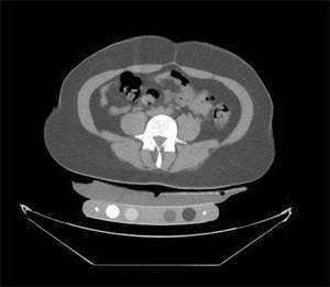
The finding indicates that "obesity does not always protect against osteoporosis," study investigator Dr. Miriam A. Bredella said in a press briefing at the annual meeting of the Radiological Society of North America. "Excessive visceral fat is not only a risk factor for heart disease and diabetes, but also for bone loss."
The study flies in the face of current thinking that obesity actually protects against osteoporosis. Previous studies suggesting a link between fat and bone health focused primarily on body mass index (BMI), which incorporates measures of muscle and bone mass and subcutaneous fat as well as visceral fat, she said. The present study zeroed in specifically on visceral fat.
Dr. Bredella noted "disturbing pictures emerging from the obesity epidemic, because the number of forearm fractures among young patients has increased dramatically over the last year, and the strongest risk factor in that group ... was actually increased body weight." This finding prompted the investigators to see whether there was a connection between osteoporosis and fat, said Dr. Bredella of Massachusetts General Hospital and Harvard Medical School, both in Boston
In the present study, 50 premenopausal women with a BMI of 19-46 kg/m2 (mean 30) underwent a magnetic resonance spectroscopy exam to assess L4 bone marrow (BM) fat, followed by quantitative computed tomography to assess trabecular bone mineral density (BMD).
The results showed a positive correlation between visceral fat and BM fat (r = 0.28) and an inverse association between visceral fat and BMD (r = –0.31) and between vertebral BM fat and BMD (r = –0.45). These results were statistically significant. There was no correlation between either subcutaneous fat (fat concentrated around the hips and thighs) or total body fat and either BM fat or BMD.
These results reveal the distinctly detrimental effect of abdominal obesity on bone health, Dr. Bredella said.
The study is among the first to explore the relationship between body fat and bone marrow fat, and the dynamic appears to be complex, she said in an interview. According to recent research, "the amount of fat within your bones could predict if you will develop a fracture independent of bone mineral density," she noted. A recent study by Dr. Bredella and her colleagues found that women with anorexia nervosa had three times the amount of bone marrow fat as did normal-weight women.
Dr. Bredella had no financial disclosures.
CHICAGO– Visceral obesity was associated with low bone mineral density in a study of premenopausal women, indicating that abdominal fat is a risk factor for osteoporosis.
The finding indicates that "obesity does not always protect against osteoporosis," study investigator Dr. Miriam A. Bredella said in a press briefing at the annual meeting of the Radiological Society of North America. "Excessive visceral fat is not only a risk factor for heart disease and diabetes, but also for bone loss."
The study flies in the face of current thinking that obesity actually protects against osteoporosis. Previous studies suggesting a link between fat and bone health focused primarily on body mass index (BMI), which incorporates measures of muscle and bone mass and subcutaneous fat as well as visceral fat, she said. The present study zeroed in specifically on visceral fat.
Dr. Bredella noted "disturbing pictures emerging from the obesity epidemic, because the number of forearm fractures among young patients has increased dramatically over the last year, and the strongest risk factor in that group ... was actually increased body weight." This finding prompted the investigators to see whether there was a connection between osteoporosis and fat, said Dr. Bredella of Massachusetts General Hospital and Harvard Medical School, both in Boston
In the present study, 50 premenopausal women with a BMI of 19-46 kg/m2 (mean 30) underwent a magnetic resonance spectroscopy exam to assess L4 bone marrow (BM) fat, followed by quantitative computed tomography to assess trabecular bone mineral density (BMD).
The results showed a positive correlation between visceral fat and BM fat (r = 0.28) and an inverse association between visceral fat and BMD (r = –0.31) and between vertebral BM fat and BMD (r = –0.45). These results were statistically significant. There was no correlation between either subcutaneous fat (fat concentrated around the hips and thighs) or total body fat and either BM fat or BMD.
These results reveal the distinctly detrimental effect of abdominal obesity on bone health, Dr. Bredella said.
The study is among the first to explore the relationship between body fat and bone marrow fat, and the dynamic appears to be complex, she said in an interview. According to recent research, "the amount of fat within your bones could predict if you will develop a fracture independent of bone mineral density," she noted. A recent study by Dr. Bredella and her colleagues found that women with anorexia nervosa had three times the amount of bone marrow fat as did normal-weight women.
Dr. Bredella had no financial disclosures.
FROM THE ANNUAL MEETING OF THE RADIOLOGICAL SOCIETY OF NORTH AMERICA
Light Exercise Best to Prevent Knee OA
CHICAGO – The wisdom about practicing everything in moderation also may hold true for knees, especially in middle-aged adults with risk factors for osteoarthritis.
Light exercise, such as walking, appears to protect against OA, but the extremes – a sedentary lifestyle or more aggressive workouts – may accelerate OA onset in at-risk individuals, according to baseline data from the Osteoarthritis Initiative, a longitudinal, multicenter, observational study funded by the National Institutes of Health.
Osteoarthritis risk can be reduced by avoiding aggressive exercise and exercising safely, said the study’s lead author, Dr. Thomas M. Link, professor of radiology and chief of musculoskeletal imaging at the University of California, San Francisco.
Among at-risk patients, light exercisers had significantly less degeneration of the cartilage surrounding the knees on magnetic resonance imaging (MRI) than did those whose exercise routines were characterized as either minimal or moderate to strenuous.
Middle-age adults, particularly those with OA risk factors, need to be "extremely careful" with their cartilage, Dr. Link said in a press briefing Nov. 29 during the annual meeting of the Radiological Society of North America. "Once cartilage is gone, it’s gone forever."
This doesn’t mean that patients should stop running or playing tennis, but it does mean they must make sure they are exercising safely to avoid sustaining an injury that could "initiate an osteoarthritis cascade, which will be very difficult to stop," he said.
Research associate Keegan K. Hovis, R.N., said the key for patients who exercise strenuously is to focus on modifiable risk factors, such as maintaining a healthy weight and strengthening the knee stabilizing muscles, including the quadriceps.
Subjects included 132 at-risk patients and 33 controls matched for age and body mass index (99 women and 66 men between the ages of 45 and 55 years, BMI range of 18-27 kg/m2). Risk factors for OA included a previous knee injury or knee surgery, a family history of total knee replacement surgery, bone spurs on the fingers, and occasional knee symptoms.
Based on responses to the leisure activity component of the Physical Activity Scale for the Elderly, study participants were stratified according to intensity of exercise habits into sedentary, light, or moderate-strenuous exercisers. Patients also were grouped by whether they engaged in frequent knee-bending activities. These activities included climbing at least 10 flights of stairs daily, lifting objects weighing more than 25 pounds, or squatting, kneeling, or deep-knee-bending for at least 30 minutes per day.
Sedentary exercisers walked less than or equal to 2 days a week for less than 2 hours per day. Light exercisers walked or participated in other forms of light exercise such as table tennis or Frisbee at least 3 days a week for less than 2 hours a day. Moderate-strenuous exercisers engaged in sports such as running, tennis, or soccer at least 3 days a week for greater than or equal to 1 hour per day.
To assess cartilage health, researchers used T2 mapping, a quantitative and qualitative MRI technique that, unlike anatomic imaging, reveals the chemical composition and structure of the cartilage (including collagen and water content) and can provide an image marker of cartilage degeneration in its earliest stages.
"T2 values can detect changes in cartilage at the molecular level, prior to irreversible changes in the structure; therefore, it has the potential to identify, with the earliest signs of degeneration, who may benefit from early treatment or behavioral intervention," said Dr. Link.
Two radiologists analyzed T2 values in five cartilage areas – patella, medial femur, medial tibia, lateral femur, and lateral tibia – and graded overall visible signs of cartilage and meniscus damage using a modified whole-organ MRI scoring method (WORMS).
Among the 132 at-risk patients, T2 values were significantly lower, indicating less cartilage degeneration, in light exercisers than sedentary and moderate-strenuous exercisers in the overall average of all compartments (43.5 vs. 44.5 and 45.0, respectively) and in the lateral tibia (38.0 vs. 39.7 and 40.2). "Light exercise was associated with more intact collagen structure and lower cartilage water content, which are indicative of healthier cartilage," said Ms. Hovis.
Moderate-strenuous exercise in women was significantly correlated with higher T2 values in the medial femur, indicating that moderate-strenuous exercise may accelerate cartilage degeneration and increase osteoarthritis risk in women who engage in more intense workouts, she reported.
Differences in cartilage health were not found in any of the three exercise groups in the control cohort. However, knee-bending activities were associated with higher T2 values in both the at-risk and control cohorts. In the at-risk cohort, those who engaged in frequent knee-bending activities had significantly higher T2 values in all compartments than those who did not (44.7 vs. 43.2) and a significantly higher WORMS score, indicating visible signs of cartilage degeneration. In the normal cohort, knee-bending activities were associated with higher T2 values in the medial femur and the medial tibia (49.9 vs. 47.0 and 35.4 vs. 32.5, respectively) with a trend toward significance in the overall average in all compartments.
"Knee-bending activity may accelerate cartilage degeneration in all individuals, but potentially to a greater degree in subjects already at risk for knee osteoarthritis," Ms. Hovis said.
The researchers plan to collect longitudinal data at 2 and 4 years and are finishing an analysis of data on strength training.
Dr. Link disclosed that he has received research support from Merck & Co. Inc.
FROM THE ANNUAL MEETING OF THE RADIOLOGICAL SOCIETY OF NORTH AMERICA