User login
Preconception Health Care
CE/CME No: CR-1408
PROGRAM OVERVIEW
Earn credit by reading this article and successfully completing the posttest and evaluation. Successful completion is defined as a cumulative score of at least 70% correct.
EDUCATIONAL OBJECTIVES
• Define preconception and interconception health care and explain how these concepts relate to primary care.
• Describe three common clinical presentations in reproductive-age women that have implications for preconception and interconception health care.
• Explain the considerations, in terms of potential pregnancies, when prescribing pharmacologic treatment for reproductive-age women.
• Discuss examples of cultural factors and beliefs that may affect preconception health counseling provided to women of reproductive age.
FACULTY
Kathleen A. Ahonen is an Assistant Professor and Colleen Quinlan is an Associate Professor at the University of Toledo College of Nursing.
The authors have no significant financial relationships to disclose.
ACCREDITATION STATEMENT
![]()
Article begins on next page >>
Because half of all pregnancies in the United States are unplanned, primary care constitutes preconception health care for women ages 15 to 44. Here are recommendations to incorporate into routine visits to improve outcomes of possible pregnancies.
Traditional interventions to improve pregnancy outcomes in the United States have focused on early, consistent prenatal care and on reducing the teenage pregnancy rate. Data indicate that early access to prenatal care has increased and that the teenage pregnancy rate has fallen.1,2

But while US rates of preterm births, low–birth-weight infants, and infant mortality have all declined since the mid-2000s,3 they remain higher than those in many other developed countries.4-6 Furthermore, there are significant differences in these rates by race and ethnicity.3
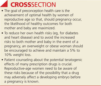
Many experts believe that waiting until a woman is pregnant to address the health needs and behaviors that adversely affect pregnancy outcomes is, at best, an incomplete solution to these problems. A more comprehensive view of women’s health care broadens this focus to stress the importance of preconception health care throughout the reproductive years. It is key to the achievement of optimal health, not only for women but also for their potential future children.7-9
The CDC defines preconception health simply as the health of women and men during their reproductive years. Preconception health care is defined as medical care during those years (for women, ages 15 to 44) that focuses on improving aspects of the patient’s health or health behaviors that can improve outcomes in the event of a pregnancy.10 The term interconception health care is also applied specifically to health care provided during the time between pregnancies.
This article highlights obesity, depression and other mood disorders, and nutritional deficiencies and reviews potential risk factors, particularly occurring very early in a pregnancy. Recommendations are offered for more effective health counseling at routine visits, with the goal of improving outcomes of potential pregnancies.
PRIMARY CARE IS PRECONCEPTION CARE
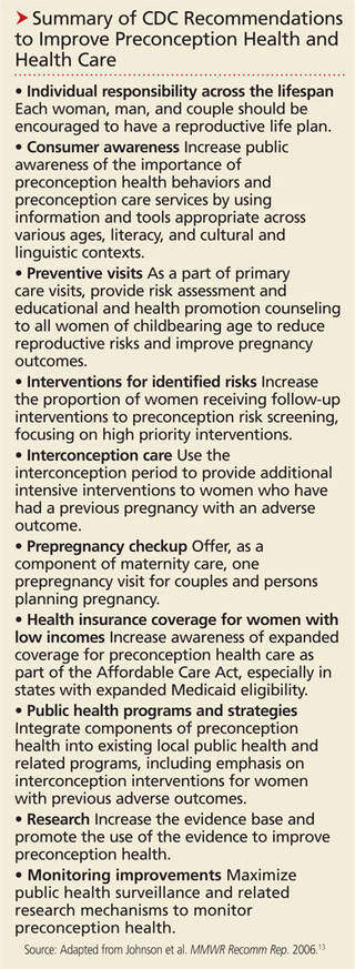
Researchers have reported that only about 50% of reproductive-age women receive counseling about health behaviors and pregnancy planning8 and that such counseling is even rarer for adolescent females.11,12 Because women of childbearing age commonly see both Ob-Gyn and family practice clinicians for health care, primary care providers are well positioned to deliver preconception health care by integrating health counseling, discussion of health behaviors, and primary and secondary prevention into patient care for reproductive-age women (see "Summary of CDC Recommendations to Improve Preconception Health and Health Care").6
On the next page: Obesity >>
OBESITY
Researchers in both the US and internationally have noted the connection between obesity and pregnancy complications such as gestational diabetes that increase the risk for poor pregnancy outcomes.13-15 Indeed, pregnancy outcomes can be altered for the better with weight loss in the preconception or interconception period.16
Weight loss is complex; it can be difficult to accomplish, and long-term maintenance also presents a challenge. Short-term weight loss is achievable, however, and can produce beneficial metabolic effects, such as a decrease in insulin resistance.17
Before weight loss attempts can be initiated, the overweight or obese patient’s weight must be acknowledged as a health problem. In one 2009 study, Callaway et al found that in their sample of 412 overweight and obese women, only 17% were advised by their health care providers to lose weight.18
Regular physical activity can facilitate the achievement and maintenance of a healthy weight. However, physical inactivity is common in nonpregnant women, especially those with higher BMIs. The CDC’s Pregnancy Risk Assessment Monitoring System (PRAMS; www.cdc.gov/prams), widely regarded as an important source of data about women’s health behaviors, reveals that nearly 40% of US women do not meet national recommendations for routine physical activity in the three months prior to pregnancy.19
Recent obesity guidelines recommend initial weight loss goals of 5% to 10% of baseline weight in six months before pregnancy.17 Some women may not achieve these goals because they are unaware of the potentially short time period between discontinuation of contraception and conception. Others, especially those older than 30, intentionally plan for short interconception intervals as part of their family planning strategy, which does not allow enough time for safe weight loss.20
Primary care clinicians should counsel these patients on the importance of a healthy weight and regular physical activity to the maintenance of optimal health. Particular emphasis should be placed on achieving weight loss slowly and safely and then maintaining it, at least for the short term. This strategy may be effective in helping a female patient achieve a lower BMI prior to pregnancy.
Bariatric surgery
Because the number of women undergoing bariatric surgery for morbid obesity is rapidly increasing,21 it is important to educate them about the specific effects of such procedures on reproductive health. A period of rapid weight loss—such as occurs after bariatric surgery—is not a time to consider pregnancy, despite the improvement in eventual pregnancy outcomes associated with a healthier BMI.
Many of these women may have been anovulatory when morbidly obese and unaware that fertility increases in the postoperative year, after a reduced BMI is achieved. If ovulation and menses have not yet normalized, a woman may become pregnant and not know it. This could result in the inadvertent exposure of the developing fetus to such teratogenic risks as alcohol, tobacco, and certain prescription drugs. While a pregnancy may be welcome, better outcomes are likely if pregnancy is avoided for at least 12 months after bariatric surgery.22 Effective contraception should be used until ovulation cycles stabilize.
Some surgical weight loss procedures, such as the Roux-en-Y gastric bypass, may alter the absorption of medication, including oral contraceptives (OCs), making the use of OCs after such procedures less than ideal. The more reliably absorbed injectable medroxyprogesterone is an option, but women who have undergone bariatric surgery often wish to avoid the associated risk for weight gain. Nonhormonal long-acting contraceptive methods not associated with weight gain, such as the copper intrauterine device, may be preferable for use in the first year postprocedure.21
On the next page: Depression and other mood disorders >>
DEPRESSION AND OTHER MOOD DISORDERS
Mood disorders include depression, bipolar disorder, and anxiety disorders. Selective serotonin reuptake inhibitors (SSRIs, such as paroxetine or sertraline) or serotonin and norepinephrine reuptake inhibitors (SNRIs, such as venlafaxine or duloxetine) are often prescribed for primary care management of depression and other mood disorders.
When SSRIs or SNRIs are not fully effective, clinicians may refer patients to mental health specialists for consultation and possible ongoing management. Women of reproductive age who receive specialty care for mood disorders are encouraged to continue their regular visits to primary care clinicians.
Medication: Risk for birth defects
Anticonvulsants, such as valproate, carbamazepine, and lamotrigine, are commonly used to treat bipolar disorder.23 When taken during the first trimester of pregnancy, these drugs pose well-documented risks to the rapidly developing fetus. Most evidence relates to the risk for neural tube defects, such as spina bifida, but other evidence suggests a risk for general cognitive impairment after prenatal valproate exposure. While the latter is based primarily on studies of women taking anti-epileptic drugs for seizure control—not psychiatric diagnoses—first-trimester risks appear to be independent of maternal seizures.23 Although folic acid supplementation decreases the incidence of neural tube defects (see discussion under “Nutritional Deficiencies"), it is unknown if such supplementation is effective in mitigating the additional risks to the fetus from exposure to anticonvulsants.
Female patients of childbearing age must be advised of the potential effects of these commonly prescribed mood-stabilizing drugs, not only as they relate to the diagnosis being treated but also regarding their possible effects on an early, undiagnosed pregnancy. Unfortunately, evidence indicates that insufficient attention is given to counseling reproductive-age women about the risks and benefits of these drugs as they relate to potential conception, at least in the context of specialty care.23 Therefore, the primary care clinician and the specialist should utilize a team approach, emphasizing careful reproductive planning to avoid pregnancy while under treatment with these drugs to ensure the best possible outcomes.
In the context of potential pregnancy, the need to manage depression and other mood disorders effectively is particularly important: Prepregnancy depressive mood has been significantly associated with preterm birth, and at least 14.5% of women experience a new episode of depression during pregnancy.24 Thus, effective treatment of mood disorders should be a priority, both as part of preconception care and during pregnancy.
Similarly, treatment strategies for postpartum depression—widely estimated to affect 10% to 20% of new mothers—must consider the potential risks of pharmacologic therapy to a fetus should the patient conceive during treatment.
On the next page: Nutritional deficiencies >>
NUTRITIONAL DEFICIENCIES
While there is widespread public awareness, at least on a basic level, of the importance of good nutrition during pregnancy, what that constitutes is not necessarily clearly understood. Even less well recognized is the importance of a woman’s nutritional status at the time of conception, at preimplantation, and during the early weeks of placental development, before pregnancy is known or confirmed. During this crucial time—three to seven weeks after the last menstrual period—an inadequate diet may result in low–birth-weight infants with lifelong health problems.25 These may include respiratory problems associated with barotrauma from ventilation at birth; neural tube defects; and orofacial clefts.25
Because of inadequate intake of fresh fruits and vegetables, many reproductive-age women in the US are deficient in vitamins A, C, B6, and E, as well as calcium, iron, zinc, magnesium, and folic acid. Although vitamin and mineral supplements are readily available, little clinical research—with the exception of folic acid—has been done on the efficacy of such supplementation.26
Until more is known, intake of these dietary components is best achieved as part of a well-balanced diet; however, this recommendation may need to be modified for African-American women. In a recent retrospective study of almost 2,500 white and African-American women who took a multivitamin supplement consistently during the month before conception, supplementation was associated with increased infant birth weight in the infants born to African-American women but not in those born to white women.26
Folic acid
In the specific case of folic acid, the crucial importance of preconception intake by reproductive-age women is hard to overstate. A well-established body of research supports supplementation to reduce the incidence of neural tube defects that may occur very early in development, before many women are aware of a pregnancy.25-27 However, it is estimated that only a minority of reproductive-age women take a regular folic acid supplement. This may be particularly true of women who are actively avoiding pregnancy and using regular contraception. Patients need to be educated that, as effective as current contraceptive methods are known to be, each method has a typical user failure rate, meaning that actual effectiveness is lower than theoretical effectiveness.
Considering that half of US pregnancies are unintended, with some occurring as a result of contraceptive method or user failures, the FDA Advisory Committee unanimously endorsed the concept of using OCs as a vehicle for folate supplementation.27 There are currently two FDA-approved OCs fortified with the equivalent of 0.4 mg of folic acid. Both contain drospirenone and therefore present a somewhat elevated risk for blood clots, especially in the first year of use.28,29 While this risk is small compared to the incidence of blood clots during pregnancy, a careful history should be taken to avoid prescribing these products to patients already at increased risk for blood clots (eg, obese women, smokers [even light smokers], those with a history of a blood clot after surgery or a motor vehicle accident). For women without risk factors, folic acid–supplemented OCs may be very beneficial should they become pregnant unintentionally or quickly after stopping contraception.
On the next page: Cultural considerations and conclusion >>
CULTURAL CONSIDERATIONS
Not all racial, ethnic, and socioeconomic groups consider health and pregnancy in the same cultural context, and nonmedical factors may affect health behaviors and sources of health counseling. Studies of women of different cultural backgrounds are illustrative.
In one study, increasing women’s evidence-based knowledge of preconception and interconception health behaviors, using group education and peer support, was shown to produce attitudinal and behavior changes in a sample of reproductive-age rural white women, especially with regard to nutrition and physical activity in the preconception period.30
Another study of primarily low-income Latina women with low levels of acculturation revealed that they had good understanding of the importance of attention to health once pregnancy is confirmed. However, they expressed much less belief in the ability of a woman to control her own preconception general health.31
A third study involving a sample of African-American women found that the women saw no clear role for preconception or interconception care through health care visits with primary care clinicians; rather, they looked to their social and cultural communities and families for such support.32
These diverse results suggest that both community-wide education and one-to-one health counseling are needed to effect improvements in health behaviors and knowledge. Subtle differences in cultural context must be recognized by health care providers who interact with a broad range of reproductive-age women.
CONCLUSION
Guidelines from the American Academy of Pediatrics, the American Congress of Obstetricians and Gynecologists, and the CDC all endorse the integration of preconception care into primary care encounters.33-36 The concept of preconception health care offers clinicians the opportunity to greatly influence the health of reproductive-age women in the primary care setting, with the potential to achieve small but clinically significant changes in health behaviors. Complications of pregnancy and poor pregnancy outcomes may be reduced when the overall health status of women of reproductive age is addressed, with awareness and mitigation of factors known to produce negative pregnancy outcomes.
There is a need, however, for ongoing research to develop effective, evidence-based strategies for use by primary care clinicians in the effort to improve women’s preconception health and, ultimately, pregnancy outcomes.
1. National Center for Health Statistics. Table 7: Prenatal care for live births, by detailed race and Hispanic origin of mother: United States, selected years 1970–2004. In: Health, United States, 2006. Report No: 2006-1232. www.ncbi.nlm.nih.gov/books/NBK21003. Accessed July 17, 2014.
2. Office of Adolescent Health, US Department of Health and Human Services. Trends in teen pregnancy and childbearing. www.hhs.gov/ash/oah/adolescent-health-topics/reproductive-health/teen-pregnancy/trends.html#_ftn6. Accessed July 17, 2014.
3. Maternal and Child Health Bureau, Health Resources and Services Administration, US Department of Health and Human Services. Child Health USA 2013. http://mchb.hrsa.gov/chusa13. Accessed July 17, 2014.
4. World Health Organization. Born Too Soon: The Global Action Report on Preterm Birth. www.who.int/pmnch/media/news/2012/201204_borntoosoon_countryranking.pdf. Accessed July 17, 2014.
5. Organization for Economic Cooperation and Development (OECD). Low birth weight infants, 2009 and change 1970-2009 (or nearest year). In: Health at a Glance 2011: OECD indicators. www.oecd.org/els/health-systems/49105858.pdf. Accessed July 17, 2014.
6. CDC. Infant mortality rates and international rankings. www.cdc.gov/nchs/data/hus/2013/016.pdf. Accessed July 17, 2014.
7. Livingood WC, Brady C, Pierce K, et al. Impact of pre-conception health care: evaluation of a social determinants focused intervention. Matern Child Health J. 2010;14:382-391.
8. Hillemeier MM, Weisman CS, Chase GA, et al. Women’s preconceptional health and use of health services: implications for preconception care. Health Serv Res. 2008;43(1):54-75.
9. Chuang CH, Weisman CS, Hillemeier MM, et al. Pregnancy intention and health behaviors: results from the Central Pennsylvania Women’s Health Study Cohort. Matern Child Health J. 2010;14:501-510.
10. CDC. Preconception health and health care. www.cdc.gov/preconcep tion/overview.html. Accessed July 17, 2014.
11. Heavey E. Don’t miss preconception care opportunities for adolescents. Am J Matern Child Nurs. 2010;35(4):213-219.
12. Hoover KW, Tao G, Berman S, Kent CK. Utilization of health services in physician offices and outpatient clinics by adolescents and young women in the United States: implications for improving access to reproductive health services. J Adolesc Health. 2010;46:324-330.
13. Johnson K, Posner SF, Biermann J. Recommendations to improve preconception health and health care—United States. MMWR Recomm Rep. 2006;55:1-23.
14. Krishnamoorthy U, Schram CM, Hill SR. Maternal obesity in pregnancy: is it time for meaningful research to inform preventive and management strategies? Brit J Obstet Gynecol. 2006;113:1134-1140.
15. Callaway LK, Prins JB, Chang AM, McIntyre HD. The prevalence and impact of overweight and obesity in an Australian obstetric population. Med J Aust. 2006;184:56-59.
16. Clark AM, Thornley B, Tomlinson L, et al. Weight loss in obese infertile women results in improvement in reproductive outcome for all forms of fertility treatment. Hum Reprod. 1998;13(6):1502-1505.
17. Jensen MD, Ryan DH, Apovian CM, et al. 2013 AHA/ACC/TOS guideline for the management of overweight and obesity in adults: a report of the American College of Cardiology/American Heart Association task force on practice guidelines and the Obesity Society. Circulation. http://circ.ahajournals.org/content/early/2013/11/11/01.cir.0000437739.71477.ee.full.pdf+html. Accessed July 17, 2014.
18. Callaway LK, O’Callaghan MJ, McIntyre HD. Barriers to addressing overweight and obesity before conception. Med J Aust. 2009;191(8):425-427.
19. Donahue SMA, Zimmerman FJ, Starr JR, Holt VL. Correlates of pre-pregnancy physical inactivity: results from the pregnancy risk assessment monitoring system. Matern Child Health J. 2010;14:235-244.
20. Gemmill A, Lindberg LD. Short interpregnancy intervals in the United States. Obstet Gynecol. 2013;122(1):64-71.
21. American College of Obstetricians and Gynecologists Committee on Obstetric Practice. Obesity in pregnancy. Obstet Gynecol. 2013;121(1):213-217.
22. Ciangura C, Corigliano N, Basdevant A, et al. Etonogestrel concentrations in morbidly obese women following Roux-en-Y gastric bypass surgery: three case reports. Contraception. 2011;84:649-651.
23. Wieck A, Rao S, Sein K, Haddad PM. A survey of antiepileptic prescribing to women of childbearing potential in psychiatry. Arch Womens Ment Health. 2007;10:83-85.
24. Gavin AR, Chae DH, Mustillo S, Kiefe CI. Prepregnancy depressive mood and preterm birth in black and white women: findings from the CARDIA study. J Womens Health. 2009;18(6):803-811.
25. Gardiner PM, Nelson L, Shellhaas CS, et al. The clinical content of preconception care: nutrition and dietary supplements. Am J Obstet Gynecol. 2008;S345-S353.
26. Burris HH, Mitchell AA, Werler MM. Periconceptional multivitamin use and infant birth weight disparities. Ann Epidemiol. 2010;20(3):233-240.
27. Taylor TN, Farkouh RA, Graham JB, et al. Potential reduction in neural tube defects associated with use of Metafolin-fortified oral contraceptives in the United States. Am J Obstet Gynecol. 2011:e1-e8.
28. Beyaz [package insert]. Wayne, NJ: Bayer HealthCare, 2010.
29. Safyral [package insert]. Wayne, NJ: Bayer HealthCare, 2010.
30. Hillemeier MM, Downs DS, Feinberg ME, et al. Improving women’s preconceptional health: findings from a randomized trial of the Strong Healthy Women intervention in the Central Pennsylvania Women’s Health Study. Womens Health Issues. 2008;18(6 suppl):S87-S96.
31. Coonrod DV, Bruce NC, Malcolm TD, et al. Knowledge and attitudes regarding preconception care in a predominantly low-income Mexican American population. Am J Obstet Gynecol. 2009;686:e1-e7.
32. Canady RB, Tiedje LB, Lauber C. Preconception care and pregnancy planning: voices of African American women. Matern Child Nurs. 2008;33(2):90-97.
33. Bellanca HK, Hunter MS. ONE KEY QUESTION: preventive reproductive health is part of high quality primary care. Contraception. 2013;88(1):3-6.
34. American Academy of Pediatrics. Preconception health care. www.healthychildren.org/English/ages-stages/prenatal/Pages/Reduce-the-Risk-of-Birth-Defects.aspx. Accessed July 17, 2014.
35. American College of Obstetricians and Gynecologists Committee on Gynecologic Practice. The importance of preconception care in the continuum of women’s health care. www.acog.org/Resources_And_Publi cations/Committee_Opinions/Committee_on_Gynecologic_Practice/The_Importance_of_Preconception_Care_in_the_Continuum_of_Wom ens_Health_Care. Accessed July 17, 2014.
36. CDC. Preconception health and health care. www.cdc.gov/preconcep tion/hcp/. Accessed July 17, 2014.
CE/CME No: CR-1408
PROGRAM OVERVIEW
Earn credit by reading this article and successfully completing the posttest and evaluation. Successful completion is defined as a cumulative score of at least 70% correct.
EDUCATIONAL OBJECTIVES
• Define preconception and interconception health care and explain how these concepts relate to primary care.
• Describe three common clinical presentations in reproductive-age women that have implications for preconception and interconception health care.
• Explain the considerations, in terms of potential pregnancies, when prescribing pharmacologic treatment for reproductive-age women.
• Discuss examples of cultural factors and beliefs that may affect preconception health counseling provided to women of reproductive age.
FACULTY
Kathleen A. Ahonen is an Assistant Professor and Colleen Quinlan is an Associate Professor at the University of Toledo College of Nursing.
The authors have no significant financial relationships to disclose.
ACCREDITATION STATEMENT
![]()
Article begins on next page >>
Because half of all pregnancies in the United States are unplanned, primary care constitutes preconception health care for women ages 15 to 44. Here are recommendations to incorporate into routine visits to improve outcomes of possible pregnancies.
Traditional interventions to improve pregnancy outcomes in the United States have focused on early, consistent prenatal care and on reducing the teenage pregnancy rate. Data indicate that early access to prenatal care has increased and that the teenage pregnancy rate has fallen.1,2
But while US rates of preterm births, low–birth-weight infants, and infant mortality have all declined since the mid-2000s,3 they remain higher than those in many other developed countries.4-6 Furthermore, there are significant differences in these rates by race and ethnicity.3
Many experts believe that waiting until a woman is pregnant to address the health needs and behaviors that adversely affect pregnancy outcomes is, at best, an incomplete solution to these problems. A more comprehensive view of women’s health care broadens this focus to stress the importance of preconception health care throughout the reproductive years. It is key to the achievement of optimal health, not only for women but also for their potential future children.7-9
The CDC defines preconception health simply as the health of women and men during their reproductive years. Preconception health care is defined as medical care during those years (for women, ages 15 to 44) that focuses on improving aspects of the patient’s health or health behaviors that can improve outcomes in the event of a pregnancy.10 The term interconception health care is also applied specifically to health care provided during the time between pregnancies.
This article highlights obesity, depression and other mood disorders, and nutritional deficiencies and reviews potential risk factors, particularly occurring very early in a pregnancy. Recommendations are offered for more effective health counseling at routine visits, with the goal of improving outcomes of potential pregnancies.
PRIMARY CARE IS PRECONCEPTION CARE
Researchers have reported that only about 50% of reproductive-age women receive counseling about health behaviors and pregnancy planning8 and that such counseling is even rarer for adolescent females.11,12 Because women of childbearing age commonly see both Ob-Gyn and family practice clinicians for health care, primary care providers are well positioned to deliver preconception health care by integrating health counseling, discussion of health behaviors, and primary and secondary prevention into patient care for reproductive-age women (see "Summary of CDC Recommendations to Improve Preconception Health and Health Care").6
On the next page: Obesity >>
OBESITY
Researchers in both the US and internationally have noted the connection between obesity and pregnancy complications such as gestational diabetes that increase the risk for poor pregnancy outcomes.13-15 Indeed, pregnancy outcomes can be altered for the better with weight loss in the preconception or interconception period.16
Weight loss is complex; it can be difficult to accomplish, and long-term maintenance also presents a challenge. Short-term weight loss is achievable, however, and can produce beneficial metabolic effects, such as a decrease in insulin resistance.17
Before weight loss attempts can be initiated, the overweight or obese patient’s weight must be acknowledged as a health problem. In one 2009 study, Callaway et al found that in their sample of 412 overweight and obese women, only 17% were advised by their health care providers to lose weight.18
Regular physical activity can facilitate the achievement and maintenance of a healthy weight. However, physical inactivity is common in nonpregnant women, especially those with higher BMIs. The CDC’s Pregnancy Risk Assessment Monitoring System (PRAMS; www.cdc.gov/prams), widely regarded as an important source of data about women’s health behaviors, reveals that nearly 40% of US women do not meet national recommendations for routine physical activity in the three months prior to pregnancy.19
Recent obesity guidelines recommend initial weight loss goals of 5% to 10% of baseline weight in six months before pregnancy.17 Some women may not achieve these goals because they are unaware of the potentially short time period between discontinuation of contraception and conception. Others, especially those older than 30, intentionally plan for short interconception intervals as part of their family planning strategy, which does not allow enough time for safe weight loss.20
Primary care clinicians should counsel these patients on the importance of a healthy weight and regular physical activity to the maintenance of optimal health. Particular emphasis should be placed on achieving weight loss slowly and safely and then maintaining it, at least for the short term. This strategy may be effective in helping a female patient achieve a lower BMI prior to pregnancy.
Bariatric surgery
Because the number of women undergoing bariatric surgery for morbid obesity is rapidly increasing,21 it is important to educate them about the specific effects of such procedures on reproductive health. A period of rapid weight loss—such as occurs after bariatric surgery—is not a time to consider pregnancy, despite the improvement in eventual pregnancy outcomes associated with a healthier BMI.
Many of these women may have been anovulatory when morbidly obese and unaware that fertility increases in the postoperative year, after a reduced BMI is achieved. If ovulation and menses have not yet normalized, a woman may become pregnant and not know it. This could result in the inadvertent exposure of the developing fetus to such teratogenic risks as alcohol, tobacco, and certain prescription drugs. While a pregnancy may be welcome, better outcomes are likely if pregnancy is avoided for at least 12 months after bariatric surgery.22 Effective contraception should be used until ovulation cycles stabilize.
Some surgical weight loss procedures, such as the Roux-en-Y gastric bypass, may alter the absorption of medication, including oral contraceptives (OCs), making the use of OCs after such procedures less than ideal. The more reliably absorbed injectable medroxyprogesterone is an option, but women who have undergone bariatric surgery often wish to avoid the associated risk for weight gain. Nonhormonal long-acting contraceptive methods not associated with weight gain, such as the copper intrauterine device, may be preferable for use in the first year postprocedure.21
On the next page: Depression and other mood disorders >>
DEPRESSION AND OTHER MOOD DISORDERS
Mood disorders include depression, bipolar disorder, and anxiety disorders. Selective serotonin reuptake inhibitors (SSRIs, such as paroxetine or sertraline) or serotonin and norepinephrine reuptake inhibitors (SNRIs, such as venlafaxine or duloxetine) are often prescribed for primary care management of depression and other mood disorders.
When SSRIs or SNRIs are not fully effective, clinicians may refer patients to mental health specialists for consultation and possible ongoing management. Women of reproductive age who receive specialty care for mood disorders are encouraged to continue their regular visits to primary care clinicians.
Medication: Risk for birth defects
Anticonvulsants, such as valproate, carbamazepine, and lamotrigine, are commonly used to treat bipolar disorder.23 When taken during the first trimester of pregnancy, these drugs pose well-documented risks to the rapidly developing fetus. Most evidence relates to the risk for neural tube defects, such as spina bifida, but other evidence suggests a risk for general cognitive impairment after prenatal valproate exposure. While the latter is based primarily on studies of women taking anti-epileptic drugs for seizure control—not psychiatric diagnoses—first-trimester risks appear to be independent of maternal seizures.23 Although folic acid supplementation decreases the incidence of neural tube defects (see discussion under “Nutritional Deficiencies"), it is unknown if such supplementation is effective in mitigating the additional risks to the fetus from exposure to anticonvulsants.
Female patients of childbearing age must be advised of the potential effects of these commonly prescribed mood-stabilizing drugs, not only as they relate to the diagnosis being treated but also regarding their possible effects on an early, undiagnosed pregnancy. Unfortunately, evidence indicates that insufficient attention is given to counseling reproductive-age women about the risks and benefits of these drugs as they relate to potential conception, at least in the context of specialty care.23 Therefore, the primary care clinician and the specialist should utilize a team approach, emphasizing careful reproductive planning to avoid pregnancy while under treatment with these drugs to ensure the best possible outcomes.
In the context of potential pregnancy, the need to manage depression and other mood disorders effectively is particularly important: Prepregnancy depressive mood has been significantly associated with preterm birth, and at least 14.5% of women experience a new episode of depression during pregnancy.24 Thus, effective treatment of mood disorders should be a priority, both as part of preconception care and during pregnancy.
Similarly, treatment strategies for postpartum depression—widely estimated to affect 10% to 20% of new mothers—must consider the potential risks of pharmacologic therapy to a fetus should the patient conceive during treatment.
On the next page: Nutritional deficiencies >>
NUTRITIONAL DEFICIENCIES
While there is widespread public awareness, at least on a basic level, of the importance of good nutrition during pregnancy, what that constitutes is not necessarily clearly understood. Even less well recognized is the importance of a woman’s nutritional status at the time of conception, at preimplantation, and during the early weeks of placental development, before pregnancy is known or confirmed. During this crucial time—three to seven weeks after the last menstrual period—an inadequate diet may result in low–birth-weight infants with lifelong health problems.25 These may include respiratory problems associated with barotrauma from ventilation at birth; neural tube defects; and orofacial clefts.25
Because of inadequate intake of fresh fruits and vegetables, many reproductive-age women in the US are deficient in vitamins A, C, B6, and E, as well as calcium, iron, zinc, magnesium, and folic acid. Although vitamin and mineral supplements are readily available, little clinical research—with the exception of folic acid—has been done on the efficacy of such supplementation.26
Until more is known, intake of these dietary components is best achieved as part of a well-balanced diet; however, this recommendation may need to be modified for African-American women. In a recent retrospective study of almost 2,500 white and African-American women who took a multivitamin supplement consistently during the month before conception, supplementation was associated with increased infant birth weight in the infants born to African-American women but not in those born to white women.26
Folic acid
In the specific case of folic acid, the crucial importance of preconception intake by reproductive-age women is hard to overstate. A well-established body of research supports supplementation to reduce the incidence of neural tube defects that may occur very early in development, before many women are aware of a pregnancy.25-27 However, it is estimated that only a minority of reproductive-age women take a regular folic acid supplement. This may be particularly true of women who are actively avoiding pregnancy and using regular contraception. Patients need to be educated that, as effective as current contraceptive methods are known to be, each method has a typical user failure rate, meaning that actual effectiveness is lower than theoretical effectiveness.
Considering that half of US pregnancies are unintended, with some occurring as a result of contraceptive method or user failures, the FDA Advisory Committee unanimously endorsed the concept of using OCs as a vehicle for folate supplementation.27 There are currently two FDA-approved OCs fortified with the equivalent of 0.4 mg of folic acid. Both contain drospirenone and therefore present a somewhat elevated risk for blood clots, especially in the first year of use.28,29 While this risk is small compared to the incidence of blood clots during pregnancy, a careful history should be taken to avoid prescribing these products to patients already at increased risk for blood clots (eg, obese women, smokers [even light smokers], those with a history of a blood clot after surgery or a motor vehicle accident). For women without risk factors, folic acid–supplemented OCs may be very beneficial should they become pregnant unintentionally or quickly after stopping contraception.
On the next page: Cultural considerations and conclusion >>
CULTURAL CONSIDERATIONS
Not all racial, ethnic, and socioeconomic groups consider health and pregnancy in the same cultural context, and nonmedical factors may affect health behaviors and sources of health counseling. Studies of women of different cultural backgrounds are illustrative.
In one study, increasing women’s evidence-based knowledge of preconception and interconception health behaviors, using group education and peer support, was shown to produce attitudinal and behavior changes in a sample of reproductive-age rural white women, especially with regard to nutrition and physical activity in the preconception period.30
Another study of primarily low-income Latina women with low levels of acculturation revealed that they had good understanding of the importance of attention to health once pregnancy is confirmed. However, they expressed much less belief in the ability of a woman to control her own preconception general health.31
A third study involving a sample of African-American women found that the women saw no clear role for preconception or interconception care through health care visits with primary care clinicians; rather, they looked to their social and cultural communities and families for such support.32
These diverse results suggest that both community-wide education and one-to-one health counseling are needed to effect improvements in health behaviors and knowledge. Subtle differences in cultural context must be recognized by health care providers who interact with a broad range of reproductive-age women.
CONCLUSION
Guidelines from the American Academy of Pediatrics, the American Congress of Obstetricians and Gynecologists, and the CDC all endorse the integration of preconception care into primary care encounters.33-36 The concept of preconception health care offers clinicians the opportunity to greatly influence the health of reproductive-age women in the primary care setting, with the potential to achieve small but clinically significant changes in health behaviors. Complications of pregnancy and poor pregnancy outcomes may be reduced when the overall health status of women of reproductive age is addressed, with awareness and mitigation of factors known to produce negative pregnancy outcomes.
There is a need, however, for ongoing research to develop effective, evidence-based strategies for use by primary care clinicians in the effort to improve women’s preconception health and, ultimately, pregnancy outcomes.
1. National Center for Health Statistics. Table 7: Prenatal care for live births, by detailed race and Hispanic origin of mother: United States, selected years 1970–2004. In: Health, United States, 2006. Report No: 2006-1232. www.ncbi.nlm.nih.gov/books/NBK21003. Accessed July 17, 2014.
2. Office of Adolescent Health, US Department of Health and Human Services. Trends in teen pregnancy and childbearing. www.hhs.gov/ash/oah/adolescent-health-topics/reproductive-health/teen-pregnancy/trends.html#_ftn6. Accessed July 17, 2014.
3. Maternal and Child Health Bureau, Health Resources and Services Administration, US Department of Health and Human Services. Child Health USA 2013. http://mchb.hrsa.gov/chusa13. Accessed July 17, 2014.
4. World Health Organization. Born Too Soon: The Global Action Report on Preterm Birth. www.who.int/pmnch/media/news/2012/201204_borntoosoon_countryranking.pdf. Accessed July 17, 2014.
5. Organization for Economic Cooperation and Development (OECD). Low birth weight infants, 2009 and change 1970-2009 (or nearest year). In: Health at a Glance 2011: OECD indicators. www.oecd.org/els/health-systems/49105858.pdf. Accessed July 17, 2014.
6. CDC. Infant mortality rates and international rankings. www.cdc.gov/nchs/data/hus/2013/016.pdf. Accessed July 17, 2014.
7. Livingood WC, Brady C, Pierce K, et al. Impact of pre-conception health care: evaluation of a social determinants focused intervention. Matern Child Health J. 2010;14:382-391.
8. Hillemeier MM, Weisman CS, Chase GA, et al. Women’s preconceptional health and use of health services: implications for preconception care. Health Serv Res. 2008;43(1):54-75.
9. Chuang CH, Weisman CS, Hillemeier MM, et al. Pregnancy intention and health behaviors: results from the Central Pennsylvania Women’s Health Study Cohort. Matern Child Health J. 2010;14:501-510.
10. CDC. Preconception health and health care. www.cdc.gov/preconcep tion/overview.html. Accessed July 17, 2014.
11. Heavey E. Don’t miss preconception care opportunities for adolescents. Am J Matern Child Nurs. 2010;35(4):213-219.
12. Hoover KW, Tao G, Berman S, Kent CK. Utilization of health services in physician offices and outpatient clinics by adolescents and young women in the United States: implications for improving access to reproductive health services. J Adolesc Health. 2010;46:324-330.
13. Johnson K, Posner SF, Biermann J. Recommendations to improve preconception health and health care—United States. MMWR Recomm Rep. 2006;55:1-23.
14. Krishnamoorthy U, Schram CM, Hill SR. Maternal obesity in pregnancy: is it time for meaningful research to inform preventive and management strategies? Brit J Obstet Gynecol. 2006;113:1134-1140.
15. Callaway LK, Prins JB, Chang AM, McIntyre HD. The prevalence and impact of overweight and obesity in an Australian obstetric population. Med J Aust. 2006;184:56-59.
16. Clark AM, Thornley B, Tomlinson L, et al. Weight loss in obese infertile women results in improvement in reproductive outcome for all forms of fertility treatment. Hum Reprod. 1998;13(6):1502-1505.
17. Jensen MD, Ryan DH, Apovian CM, et al. 2013 AHA/ACC/TOS guideline for the management of overweight and obesity in adults: a report of the American College of Cardiology/American Heart Association task force on practice guidelines and the Obesity Society. Circulation. http://circ.ahajournals.org/content/early/2013/11/11/01.cir.0000437739.71477.ee.full.pdf+html. Accessed July 17, 2014.
18. Callaway LK, O’Callaghan MJ, McIntyre HD. Barriers to addressing overweight and obesity before conception. Med J Aust. 2009;191(8):425-427.
19. Donahue SMA, Zimmerman FJ, Starr JR, Holt VL. Correlates of pre-pregnancy physical inactivity: results from the pregnancy risk assessment monitoring system. Matern Child Health J. 2010;14:235-244.
20. Gemmill A, Lindberg LD. Short interpregnancy intervals in the United States. Obstet Gynecol. 2013;122(1):64-71.
21. American College of Obstetricians and Gynecologists Committee on Obstetric Practice. Obesity in pregnancy. Obstet Gynecol. 2013;121(1):213-217.
22. Ciangura C, Corigliano N, Basdevant A, et al. Etonogestrel concentrations in morbidly obese women following Roux-en-Y gastric bypass surgery: three case reports. Contraception. 2011;84:649-651.
23. Wieck A, Rao S, Sein K, Haddad PM. A survey of antiepileptic prescribing to women of childbearing potential in psychiatry. Arch Womens Ment Health. 2007;10:83-85.
24. Gavin AR, Chae DH, Mustillo S, Kiefe CI. Prepregnancy depressive mood and preterm birth in black and white women: findings from the CARDIA study. J Womens Health. 2009;18(6):803-811.
25. Gardiner PM, Nelson L, Shellhaas CS, et al. The clinical content of preconception care: nutrition and dietary supplements. Am J Obstet Gynecol. 2008;S345-S353.
26. Burris HH, Mitchell AA, Werler MM. Periconceptional multivitamin use and infant birth weight disparities. Ann Epidemiol. 2010;20(3):233-240.
27. Taylor TN, Farkouh RA, Graham JB, et al. Potential reduction in neural tube defects associated with use of Metafolin-fortified oral contraceptives in the United States. Am J Obstet Gynecol. 2011:e1-e8.
28. Beyaz [package insert]. Wayne, NJ: Bayer HealthCare, 2010.
29. Safyral [package insert]. Wayne, NJ: Bayer HealthCare, 2010.
30. Hillemeier MM, Downs DS, Feinberg ME, et al. Improving women’s preconceptional health: findings from a randomized trial of the Strong Healthy Women intervention in the Central Pennsylvania Women’s Health Study. Womens Health Issues. 2008;18(6 suppl):S87-S96.
31. Coonrod DV, Bruce NC, Malcolm TD, et al. Knowledge and attitudes regarding preconception care in a predominantly low-income Mexican American population. Am J Obstet Gynecol. 2009;686:e1-e7.
32. Canady RB, Tiedje LB, Lauber C. Preconception care and pregnancy planning: voices of African American women. Matern Child Nurs. 2008;33(2):90-97.
33. Bellanca HK, Hunter MS. ONE KEY QUESTION: preventive reproductive health is part of high quality primary care. Contraception. 2013;88(1):3-6.
34. American Academy of Pediatrics. Preconception health care. www.healthychildren.org/English/ages-stages/prenatal/Pages/Reduce-the-Risk-of-Birth-Defects.aspx. Accessed July 17, 2014.
35. American College of Obstetricians and Gynecologists Committee on Gynecologic Practice. The importance of preconception care in the continuum of women’s health care. www.acog.org/Resources_And_Publi cations/Committee_Opinions/Committee_on_Gynecologic_Practice/The_Importance_of_Preconception_Care_in_the_Continuum_of_Wom ens_Health_Care. Accessed July 17, 2014.
36. CDC. Preconception health and health care. www.cdc.gov/preconcep tion/hcp/. Accessed July 17, 2014.

Is Your Patient at High Risk for Breast Cancer?
Some 200,000 women in the United States are diagnosed with breast cancer each year. Among them, 15% to 20% have a family history of breast cancer and/or other cancers, and an additional 5% to 10% have a hereditary form of the disease.1 Although great strides are being taken in the early detection and treatment of breast cancer, this disease remains the second leading cause of cancer deaths among women.
Equally important to early detection and improved treatment options is an understanding of factors that increase women’s risk for breast cancer, and the identification and management of women at increased risk—as well as identifying women who should be referred for high-risk evaluation and management. Interventions that limit the risk, whether offered by the primary care provider or a breast cancer specialist, can ultimately prevent some breast cancers from developing.
Risk assessment for breast cancer and an overview of high-risk evaluation and management provide the basis for this article.
Risk Factors
A variety of factors—some modifiable, some not—increase a woman’s risk for breast cancer (see Table 12). Factors associated with particularly increased risk include:
• Family history of breast cancer in a first-degree relative or of breast or ovarian cancer in two or more close relatives1-3
• Prior breast biopsy findings that revealed atypical ductal hyperplasia (ADH), atypical lobular hyperplasia (ALH), or lobular carcinoma in situ (LCIS)
• Personal or family history of a BRCA1 or BRCA2 mutation
• Having undergone radiation to the chest wall between ages 10 and 30
• Personal or family history of other rare hereditary breast cancer syndromes,4 such as Cowden syndrome, Li-Fraumeni syndrome, or Peutz-Jeghers syndrome.5-7
To understand high-risk factors in the overall context of breast cancer, it is important to understand the differences between sporadic, familial, and hereditary cancers. The majority of breast cancers, approximately 70%, occur sporadically.8 Sporadic cancers may be caused by random events occurring among or within cells, radiation exposure, or environmental or other unknown factors.
Among breast cancers, 15% to 20% are familial8,9—ie, a recognized pattern of cancers occurs in the patient’s family, but affected members have tested negative for known genetic mutations. Familial cancers may be influenced by not-yet-identified genetic mutations or by environmental factors. As families usually share the same environment, dietary habits, and lifestyles, there may be an association between those factors and families with a cancer history.
Anyone who has a personal or family history of a mutation in either of the breast or ovarian cancer susceptibility genes, BRCA1 and BRCA2, has a significant risk for breast cancer. These are hereditary autosomal dominant mutations, inherited from an affected parent. Hereditary cancer syndromes, such as those associated with BRCA1 or BRCA2, are relatively rare. Only 5% to 10% of breast cancers are considered hereditary; of those, approximately 80% are related to mutations in BRCA1 or BRCA2.10
Several “red flags” suggest the possibility of a hereditary type of breast cancer syndrome. Genetic risk is primarily identified through thorough history taking, which must address both the maternal and paternal sides of the family—over three generations, if possible.10 Table 210-14 identifies these red flags.
Atypical cells identified on breast biopsy also increase breast cancer risk. The presence of ADH or ALH increases a woman’s risk four to five times higher than the average.2 LCIS is associated with a 10-fold increase in breast cancer risk.9 The overall incidence of breast cancer in women with LCIS is estimated at 22.3%.3
Radiation exposure to the chest wall, particularly when administered between ages 10 and 30, also increases the risk for breast cancer. This usually occurs in female patients who have undergone mantle-field radiation treatment for another cancer, such as Hodgkin’s disease or non-Hodgkin’s lymphoma.15,16 Risk associated with this type of treatment can be as great as 12 times the normal risk. Patients treated before or during adolescence appear to be at highest risk.2 Persons exposed to other types of radiation (eg, survivors of atomic weapons) also have increased breast cancer risk.1
Rare genetic syndromes not associated with BRCA1 or BRCA2 have been associated with breast cancer as well. Cowden, Li-Fraumeni, and Peutz-Jeghers syndromes are all known hereditary breast cancer syndromes.4-7 Although these syndromes account for less than 1% of all breast cancers,10 it is important to be able to identify a patient who is at risk for harboring a genetic mutation that causes one of these syndromes—and to understand how to manage a woman who is already affected.
Cowden syndrome is an autosomal dominant disorder caused by a mutation in the PTEN gene.4,17 This syndrome carries a 25% to 50% risk for breast cancer.18 Other findings associated with Cowden syndrome include facial or buccal lesions, fibrocystic breast disease, benign thyroid conditions (eg, goiter), nonmedullary thyroid cancer, endometrial cancer, macrocephaly, uterine fibroids, and gastrointestinal hamartomas.3,10
Li-Fraumeni syndrome is a highly penetrant, autosomal dominant disorder caused by a mutation in the TP53 gene.4,6,19,20 Affected women’s overall cancer risk is 50% by age 35 and 90% lifelong. Breast cancer risk is estimated at 50% by age 50.3 Multiple primary cancers, including early-onset breast cancer (ie, before age 40), sarcoma, leukemia, childhood brain tumors, and adrenocortical carcinoma, are all associated with Li-Fraumeni.6,10,19,21
Peutz-Jeghers syndrome is also an autosomal dominant condition, caused by a mutation in the STK11 gene.3,22,23 The mean age of breast cancer diagnosis in affected women is 44. Hallmark features of Peutz-Jeghers include gastrointestinal hamartomas (often discovered during childhood24) and cancers of the colon, small bowel, pancreas, uterus, thyroid, lung, and breast. The most commonly reported malignancies in patients with Peutz-Jeghers syndrome are breast cancer and colon cancer. Associated phenotypic features include pigmented spots on the lips, buccal mucosa, and skin.10,24
Risk Assessment Tools
Women with any of the identified high-risk factors should be referred to a provider experienced in high-risk breast cancer assessment and management. Many comprehensive breast centers are adding high-risk programs to their array of services. Researching those programs in your area will facilitate the process of referral for women in your practice who are identified as high risk.
In efforts to identify patients at high risk for breast cancer, the importance of thorough history taking cannot be overstated. It is imperative that both the maternal and paternal sides of the family be assessed, as genetic mutations can be acquired through the patient’s mother or father.10
A variety of tools to assess breast cancer risk are available. The one most commonly used is the Gail risk assessment model,25 which was developed at the National Cancer Institute. A modified version, validated during the Breast Cancer Prevention Trial,26 calculates five-year and lifetime risk based on the following criteria:
• Current age
• Age at menarche
• Age at first live birth
• Race
• Number of first-degree relatives with a history of breast cancer
• Number of previous breast biopsies
• History of atypical cells on previous breast biopsy.
The Gail model has certain limitations. It cannot be used to assess women younger than 3527 or older than 85. Also, age at breast cancer diagnosis, non–first-degree relatives with breast cancer, paternal cancer history, ovarian cancer history, and ethnicity are not included among its considerations.3
The Claus model28 assesses risk in women with a family history of breast cancer more accurately than the Gail model, but it does not incorporate male breast cancer, ovarian cancer, or ethnicity.9
Several models are available to predict the risk for carrying a BRCA1 or BRCA2 mutation. The Myriad Genetic Laboratories database,29 the BRCAPRO model,30,31 the Manchester scoring system,32,33 the Tyrer-Duffy-Cuzick (IBIS) model,34 and the BOADICEA model35 all provide probability data, but each has its limitations. Breast specialists who provide high-risk assessment services calculate risk using a variety of models and choosing the most appropriate model(s) for each patient.36,37 Women with a high probability of carrying a mutation, based on history and findings from the risk assessment model, are referred for genetics counseling and testing.
Reducing the Risk
Risk reduction is an integral component of high-risk breast cancer management, and several strategies are currently recommended, including these:
• Exercise for 45 to 60 minutes at least five times per week.2,38 In women who engage in at least five hours of vigorous exercise each week, a 0.62 relative risk for breast cancer has been reported.38,39
• Limit alcohol intake to fewer than one to two drinks per day. A number of studies have documented a 30% to 50% increase in the incidence of breast cancer among women who consume alcohol in greater quantities.38,40
• Avoid obesity. Compared with women who have maintained their weight since age 18, those who gain 55 lb or more by menopause onset have a 1.45 relative risk for breast cancer.38,41
• Breastfeed infants. Lactation for two years or longer decreases the lifetime risk for breast cancer by 50%.3
• Follow a low-fat, high-fiber diet, rich in fresh fruits and vegetables. Increasing fruit and vegetable intake by even one serving per day has been associated with a 9% decrease in breast cancer incidence.38,42
Chemoprevention for At-Risk Women
Another risk reduction strategy for women at particularly high risk for breast cancer is chemoprevention. Currently, two medications, tamoxifen and raloxifene, are FDA approved for breast cancer risk reduction in women at high risk. These agents are offered to women with specific high-risk factors, including:
• Findings of LCIS, ADH, or ALH on previous breast biopsy
• A five-year probability of breast cancer exceeding 1.7%, based on a validated risk assessment model (eg, the Gail model, BRCAPRO)
• Presence of the BRCA1 or BRCA2 mutation.
Tamoxifen is a selective estrogen receptor modulator (SERM) that blocks the effects of estrogen on breast tissue.43 Often referred to as an “antiestrogen,” it is approved for use in both premenopausal and postmenopausal women. In the Breast Cancer Prevention Trial,44 women at high risk for breast cancer who took tamoxifen for five years had about a 50% risk reduction for invasive breast cancer and a 30% risk reduction in noninvasive breast cancer. The usual dose of tamoxifen is 20 mg/d.
Adverse effects associated with tamoxifen use, however, include blood clots, stroke, and uterine cancer. Women should not take tamoxifen if they have a history of cataracts, are current hormone therapy users, are planning a pregnancy or have the potential for becoming pregnant, or have a history of stroke, deep venous thrombosis, or pulmonary embolus. Less serious adverse effects include menopausal symptoms, menstrual irregularities, headache, fatigue, nausea, and skin irritation.43
Raloxifene, though more commonly prescribed to prevent and treat osteoporosis, has been shown to reduce the risk for invasive breast cancer by 56% to 72%, compared with placebo45,46; it exerts estrogenic effects on bone and antiestrogenic effects on breast and endometrial tissue.47 This SERM is approved for breast cancer risk reduction only in postmenopausal women.48 Recommended use is 60 mg/d for five years.
Although raloxifene provides a risk reduction benefit comparable to that of tamoxifen against invasive breast cancer (ie, incidence rates of 4.4 and 4.3, respectively, per 1,000 women per year49), it does not appear to reduce the risk for noninvasive breast cancer (ie, ductal carcinoma in situ).50 Potential major complications attributed to raloxifene use include blood clots, stroke, and uterine cancer, although the risk for uterine cancer is lower than with tamoxifen use.49 Minor adverse effects include leg cramps, menopausal symptoms, edema of the extremities, and flulike symptoms. Contraindications are comparable to those associated with tamoxifen.48
Screening Recommendations
Combined with mammography and breast ultrasound, the use of MRI to screen high-risk women is now being recommended, according to guidelines published in March 2007 by the American Cancer Society.20 Patient factors suggesting greatest benefit from annual MRI screening (combined with mammography) are listed in Table 3.4,20,51-55
In addition to patient-driven reduction strategies and the provider-initiated interventions for surveillance and management, monthly breast self-examination is encouraged, as are clinical breast examinations every six months.
Even with the most aggressive risk reduction program, not all breast cancers can be prevented. A key objective of high-risk screening and management is to identify patients with breast cancer at the earliest possible stage so that a cure is more likely to be achieved.
Conclusion
Identifying and screening women at high risk for breast cancer are essential skills for all primary care providers. Maintaining a comprehensive list of referral sources for high-risk management and genetic counseling services in your area will allow you to partner with other professionals to provide patients with the best possible care. Women feel empowered by education, particularly the newly acquired knowledge about breast cancer risk reduction—and reassured, knowing that their providers are interested and well informed in this complex area of women’s health.
1. National Cancer Institute. Genetics of breast and ovarian cancer (2008). www.cancer.gov/cancertopics/pdq/genetics/breast-and-ovarian. Accessed September 23, 2008.
2. American Cancer Society. Detailed Guide: Breast Cancer. What are the risk factors for breast cancer? 2007. www.cancer.org/docroot/CRI/content/CRI_2_4_2X_What_are_the_risk_factors_for_breast_cancer_5.asp. Accessed September 23, 2008.
3. Vogel V. Handbook of Breast Cancer Risk Assessment. Sudbury, MA: Jones and Bartlett; 2004.
4. Garber JE, Offit K. Hereditary cancer predisposition syndromes. J Clin Oncol. 2005;23(2):276-292.
5. Kelly P. Hereditary breast cancer considering Cowden syndrome: a case study. Cancer Nurs. 2003;26(5):370-375.
6. Li FP, Fraumeni JF Jr. Soft-tissue sarcomas, breast cancer, and other neoplasms: a familial syndrome? Ann Intern Med. 1969;71(4):747-752.
7. Giardiello FM, Welsh SB, Hamilton SR, et al. Increased risk of cancer in the Peutz-Jeghers syndrome. N Engl J Med. 1987;316(24):1511-1514.
8. Myriad Genetic Laboratories. Making Informed Decisions: Testing and Management for Hereditary Breast and Ovarian Cancer [video]. www.myriadtests.com/breast-cancer-patient-video.htm. Accessed September 23, 2008.
9. Korde LA, Calzone KA, Zujewski J. Assessing breast cancer risk: genetic factors are not the whole story. Postgrad Med. 2004;116(4):6-8, 11-14, 19-20.
10. Thull DL, Vogel VG. Recognition and management of hereditary breast cancer syndromes. Oncologist. 2004;9(1):13-24.
11. Breast Cancer Linkage Consortium. Cancer risks in BRCA2 mutation carriers. J Natl Cancer Inst. 1999;91(15):1310-1316.
12. Krainer M, Silva-Arrieta S, FitzGerald MG, et al. Differential contributions of BRCA1 and BRCA2 to early-onset breast cancer. N Engl J Med. 1997;336(20):1416-1421.
13. FitzGerald MG, MacDonald DH, Krainer M, et al. Germ-line BRCA1 mutations in Jewish and non-Jewish women with early-onset breast cancer. N Engl J Med. 1996;334(3):143-149.
14. Malone KE, Daling JR, Neal C, et al. Frequency of BRCA1/BRCA2 mutations in a population-based sample of young breast carcinoma cases. Cancer. 2000;88(6):1393-1402.
15. Travis LB, Hill D, Dores GM, et al. Cumulative absolute breast cancer risk for young women treated for Hodgkin lymphoma. J Natl Cancer Inst. 2005;97(19):1428-1437.
16. Longo DL. Radiation therapy in Hodgkin disease: why risk a Pyrrhic victory? [editorial]. J Natl Cancer Inst. 2005;97(19): 1394-1395.
17. Nelen MR, Padberg GW, Peeters EA, et al. Localization of the gene for Cowden disease to chromosome 10q22-23. Nat Genet. 1996;13(1):114-116.
18. Eng C. Genetics of Cowden syndrome: through the looking glass of oncology. Int J Oncol. 1998;12(3):701-710.
19. Li FP, Fraumeni JF Jr, Mulvihill JJ, et al. A cancer family syndrome in twenty-four kindreds. Cancer Res. 1988;48(18):5358-5362.
20. Saslow D, Boetes C, Burke W, et al; American Cancer Society Breast Cancer Advisory Group. American Cancer Society guidelines for breast screening with MRI as an adjunct to mammography. CA Cancer J Clin. 2007;57(2):75-89.
21. Malkin D, Li FP, Strong LC, et al. Germ line p53 mutations in a familial syndrome of breast cancer, sarcomas, and other neoplasms. Science. 1990;250(4985):1233-1238.
22. Schumacher V, Vogel T, Leube B, et al. STK11 genotyping and cancer risk in Peutz-Jeghers syndrome. J Med Genet. 2005;42(5):428-435.
23. Boardman LA, Thibodeau SN, Schaid DJ, et al. Increased risk for cancer in patients with the Peutz-Jeghers syndrome. Ann Intern Med. 1998;128(11):896-899.
24. Vasen HF. Clinical diagnosis and management of hereditary colorectal cancer syndromes. J Clin Oncol. 2000;18(21 Suppl):81S-92S.
25. Gail MH, Brinton LA, Byar DP, et al. Projecting individualized probabilities of developing breast cancer for white females who are being examined annually. J Natl Cancer Inst. 1989;81(24):1879-1886.
26. Costantino JP, Gail MH, Pee D, et al. Validation studies for models projecting the risk of invasive and total breast cancer incidence. J Natl Cancer Inst. 1999;91(18):1541-1548.
27. Rockhill B, Spiegelman D, Byrne C, et al. Validation of the Gail et al. model of breast cancer risk prediction and implications for chemoprevention. J Natl Cancer Inst. 2001;93(5): 358-366.
28. Claus EB, Risch N, Thompson WD. Autosomal dominant inheritance of early-onset breast cancer: implications for risk prediction. Cancer. 1994;73(3):643-651.
29. Easton DF, Deffenbaugh AM, Pruss D, et al. A systematic genetic assessment of 1,433 sequence variants of unknown clinical significance in the BRCA1 and BRCA2 breast cancer–predisposition genes. Am J Hum Genet. 2007;81(5):873-883.
30. Parmigiani G, Berry D, Aguilar O. Determining carrier probabilities for breast cancer–susceptibility genes BRCA1 and BRCA2. Am J Hum Genet. 1998;62(1):145-158.
31. Berry DA, Iversen ES Jr, Gudbjartsson DF, et al. BRCAPRO validation, sensitivity of genetic testing of BRCA1/BRCA2, and prevalence of other breast cancer susceptibility genes. J Clin Oncol. 2002;20(11):2701-2712.
32. Evans DG, Eccles DM, Rahman N, et al. A new scoring system for the chances of identifying a BRCA1/2 mutation outperforms existing models including BRCAPRO. J Med Genet. 2004;41(6):474-480.
33. Evans DG, Lalloo F, Wallace A, Rahman N. Update on the Manchester Scoring System for BRCA1 and BRCA2 testing.
J Med Genet. 2005;42(7):e39.
34. Tyrer J, Duffy SW, Cuzick J. A breast cancer prediction model incorporating familial and personal risk factors. Stat Med. 2004;23(7):1111-1130.
35. Antoniou AC, Pharaoh PP, Smith P, Easton DF. The BOADICEA model of genetic susceptibility to breast and ovarian cancer. Br J Cancer. 2004;91(8):1580-1590.
36. Antoniou AC, Hardy R, Walker L, et al. Predicting the likelihood of carrying a BRCA1 or BRCA2 mutation: validation of BOADICEA, BRCAPRO, IBIS, Myriad and the Manchester scoring system using data from UK genetics clinics. J Med Genet. 2008;45(7):425-431.
37. Thirthagiri E, Lee SY, Kang P, et al. Evaluation of BRCA1 and BRCA2 mutations and risk-prediction models in a typical Asian country (Malaysia) with a relatively low incidence of breast cancer. Breast Cancer Res. 2008;10(4):R59.
38. National Comprehensive Cancer Network. NCCN Clinical Practice Guidelines in Oncology.™ Breast Cancer Risk Reduction. V.I.2008. www.nccn.org/professionals/physician_gls/PDF/breast_risk.pdf. Accessed September 23, 2008.
39. Tehard B, Friedenreich CM, Oppert JM, Clavel-Chapelon F. Effect of physical activity on women at increased risk of breast cancer: results from the E3N cohort study. Cancer Epidemiol Biomarkers Prev. 2006;15(1):57-64.
40. Terry MB, Zhang FF, Kabat G, et al. Lifetime alcohol intake and breast cancer risk. Ann Epidemiol. 2006;16(3):230-240.
41. Eliassen AH, Colditz GA, Rosner B, et al. Adult weight change and risk of postmenopausal breast cancer. JAMA. 2006;296(2):193-201.
42. Prentice RL, Caan B, Chlebowski RT, et al. Low-fat dietary pattern and risk of invasive breast cancer: the Women’s Health Initiative Randomized Controlled Dietary Modification Trial. JAMA. 2006;295(6):629-642.
43. US Food and Drug Administration, Center for Drug Evaluation and Research. Tamoxifen information. www.fda.gov/cder/news/tamoxifen/default.htm. Accessed September 23, 2008.
44. Fisher B, Costantino JP, Wickerham DL, et al. Tamoxifen for prevention of breast cancer: report of the National Surgical Adjuvant Breast and Bowel Project P-1 Study. J Natl Cancer Inst. 1998;90(18):1371-1388.
45. Cauley JA, Norton L, Lippman ME, et al. Continued breast cancer risk reduction in postmenopausal women treated with raloxifene: 4-year results from the MORE trial: Multiple Outcomes of Raloxifene Evaluation. Breast Cancer Res Treat. 2001;65(2):125-134.
46. Martino S, Cauley JA, Barrett-Connor E, et al; CORE In—vestigators. Continuing outcomes relevant to Evista: breast cancer incidence in postmenopausal osteoporotic women in a randomized trial of raloxifene. J Natl Cancer Inst. 2004;96(23):1751-1761.
47. Cummings SR, Eckert S, Krueger KA, et al. The effect of raloxifene on risk of breast cancer in postmenopausal women: results from the MORE randomized trial: Multiple Outcomes of Raloxifene Evaluation [erratum in: JAMA. 1999;282(22):2124]. JAMA. 1999;281(23):2189-2197.
48. US Food and Drug Administration, Center for Drug Evaluation and Research. FDA approves new uses for Evista (raloxifene hydrochloride). www.fda.gov/cder/Offices/OODP/whatsnew/raloxifene.htm. Accessed September 23, 2008.
49. Vogel VG, Costantino JP, Wickerham DL, et al; National Surgical Adjuvant Breast and Bowel Project (NSABP). Effects of tamoxifen vs raloxifene on the risk of developing invasive breast cancer and other disease outcomes: the NSABP Study of Tamoxifen and Raloxifene (STAR) P-2 trial. JAMA. 2006;295(23):2727-2741.
50. Grady D, Cauley JA, Geiger MJ, et al; Raloxifene Use for the Heart Trial Investigators. Reduced incidence of invasive breast cancer with raloxifene among women at increased coronary risk. J Natl Cancer Inst. 2008;100(12):854-861.
51. Kriege M, Brekelmans CT, Boetes C, et al; Magnetic Resonance Imaging Screening Study Group. Efficacy of MRI and mammography for breast-cancer screening in women with a familial or genetic predisposition. N Engl J Med. 2004;351(5):427-437.
52. Leach MO, Boggis CR, Dixon AK, et al; MARIBS Study Group. Screening with magnetic resonance imaging and mammography of a UK population at high familial risk of breast cancer: a prospective multicentre cohort study (MARIBS). Lancet. 2005;365(9473):1769-1778.
53. Lehman CD, Blume JD, Weatherall P, et al; International Breast MRI Consortium Working Group. Screening women at high risk for breast cancer with mammography and magnetic resonance imaging. Cancer. 2005;103(9):1898-1905.
54. Sardanelli F, Podo F. Breast MR imaging in women at high-risk of breast cancer: is something changing in early breast cancer detection? Eur Radiol. 2007;17(4):873-887.
55. Warner E, Plewes DB, Hill KA, et al. Surveillance of BRCA1 and BRCA2 mutation carriers with magnetic resonance imaging, ultrasound, mammography, and clinical breast examination. JAMA. 2004;292(11):1317-1325.
Some 200,000 women in the United States are diagnosed with breast cancer each year. Among them, 15% to 20% have a family history of breast cancer and/or other cancers, and an additional 5% to 10% have a hereditary form of the disease.1 Although great strides are being taken in the early detection and treatment of breast cancer, this disease remains the second leading cause of cancer deaths among women.
Equally important to early detection and improved treatment options is an understanding of factors that increase women’s risk for breast cancer, and the identification and management of women at increased risk—as well as identifying women who should be referred for high-risk evaluation and management. Interventions that limit the risk, whether offered by the primary care provider or a breast cancer specialist, can ultimately prevent some breast cancers from developing.
Risk assessment for breast cancer and an overview of high-risk evaluation and management provide the basis for this article.
Risk Factors
A variety of factors—some modifiable, some not—increase a woman’s risk for breast cancer (see Table 12). Factors associated with particularly increased risk include:
• Family history of breast cancer in a first-degree relative or of breast or ovarian cancer in two or more close relatives1-3
• Prior breast biopsy findings that revealed atypical ductal hyperplasia (ADH), atypical lobular hyperplasia (ALH), or lobular carcinoma in situ (LCIS)
• Personal or family history of a BRCA1 or BRCA2 mutation
• Having undergone radiation to the chest wall between ages 10 and 30
• Personal or family history of other rare hereditary breast cancer syndromes,4 such as Cowden syndrome, Li-Fraumeni syndrome, or Peutz-Jeghers syndrome.5-7
To understand high-risk factors in the overall context of breast cancer, it is important to understand the differences between sporadic, familial, and hereditary cancers. The majority of breast cancers, approximately 70%, occur sporadically.8 Sporadic cancers may be caused by random events occurring among or within cells, radiation exposure, or environmental or other unknown factors.
Among breast cancers, 15% to 20% are familial8,9—ie, a recognized pattern of cancers occurs in the patient’s family, but affected members have tested negative for known genetic mutations. Familial cancers may be influenced by not-yet-identified genetic mutations or by environmental factors. As families usually share the same environment, dietary habits, and lifestyles, there may be an association between those factors and families with a cancer history.
Anyone who has a personal or family history of a mutation in either of the breast or ovarian cancer susceptibility genes, BRCA1 and BRCA2, has a significant risk for breast cancer. These are hereditary autosomal dominant mutations, inherited from an affected parent. Hereditary cancer syndromes, such as those associated with BRCA1 or BRCA2, are relatively rare. Only 5% to 10% of breast cancers are considered hereditary; of those, approximately 80% are related to mutations in BRCA1 or BRCA2.10
Several “red flags” suggest the possibility of a hereditary type of breast cancer syndrome. Genetic risk is primarily identified through thorough history taking, which must address both the maternal and paternal sides of the family—over three generations, if possible.10 Table 210-14 identifies these red flags.
Atypical cells identified on breast biopsy also increase breast cancer risk. The presence of ADH or ALH increases a woman’s risk four to five times higher than the average.2 LCIS is associated with a 10-fold increase in breast cancer risk.9 The overall incidence of breast cancer in women with LCIS is estimated at 22.3%.3
Radiation exposure to the chest wall, particularly when administered between ages 10 and 30, also increases the risk for breast cancer. This usually occurs in female patients who have undergone mantle-field radiation treatment for another cancer, such as Hodgkin’s disease or non-Hodgkin’s lymphoma.15,16 Risk associated with this type of treatment can be as great as 12 times the normal risk. Patients treated before or during adolescence appear to be at highest risk.2 Persons exposed to other types of radiation (eg, survivors of atomic weapons) also have increased breast cancer risk.1
Rare genetic syndromes not associated with BRCA1 or BRCA2 have been associated with breast cancer as well. Cowden, Li-Fraumeni, and Peutz-Jeghers syndromes are all known hereditary breast cancer syndromes.4-7 Although these syndromes account for less than 1% of all breast cancers,10 it is important to be able to identify a patient who is at risk for harboring a genetic mutation that causes one of these syndromes—and to understand how to manage a woman who is already affected.
Cowden syndrome is an autosomal dominant disorder caused by a mutation in the PTEN gene.4,17 This syndrome carries a 25% to 50% risk for breast cancer.18 Other findings associated with Cowden syndrome include facial or buccal lesions, fibrocystic breast disease, benign thyroid conditions (eg, goiter), nonmedullary thyroid cancer, endometrial cancer, macrocephaly, uterine fibroids, and gastrointestinal hamartomas.3,10
Li-Fraumeni syndrome is a highly penetrant, autosomal dominant disorder caused by a mutation in the TP53 gene.4,6,19,20 Affected women’s overall cancer risk is 50% by age 35 and 90% lifelong. Breast cancer risk is estimated at 50% by age 50.3 Multiple primary cancers, including early-onset breast cancer (ie, before age 40), sarcoma, leukemia, childhood brain tumors, and adrenocortical carcinoma, are all associated with Li-Fraumeni.6,10,19,21
Peutz-Jeghers syndrome is also an autosomal dominant condition, caused by a mutation in the STK11 gene.3,22,23 The mean age of breast cancer diagnosis in affected women is 44. Hallmark features of Peutz-Jeghers include gastrointestinal hamartomas (often discovered during childhood24) and cancers of the colon, small bowel, pancreas, uterus, thyroid, lung, and breast. The most commonly reported malignancies in patients with Peutz-Jeghers syndrome are breast cancer and colon cancer. Associated phenotypic features include pigmented spots on the lips, buccal mucosa, and skin.10,24
Risk Assessment Tools
Women with any of the identified high-risk factors should be referred to a provider experienced in high-risk breast cancer assessment and management. Many comprehensive breast centers are adding high-risk programs to their array of services. Researching those programs in your area will facilitate the process of referral for women in your practice who are identified as high risk.
In efforts to identify patients at high risk for breast cancer, the importance of thorough history taking cannot be overstated. It is imperative that both the maternal and paternal sides of the family be assessed, as genetic mutations can be acquired through the patient’s mother or father.10
A variety of tools to assess breast cancer risk are available. The one most commonly used is the Gail risk assessment model,25 which was developed at the National Cancer Institute. A modified version, validated during the Breast Cancer Prevention Trial,26 calculates five-year and lifetime risk based on the following criteria:
• Current age
• Age at menarche
• Age at first live birth
• Race
• Number of first-degree relatives with a history of breast cancer
• Number of previous breast biopsies
• History of atypical cells on previous breast biopsy.
The Gail model has certain limitations. It cannot be used to assess women younger than 3527 or older than 85. Also, age at breast cancer diagnosis, non–first-degree relatives with breast cancer, paternal cancer history, ovarian cancer history, and ethnicity are not included among its considerations.3
The Claus model28 assesses risk in women with a family history of breast cancer more accurately than the Gail model, but it does not incorporate male breast cancer, ovarian cancer, or ethnicity.9
Several models are available to predict the risk for carrying a BRCA1 or BRCA2 mutation. The Myriad Genetic Laboratories database,29 the BRCAPRO model,30,31 the Manchester scoring system,32,33 the Tyrer-Duffy-Cuzick (IBIS) model,34 and the BOADICEA model35 all provide probability data, but each has its limitations. Breast specialists who provide high-risk assessment services calculate risk using a variety of models and choosing the most appropriate model(s) for each patient.36,37 Women with a high probability of carrying a mutation, based on history and findings from the risk assessment model, are referred for genetics counseling and testing.
Reducing the Risk
Risk reduction is an integral component of high-risk breast cancer management, and several strategies are currently recommended, including these:
• Exercise for 45 to 60 minutes at least five times per week.2,38 In women who engage in at least five hours of vigorous exercise each week, a 0.62 relative risk for breast cancer has been reported.38,39
• Limit alcohol intake to fewer than one to two drinks per day. A number of studies have documented a 30% to 50% increase in the incidence of breast cancer among women who consume alcohol in greater quantities.38,40
• Avoid obesity. Compared with women who have maintained their weight since age 18, those who gain 55 lb or more by menopause onset have a 1.45 relative risk for breast cancer.38,41
• Breastfeed infants. Lactation for two years or longer decreases the lifetime risk for breast cancer by 50%.3
• Follow a low-fat, high-fiber diet, rich in fresh fruits and vegetables. Increasing fruit and vegetable intake by even one serving per day has been associated with a 9% decrease in breast cancer incidence.38,42
Chemoprevention for At-Risk Women
Another risk reduction strategy for women at particularly high risk for breast cancer is chemoprevention. Currently, two medications, tamoxifen and raloxifene, are FDA approved for breast cancer risk reduction in women at high risk. These agents are offered to women with specific high-risk factors, including:
• Findings of LCIS, ADH, or ALH on previous breast biopsy
• A five-year probability of breast cancer exceeding 1.7%, based on a validated risk assessment model (eg, the Gail model, BRCAPRO)
• Presence of the BRCA1 or BRCA2 mutation.
Tamoxifen is a selective estrogen receptor modulator (SERM) that blocks the effects of estrogen on breast tissue.43 Often referred to as an “antiestrogen,” it is approved for use in both premenopausal and postmenopausal women. In the Breast Cancer Prevention Trial,44 women at high risk for breast cancer who took tamoxifen for five years had about a 50% risk reduction for invasive breast cancer and a 30% risk reduction in noninvasive breast cancer. The usual dose of tamoxifen is 20 mg/d.
Adverse effects associated with tamoxifen use, however, include blood clots, stroke, and uterine cancer. Women should not take tamoxifen if they have a history of cataracts, are current hormone therapy users, are planning a pregnancy or have the potential for becoming pregnant, or have a history of stroke, deep venous thrombosis, or pulmonary embolus. Less serious adverse effects include menopausal symptoms, menstrual irregularities, headache, fatigue, nausea, and skin irritation.43
Raloxifene, though more commonly prescribed to prevent and treat osteoporosis, has been shown to reduce the risk for invasive breast cancer by 56% to 72%, compared with placebo45,46; it exerts estrogenic effects on bone and antiestrogenic effects on breast and endometrial tissue.47 This SERM is approved for breast cancer risk reduction only in postmenopausal women.48 Recommended use is 60 mg/d for five years.
Although raloxifene provides a risk reduction benefit comparable to that of tamoxifen against invasive breast cancer (ie, incidence rates of 4.4 and 4.3, respectively, per 1,000 women per year49), it does not appear to reduce the risk for noninvasive breast cancer (ie, ductal carcinoma in situ).50 Potential major complications attributed to raloxifene use include blood clots, stroke, and uterine cancer, although the risk for uterine cancer is lower than with tamoxifen use.49 Minor adverse effects include leg cramps, menopausal symptoms, edema of the extremities, and flulike symptoms. Contraindications are comparable to those associated with tamoxifen.48
Screening Recommendations
Combined with mammography and breast ultrasound, the use of MRI to screen high-risk women is now being recommended, according to guidelines published in March 2007 by the American Cancer Society.20 Patient factors suggesting greatest benefit from annual MRI screening (combined with mammography) are listed in Table 3.4,20,51-55
In addition to patient-driven reduction strategies and the provider-initiated interventions for surveillance and management, monthly breast self-examination is encouraged, as are clinical breast examinations every six months.
Even with the most aggressive risk reduction program, not all breast cancers can be prevented. A key objective of high-risk screening and management is to identify patients with breast cancer at the earliest possible stage so that a cure is more likely to be achieved.
Conclusion
Identifying and screening women at high risk for breast cancer are essential skills for all primary care providers. Maintaining a comprehensive list of referral sources for high-risk management and genetic counseling services in your area will allow you to partner with other professionals to provide patients with the best possible care. Women feel empowered by education, particularly the newly acquired knowledge about breast cancer risk reduction—and reassured, knowing that their providers are interested and well informed in this complex area of women’s health.
1. National Cancer Institute. Genetics of breast and ovarian cancer (2008). www.cancer.gov/cancertopics/pdq/genetics/breast-and-ovarian. Accessed September 23, 2008.
2. American Cancer Society. Detailed Guide: Breast Cancer. What are the risk factors for breast cancer? 2007. www.cancer.org/docroot/CRI/content/CRI_2_4_2X_What_are_the_risk_factors_for_breast_cancer_5.asp. Accessed September 23, 2008.
3. Vogel V. Handbook of Breast Cancer Risk Assessment. Sudbury, MA: Jones and Bartlett; 2004.
4. Garber JE, Offit K. Hereditary cancer predisposition syndromes. J Clin Oncol. 2005;23(2):276-292.
5. Kelly P. Hereditary breast cancer considering Cowden syndrome: a case study. Cancer Nurs. 2003;26(5):370-375.
6. Li FP, Fraumeni JF Jr. Soft-tissue sarcomas, breast cancer, and other neoplasms: a familial syndrome? Ann Intern Med. 1969;71(4):747-752.
7. Giardiello FM, Welsh SB, Hamilton SR, et al. Increased risk of cancer in the Peutz-Jeghers syndrome. N Engl J Med. 1987;316(24):1511-1514.
8. Myriad Genetic Laboratories. Making Informed Decisions: Testing and Management for Hereditary Breast and Ovarian Cancer [video]. www.myriadtests.com/breast-cancer-patient-video.htm. Accessed September 23, 2008.
9. Korde LA, Calzone KA, Zujewski J. Assessing breast cancer risk: genetic factors are not the whole story. Postgrad Med. 2004;116(4):6-8, 11-14, 19-20.
10. Thull DL, Vogel VG. Recognition and management of hereditary breast cancer syndromes. Oncologist. 2004;9(1):13-24.
11. Breast Cancer Linkage Consortium. Cancer risks in BRCA2 mutation carriers. J Natl Cancer Inst. 1999;91(15):1310-1316.
12. Krainer M, Silva-Arrieta S, FitzGerald MG, et al. Differential contributions of BRCA1 and BRCA2 to early-onset breast cancer. N Engl J Med. 1997;336(20):1416-1421.
13. FitzGerald MG, MacDonald DH, Krainer M, et al. Germ-line BRCA1 mutations in Jewish and non-Jewish women with early-onset breast cancer. N Engl J Med. 1996;334(3):143-149.
14. Malone KE, Daling JR, Neal C, et al. Frequency of BRCA1/BRCA2 mutations in a population-based sample of young breast carcinoma cases. Cancer. 2000;88(6):1393-1402.
15. Travis LB, Hill D, Dores GM, et al. Cumulative absolute breast cancer risk for young women treated for Hodgkin lymphoma. J Natl Cancer Inst. 2005;97(19):1428-1437.
16. Longo DL. Radiation therapy in Hodgkin disease: why risk a Pyrrhic victory? [editorial]. J Natl Cancer Inst. 2005;97(19): 1394-1395.
17. Nelen MR, Padberg GW, Peeters EA, et al. Localization of the gene for Cowden disease to chromosome 10q22-23. Nat Genet. 1996;13(1):114-116.
18. Eng C. Genetics of Cowden syndrome: through the looking glass of oncology. Int J Oncol. 1998;12(3):701-710.
19. Li FP, Fraumeni JF Jr, Mulvihill JJ, et al. A cancer family syndrome in twenty-four kindreds. Cancer Res. 1988;48(18):5358-5362.
20. Saslow D, Boetes C, Burke W, et al; American Cancer Society Breast Cancer Advisory Group. American Cancer Society guidelines for breast screening with MRI as an adjunct to mammography. CA Cancer J Clin. 2007;57(2):75-89.
21. Malkin D, Li FP, Strong LC, et al. Germ line p53 mutations in a familial syndrome of breast cancer, sarcomas, and other neoplasms. Science. 1990;250(4985):1233-1238.
22. Schumacher V, Vogel T, Leube B, et al. STK11 genotyping and cancer risk in Peutz-Jeghers syndrome. J Med Genet. 2005;42(5):428-435.
23. Boardman LA, Thibodeau SN, Schaid DJ, et al. Increased risk for cancer in patients with the Peutz-Jeghers syndrome. Ann Intern Med. 1998;128(11):896-899.
24. Vasen HF. Clinical diagnosis and management of hereditary colorectal cancer syndromes. J Clin Oncol. 2000;18(21 Suppl):81S-92S.
25. Gail MH, Brinton LA, Byar DP, et al. Projecting individualized probabilities of developing breast cancer for white females who are being examined annually. J Natl Cancer Inst. 1989;81(24):1879-1886.
26. Costantino JP, Gail MH, Pee D, et al. Validation studies for models projecting the risk of invasive and total breast cancer incidence. J Natl Cancer Inst. 1999;91(18):1541-1548.
27. Rockhill B, Spiegelman D, Byrne C, et al. Validation of the Gail et al. model of breast cancer risk prediction and implications for chemoprevention. J Natl Cancer Inst. 2001;93(5): 358-366.
28. Claus EB, Risch N, Thompson WD. Autosomal dominant inheritance of early-onset breast cancer: implications for risk prediction. Cancer. 1994;73(3):643-651.
29. Easton DF, Deffenbaugh AM, Pruss D, et al. A systematic genetic assessment of 1,433 sequence variants of unknown clinical significance in the BRCA1 and BRCA2 breast cancer–predisposition genes. Am J Hum Genet. 2007;81(5):873-883.
30. Parmigiani G, Berry D, Aguilar O. Determining carrier probabilities for breast cancer–susceptibility genes BRCA1 and BRCA2. Am J Hum Genet. 1998;62(1):145-158.
31. Berry DA, Iversen ES Jr, Gudbjartsson DF, et al. BRCAPRO validation, sensitivity of genetic testing of BRCA1/BRCA2, and prevalence of other breast cancer susceptibility genes. J Clin Oncol. 2002;20(11):2701-2712.
32. Evans DG, Eccles DM, Rahman N, et al. A new scoring system for the chances of identifying a BRCA1/2 mutation outperforms existing models including BRCAPRO. J Med Genet. 2004;41(6):474-480.
33. Evans DG, Lalloo F, Wallace A, Rahman N. Update on the Manchester Scoring System for BRCA1 and BRCA2 testing.
J Med Genet. 2005;42(7):e39.
34. Tyrer J, Duffy SW, Cuzick J. A breast cancer prediction model incorporating familial and personal risk factors. Stat Med. 2004;23(7):1111-1130.
35. Antoniou AC, Pharaoh PP, Smith P, Easton DF. The BOADICEA model of genetic susceptibility to breast and ovarian cancer. Br J Cancer. 2004;91(8):1580-1590.
36. Antoniou AC, Hardy R, Walker L, et al. Predicting the likelihood of carrying a BRCA1 or BRCA2 mutation: validation of BOADICEA, BRCAPRO, IBIS, Myriad and the Manchester scoring system using data from UK genetics clinics. J Med Genet. 2008;45(7):425-431.
37. Thirthagiri E, Lee SY, Kang P, et al. Evaluation of BRCA1 and BRCA2 mutations and risk-prediction models in a typical Asian country (Malaysia) with a relatively low incidence of breast cancer. Breast Cancer Res. 2008;10(4):R59.
38. National Comprehensive Cancer Network. NCCN Clinical Practice Guidelines in Oncology.™ Breast Cancer Risk Reduction. V.I.2008. www.nccn.org/professionals/physician_gls/PDF/breast_risk.pdf. Accessed September 23, 2008.
39. Tehard B, Friedenreich CM, Oppert JM, Clavel-Chapelon F. Effect of physical activity on women at increased risk of breast cancer: results from the E3N cohort study. Cancer Epidemiol Biomarkers Prev. 2006;15(1):57-64.
40. Terry MB, Zhang FF, Kabat G, et al. Lifetime alcohol intake and breast cancer risk. Ann Epidemiol. 2006;16(3):230-240.
41. Eliassen AH, Colditz GA, Rosner B, et al. Adult weight change and risk of postmenopausal breast cancer. JAMA. 2006;296(2):193-201.
42. Prentice RL, Caan B, Chlebowski RT, et al. Low-fat dietary pattern and risk of invasive breast cancer: the Women’s Health Initiative Randomized Controlled Dietary Modification Trial. JAMA. 2006;295(6):629-642.
43. US Food and Drug Administration, Center for Drug Evaluation and Research. Tamoxifen information. www.fda.gov/cder/news/tamoxifen/default.htm. Accessed September 23, 2008.
44. Fisher B, Costantino JP, Wickerham DL, et al. Tamoxifen for prevention of breast cancer: report of the National Surgical Adjuvant Breast and Bowel Project P-1 Study. J Natl Cancer Inst. 1998;90(18):1371-1388.
45. Cauley JA, Norton L, Lippman ME, et al. Continued breast cancer risk reduction in postmenopausal women treated with raloxifene: 4-year results from the MORE trial: Multiple Outcomes of Raloxifene Evaluation. Breast Cancer Res Treat. 2001;65(2):125-134.
46. Martino S, Cauley JA, Barrett-Connor E, et al; CORE In—vestigators. Continuing outcomes relevant to Evista: breast cancer incidence in postmenopausal osteoporotic women in a randomized trial of raloxifene. J Natl Cancer Inst. 2004;96(23):1751-1761.
47. Cummings SR, Eckert S, Krueger KA, et al. The effect of raloxifene on risk of breast cancer in postmenopausal women: results from the MORE randomized trial: Multiple Outcomes of Raloxifene Evaluation [erratum in: JAMA. 1999;282(22):2124]. JAMA. 1999;281(23):2189-2197.
48. US Food and Drug Administration, Center for Drug Evaluation and Research. FDA approves new uses for Evista (raloxifene hydrochloride). www.fda.gov/cder/Offices/OODP/whatsnew/raloxifene.htm. Accessed September 23, 2008.
49. Vogel VG, Costantino JP, Wickerham DL, et al; National Surgical Adjuvant Breast and Bowel Project (NSABP). Effects of tamoxifen vs raloxifene on the risk of developing invasive breast cancer and other disease outcomes: the NSABP Study of Tamoxifen and Raloxifene (STAR) P-2 trial. JAMA. 2006;295(23):2727-2741.
50. Grady D, Cauley JA, Geiger MJ, et al; Raloxifene Use for the Heart Trial Investigators. Reduced incidence of invasive breast cancer with raloxifene among women at increased coronary risk. J Natl Cancer Inst. 2008;100(12):854-861.
51. Kriege M, Brekelmans CT, Boetes C, et al; Magnetic Resonance Imaging Screening Study Group. Efficacy of MRI and mammography for breast-cancer screening in women with a familial or genetic predisposition. N Engl J Med. 2004;351(5):427-437.
52. Leach MO, Boggis CR, Dixon AK, et al; MARIBS Study Group. Screening with magnetic resonance imaging and mammography of a UK population at high familial risk of breast cancer: a prospective multicentre cohort study (MARIBS). Lancet. 2005;365(9473):1769-1778.
53. Lehman CD, Blume JD, Weatherall P, et al; International Breast MRI Consortium Working Group. Screening women at high risk for breast cancer with mammography and magnetic resonance imaging. Cancer. 2005;103(9):1898-1905.
54. Sardanelli F, Podo F. Breast MR imaging in women at high-risk of breast cancer: is something changing in early breast cancer detection? Eur Radiol. 2007;17(4):873-887.
55. Warner E, Plewes DB, Hill KA, et al. Surveillance of BRCA1 and BRCA2 mutation carriers with magnetic resonance imaging, ultrasound, mammography, and clinical breast examination. JAMA. 2004;292(11):1317-1325.