User login
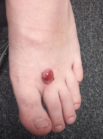
Two months ago, a tender lesion manifested on the dorsum of a 14-year-old boy’s right foot. Before it appeared, there was a tiny papule in the same spot; the boy’s mother says they thought it was a wart and attempted to remove it. As a result, the papule bled and became inflamed before quickly growing to its present form.
Besides being tender to touch, the lesion bleeds with minimal trauma. Attempts to remove it—using silver nitrate, liquid nitrogen, and triple-antibiotic cream—have all failed to have any positive impact.
The patient is reportedly otherwise healthy. He takes no medications of any kind.

EXAMINATION
The lesion is a 2-cm, domelike, red shiny nodule located on the mid-dorsum of the right foot. It is attached to the underlying skin by a thick sessile base. There is no surrounding erythema or other skin changes.
The site is anesthetized with local infiltrate of 1% lidocaine with epinephrine before deep shave biopsy is used to remove the lesion. The base is curetted, then cauterized for hemostasis.
PATHOLOGY
Microscopic examination shows a tangle of capillaries arranged in lobules separated by septae of connective tissue. A brisk inflammatory reaction and faint erosions are seen on the lesion’s surface.
What is the diagnosis?
DISCUSSION
First described by Ponce and Dor in 1897, pyogenic granuloma (PG) was thought to represent a kind of infection but turned out to be neither infectious nor granulomatous. It is now understood to represent the body’s attempt to heal a wound or other trauma (albeit this process goes awry for unknown reasons).
PG has a range of morphologic presentations; this case, with a berry-like friable papule, is fairly typical. PG is common on fingertips, lips, and faces (in children) and can even appear on gingival mucosae (particularly in first-trimester pregnancy). Other common areas of manifestation include ingrown toenails (lesion forms where the distal edge of the nail cuts into the lateral aspect of the nail bed) and the umbilical stump of newborns. An extremely common scenario is the child who cannot leave a wart or skin tag alone, picking and squeezing the lesion repeatedly until a PG forms.
PG is reactive in nature and not neoplastic. It is essentially vascular (other names for it include lobular capillary hemangioma and eruptive hemangioma), and the amount of blood that can flow from these lesions tends to frighten patients and parents.
While PG is completely benign, it is part of a differential of “look-alikes” that include nodular melanoma and metastatic cancer. For this reason, once removed, PGs are always sent for pathologic examination.
Removal should be done by deep shave, followed by electrodessication and curettage to prevent recurrence. Cryosurgery or silver nitrate application, though frequently done, is seldom effective. Significantly, neither yields a definitive diagnosis.
Certain drugs are associated with PG formation. These include isotretinoin, some chemotherapy drugs, and retroviral medications.
TAKE-HOME LEARNING POINTS
• Pyogenic granuloma (PG) is neither pyogenic nor granulomatous and is instead a reactive process.
• PG represents the body’s incomplete attempt to heal a wound.
• The trauma is often repetitive, particularly in PG associated with ingrown toenails or digitally manipulated by the patient.
• A berry-like appearance, sudden growth, and friability are all characteristic of PG.
• PG, though extremely common, needs to be removed to provide relief and to establish the lesion’s benign nature by pathologic examination.
Two months ago, a tender lesion manifested on the dorsum of a 14-year-old boy’s right foot. Before it appeared, there was a tiny papule in the same spot; the boy’s mother says they thought it was a wart and attempted to remove it. As a result, the papule bled and became inflamed before quickly growing to its present form.
Besides being tender to touch, the lesion bleeds with minimal trauma. Attempts to remove it—using silver nitrate, liquid nitrogen, and triple-antibiotic cream—have all failed to have any positive impact.
The patient is reportedly otherwise healthy. He takes no medications of any kind.
EXAMINATION
The lesion is a 2-cm, domelike, red shiny nodule located on the mid-dorsum of the right foot. It is attached to the underlying skin by a thick sessile base. There is no surrounding erythema or other skin changes.
The site is anesthetized with local infiltrate of 1% lidocaine with epinephrine before deep shave biopsy is used to remove the lesion. The base is curetted, then cauterized for hemostasis.
PATHOLOGY
Microscopic examination shows a tangle of capillaries arranged in lobules separated by septae of connective tissue. A brisk inflammatory reaction and faint erosions are seen on the lesion’s surface.
What is the diagnosis?
DISCUSSION
First described by Ponce and Dor in 1897, pyogenic granuloma (PG) was thought to represent a kind of infection but turned out to be neither infectious nor granulomatous. It is now understood to represent the body’s attempt to heal a wound or other trauma (albeit this process goes awry for unknown reasons).
PG has a range of morphologic presentations; this case, with a berry-like friable papule, is fairly typical. PG is common on fingertips, lips, and faces (in children) and can even appear on gingival mucosae (particularly in first-trimester pregnancy). Other common areas of manifestation include ingrown toenails (lesion forms where the distal edge of the nail cuts into the lateral aspect of the nail bed) and the umbilical stump of newborns. An extremely common scenario is the child who cannot leave a wart or skin tag alone, picking and squeezing the lesion repeatedly until a PG forms.
PG is reactive in nature and not neoplastic. It is essentially vascular (other names for it include lobular capillary hemangioma and eruptive hemangioma), and the amount of blood that can flow from these lesions tends to frighten patients and parents.
While PG is completely benign, it is part of a differential of “look-alikes” that include nodular melanoma and metastatic cancer. For this reason, once removed, PGs are always sent for pathologic examination.
Removal should be done by deep shave, followed by electrodessication and curettage to prevent recurrence. Cryosurgery or silver nitrate application, though frequently done, is seldom effective. Significantly, neither yields a definitive diagnosis.
Certain drugs are associated with PG formation. These include isotretinoin, some chemotherapy drugs, and retroviral medications.
TAKE-HOME LEARNING POINTS
• Pyogenic granuloma (PG) is neither pyogenic nor granulomatous and is instead a reactive process.
• PG represents the body’s incomplete attempt to heal a wound.
• The trauma is often repetitive, particularly in PG associated with ingrown toenails or digitally manipulated by the patient.
• A berry-like appearance, sudden growth, and friability are all characteristic of PG.
• PG, though extremely common, needs to be removed to provide relief and to establish the lesion’s benign nature by pathologic examination.