User login
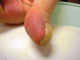
LAS VEGAS – A 39-year-old male patient presented with a 5-year history of an isolated, nonpainful, nonfluctuant left thumbnail deformity. He had no other skin findings, and no other known medical problems. His thumbnail was distinctly curved, with what appeared to be a slight pinkish growth underneath the nail. The nail was avulsed, revealing a readily visible and protruding tumor from the nail bed, extending from underneath the nail matrix to the level of the distal phalanx, with only a small portion of the nail bed not affected.
Digital myxomas are rare neoplasms, and the subungual location is rarer still, according to Dr. Miriam S. Bettencourt.
There have been just four mentions of subungual myxoma in the medical literature since 1982, all of which were case reports. Surgical excision is usually curative, without the need for repeat surgery. One previous case report described a midlateral approach that spared the nail apparatus and demonstrated remodeling of the nail despite a void left under the nail matrix after excision of the tumor (J. Hand Surg. Am. 1998;23:178-80).
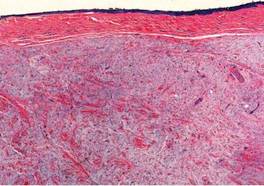
In this case, the patient’s histology revealed a large, circumscribed nonencapsulated nodular deposition of myxoid material, associated with a slight increase in spindle-shaped fibroblasts and thin intervening fibrocollagenous tissue underneath the nail bed epithelium. Scattered mast cells were visible within the lesion.
The tumor was sharply excised and the nail bed was roughly debrided using a small curette. A piece of tinfoil from a suture pack was fashioned into a pseudo nail shape and sutured beneath the patient’s cuticle in order to maintain the opening of the nail matrix during the excision, said Dr. Bettencourt, who presented the case at the Las Vegas Dermatology Seminar, sponsored by Skin Disease Education Foundation (SDEF).
Differential diagnosis included subungual glomus tumor, which causes a bluish-red discoloration of the nail plate and, unlike myxoma, is painful and temperature-sensitive. Onychomatricoma can cause a nail curvature similar to that of myxoma. However, this rare benign tumor of the nail matrix causes a yellowish discoloration and splinter hemorrhages, which are not seen with myxoma.
Fibromas in the digits, including periungual fibroma (a marker of tuberous sclerosis), acquired digital fibrokeratoma, dermatofibroma, and recurrent digital fibrous tumors of childhood may also be considered in the differential diagnosis. So can myxoid cysts, for which there are three presentations in the nail apparatus: one related to the interphalangeal joint, a second having no connection with joints, and a third submatrical type. Patients with myxoid cysts tend to be female and middle-age or older. Squamous cell carcinomas, the most common of subungual malignancies, should also be considered, said Dr. Bettencourt, of the University of Nevada, Las Vegas.
When a subungual myxoma is identified, it is important to investigate the patient for signs of myxoma syndrome, the name of a group of diseases characterized by atrial and cutaneous myxomas, as well as lentigines.
The patient has not presented with any nail deformity or recurrence in the year since surgical recovery, she noted.
"Dermatologists need to and care for patients with nail problems, in place of having the patient be seen solely by a podiatrist, since nail diseases are part of our specialty," Dr. Bettencourt concluded.
She reported having no disclosures. SDEF and this news organization are owned by Elsevier.
LAS VEGAS – A 39-year-old male patient presented with a 5-year history of an isolated, nonpainful, nonfluctuant left thumbnail deformity. He had no other skin findings, and no other known medical problems. His thumbnail was distinctly curved, with what appeared to be a slight pinkish growth underneath the nail. The nail was avulsed, revealing a readily visible and protruding tumor from the nail bed, extending from underneath the nail matrix to the level of the distal phalanx, with only a small portion of the nail bed not affected.
Digital myxomas are rare neoplasms, and the subungual location is rarer still, according to Dr. Miriam S. Bettencourt.
There have been just four mentions of subungual myxoma in the medical literature since 1982, all of which were case reports. Surgical excision is usually curative, without the need for repeat surgery. One previous case report described a midlateral approach that spared the nail apparatus and demonstrated remodeling of the nail despite a void left under the nail matrix after excision of the tumor (J. Hand Surg. Am. 1998;23:178-80).
In this case, the patient’s histology revealed a large, circumscribed nonencapsulated nodular deposition of myxoid material, associated with a slight increase in spindle-shaped fibroblasts and thin intervening fibrocollagenous tissue underneath the nail bed epithelium. Scattered mast cells were visible within the lesion.
The tumor was sharply excised and the nail bed was roughly debrided using a small curette. A piece of tinfoil from a suture pack was fashioned into a pseudo nail shape and sutured beneath the patient’s cuticle in order to maintain the opening of the nail matrix during the excision, said Dr. Bettencourt, who presented the case at the Las Vegas Dermatology Seminar, sponsored by Skin Disease Education Foundation (SDEF).
Differential diagnosis included subungual glomus tumor, which causes a bluish-red discoloration of the nail plate and, unlike myxoma, is painful and temperature-sensitive. Onychomatricoma can cause a nail curvature similar to that of myxoma. However, this rare benign tumor of the nail matrix causes a yellowish discoloration and splinter hemorrhages, which are not seen with myxoma.
Fibromas in the digits, including periungual fibroma (a marker of tuberous sclerosis), acquired digital fibrokeratoma, dermatofibroma, and recurrent digital fibrous tumors of childhood may also be considered in the differential diagnosis. So can myxoid cysts, for which there are three presentations in the nail apparatus: one related to the interphalangeal joint, a second having no connection with joints, and a third submatrical type. Patients with myxoid cysts tend to be female and middle-age or older. Squamous cell carcinomas, the most common of subungual malignancies, should also be considered, said Dr. Bettencourt, of the University of Nevada, Las Vegas.
When a subungual myxoma is identified, it is important to investigate the patient for signs of myxoma syndrome, the name of a group of diseases characterized by atrial and cutaneous myxomas, as well as lentigines.
The patient has not presented with any nail deformity or recurrence in the year since surgical recovery, she noted.
"Dermatologists need to and care for patients with nail problems, in place of having the patient be seen solely by a podiatrist, since nail diseases are part of our specialty," Dr. Bettencourt concluded.
She reported having no disclosures. SDEF and this news organization are owned by Elsevier.