User login
ANSWER
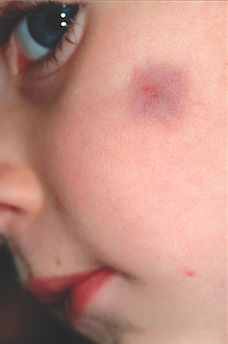
The correct answer is pilomatricoma (choice “a”), also know as calcifying epithelioma of Malherbe. See discussion.
Dermatofibromas (choice “b”) can be dark, especially in darker-skinned individuals, but are rare in children and even more so on the face.
Sebaceous cysts (choice “c”) are quite common on the face and can develop in children but are more likely to have a fluctuant feel rather than a firm one. They are also unlikely to be hyperpigmented and will usually involve an overlying punctum (comedone) on the surface.
Spitz tumors (choice “d”) can range from red to black and papular to macular. They tend to be intradermal in terms of vertical placement and are quite unlikely to be subcutaneous and indurated. They belong in the differential diagnosis because of their color and the fact that they are usually seen on children.
DISCUSSION
Pilomatricomas are uncommon but not at all rare. They usually manifest on the head, neck, or upper extremities of children as bluish subcutaneous masses devoid of overlying skin changes (eg, atrophy, comedones, or breaks in the skin). Though not seen in this case, the deep lesion often “tents” the overlying skin in one or more foci, a phenomenon termed the tent sign.
Although the exact reason they appear is not known, it is clear that they derive histologically from hair matrix cells. Quite rarely, they can undergo malignant transformation. They can also be quite a bit larger than this one and can present in multiples, with color ranging from deep blue (as in this case) to pinkish brown.
TREATMENT
For the purposes of positive identification and cosmesis, pilomatricomas are usually excised. At that time, identifying features such as the lack of an organized cyst wall and the granular, calcified contents are typically seen.
ANSWER
The correct answer is pilomatricoma (choice “a”), also know as calcifying epithelioma of Malherbe. See discussion.
Dermatofibromas (choice “b”) can be dark, especially in darker-skinned individuals, but are rare in children and even more so on the face.
Sebaceous cysts (choice “c”) are quite common on the face and can develop in children but are more likely to have a fluctuant feel rather than a firm one. They are also unlikely to be hyperpigmented and will usually involve an overlying punctum (comedone) on the surface.
Spitz tumors (choice “d”) can range from red to black and papular to macular. They tend to be intradermal in terms of vertical placement and are quite unlikely to be subcutaneous and indurated. They belong in the differential diagnosis because of their color and the fact that they are usually seen on children.
DISCUSSION
Pilomatricomas are uncommon but not at all rare. They usually manifest on the head, neck, or upper extremities of children as bluish subcutaneous masses devoid of overlying skin changes (eg, atrophy, comedones, or breaks in the skin). Though not seen in this case, the deep lesion often “tents” the overlying skin in one or more foci, a phenomenon termed the tent sign.
Although the exact reason they appear is not known, it is clear that they derive histologically from hair matrix cells. Quite rarely, they can undergo malignant transformation. They can also be quite a bit larger than this one and can present in multiples, with color ranging from deep blue (as in this case) to pinkish brown.
TREATMENT
For the purposes of positive identification and cosmesis, pilomatricomas are usually excised. At that time, identifying features such as the lack of an organized cyst wall and the granular, calcified contents are typically seen.

A 6-year-old boy is brought in by his mother for evaluation of a lesion that has been present on his face, with little change, for about two years. Though asymptomatic, the lesion is nonetheless worrisome, since the boy’s pediatrician isn’t sure what it is. The boy is in good health otherwise. His mother’s expectation is that the lesion will be removed at this initial visit to dermatology, since this is the reason she consulted the pediatrician in the first place. The 2-cm left facial lesion can be seen from across the room; it is quite large, round, and dark blue. While no changes are seen on the surface of the skin, palpation reveals definite, deep induration. No punctum can be seen on the overlying surface, and there is no palpable adenopathy in the area.