User login
ANSWER
The one incorrect statement above is choice “d.” Although most patients call them age spots, age is, in fact, only one factor in the development of such lesions. All the other statements are true and are discussed below.
DISCUSSION
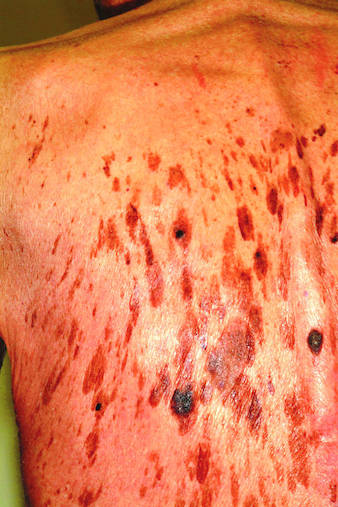
Seborrheic keratoses (SKs) are the most common benign skin lesions seen in older patients—but they’re also commonly seen on the skin of people in their 20s and 30s, so age is only one factor favoring their appearance. Heredity is the main cause in about 50% of cases. One of the most common dermatologic diagnoses, they are responsible for a large percentage of referrals to dermatology. As with this patient, SKs appear to be related to sun exposure in a significant percentage of patients, even though they have no malignant potential themselves.
Becoming thoroughly familiar with the myriad morphologic presentations of SKs is essential in learning dermatology. Their color ranges from gray to pink, tan to brown, and even black. Fortunately, they’re usually distinctly epidermal, with a “stuck-on” look that helps greatly in distinguishing them from malignant lesions. In oilier areas of the body, such as on the face, they can become much less scaly and much darker, making them difficult to distinguish from pigmented basal cell carcinoma or even melanoma in some cases; biopsy is thus necessary. Indeed, the “secret” to learning how to recognize SKs in all their varied presentations is the routine performance of punch biopsies.
SKs, when they’re as numerous as on this patient, can easily obscure a melanoma and can even coexist with the latter in the same location. This is especially true in high-risk patients (ie, those with fair, sun-damaged skin) who tan poorly. Fortunately, this patient had, by history and exam, type IV skin (with type I being the most fair, and type VI being the darkest) and was therefore at relatively low risk for skin cancer.
But on empirical grounds alone, one could correctly surmise that the mere presence of so many similar lesions, often present for years without change, effectively rules out skin cancer. The latter is far more likely to appear as a solitary lesion, as well as to be dynamic in nature (ie, to grow and/or change over time).
ANSWER
The one incorrect statement above is choice “d.” Although most patients call them age spots, age is, in fact, only one factor in the development of such lesions. All the other statements are true and are discussed below.
DISCUSSION
Seborrheic keratoses (SKs) are the most common benign skin lesions seen in older patients—but they’re also commonly seen on the skin of people in their 20s and 30s, so age is only one factor favoring their appearance. Heredity is the main cause in about 50% of cases. One of the most common dermatologic diagnoses, they are responsible for a large percentage of referrals to dermatology. As with this patient, SKs appear to be related to sun exposure in a significant percentage of patients, even though they have no malignant potential themselves.
Becoming thoroughly familiar with the myriad morphologic presentations of SKs is essential in learning dermatology. Their color ranges from gray to pink, tan to brown, and even black. Fortunately, they’re usually distinctly epidermal, with a “stuck-on” look that helps greatly in distinguishing them from malignant lesions. In oilier areas of the body, such as on the face, they can become much less scaly and much darker, making them difficult to distinguish from pigmented basal cell carcinoma or even melanoma in some cases; biopsy is thus necessary. Indeed, the “secret” to learning how to recognize SKs in all their varied presentations is the routine performance of punch biopsies.
SKs, when they’re as numerous as on this patient, can easily obscure a melanoma and can even coexist with the latter in the same location. This is especially true in high-risk patients (ie, those with fair, sun-damaged skin) who tan poorly. Fortunately, this patient had, by history and exam, type IV skin (with type I being the most fair, and type VI being the darkest) and was therefore at relatively low risk for skin cancer.
But on empirical grounds alone, one could correctly surmise that the mere presence of so many similar lesions, often present for years without change, effectively rules out skin cancer. The latter is far more likely to appear as a solitary lesion, as well as to be dynamic in nature (ie, to grow and/or change over time).

A 71-year-old man is urgently referred to dermatology by his primary care provider at the request of the patient’s children. These adult children recently noticed changes to the skin on their father’s back. On the day of the appointment, two of his children—a son and a daughter—bring the man in and already have his shirt off when you enter the room. Before you can evaluate the patient, his children express their anxiety about the lesions: “They’re so dark, and there are so many of them! We’re really concerned about this being skin cancer.” Examination reveals relatively dark skin with little evidence of sun damage, although the patient reports spending much of his life outdoors. When he is exposed to sun, he says, he tans relatively easily, holding the tan for weeks and very seldom burning. The lesions number easily in the hundreds and are tan to brown, papular, and nodular. Many coalesce into large plaques, with linear and in some cases digitate configuration. They virtually cover his back. On palpation, the surfaces of these lesions are epidermal, dry, and rough, with a warty texture. Lesions are also seen on his face, chest, and arms, though they are far fewer in number. Interestingly, both children have numerous similar lesions that can be easily seen.