User login
SAN FRANCISCO – A study of cases at seven academic pediatric dermatology centers has more precisely described the cutaneous eruptions caused by a severe form of hand, foot, and mouth disease in U.S. children.
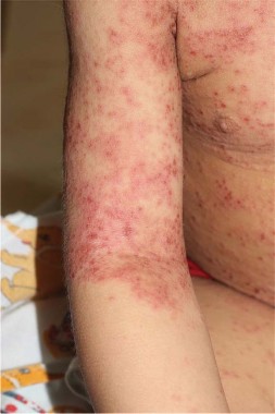
Dermatologists don’t see many cases of hand, foot, and mouth disease because these usually are routine viral infections managed by pediatricians. In the spring of 2012, however, Dr. Erin Mathes and her associates started seeing children with the fevers and GI symptoms typical of hand, foot, and mouth disease, but with a very severe rash with severe blisters, vesicles, and erosions instead of the milder papulovesicles of the hands, feet, buttocks, and mouth seen with the classic disease.
"We were left scratching our heads wondering exactly what was going on" until the Centers for Disease Control and Prevention reported an outbreak of severe hand, foot, and mouth disease associated with coxsackievirus A6 (CVA6) that also had been reported in several other countries (MMWR 2012;61:213-4).

"These reports said that the rash was severe, but they didn’t characterize it very carefully," so she and her colleagues across the country set out to describe the cutaneous manifestations in U.S. patients more specifically, said Dr. Mathes of the University of California, San Francisco.
The 80 cases they reviewed were treated between July 2011 and June 2012 at seven pediatric academic centers. Seventeen were caused by CVA6 infection that was confirmed by polymerase chain reaction (PCR) testing and 63 cases met predefined clinical criteria for inclusion as likely CVA6 infection, she reported at the annual meeting of the Pacific Dermatologic Association. Patients ranged in age from 4 months to 16 years.
Nearly all of the patients had vesiculobullous and erosive eruptions (99% of patients). "Some of the smaller vesicles were more widespread than in classic hand, foot, and mouth disease. We saw a striking perioral distribution," she said. "And then some of our younger patients had quite large blisters," such as a 4-cm blister on the foot of a 4-month-old baby (Pediatrics 2013;132:e149-57 [doi: 10.1542/peds.2012-3175]).
The most interesting finding was an eczema herpeticum–like rash in 55% of patients. From a distance, all of the patients looked like they had eczema herpeticum – erosions and vesicles in areas that previously were affected by atopic dermatitis but closer inspection showed that they weren’t as monomorphous or as "punched out" as erosions from eczema herpeticum. "So, there are some morphologic clues that this is not eczema herpeticum," she said. Most patients tested negative for herpes simplex virus (HSV).
CVA6 now can be added to the short list of HSV-1, varicella, and coxsackievirus A16 as viruses that have been reported to cause a Kaposi’s varicelliform eruption, in which preexisting dermatitis becomes superinfected with virus. Dr. Mathes proposed that the CVA6 eruption be called "eczema coxsackium." "We like this [term] because it brings to mind eczema herpeticum and yet indicates that the virus is coxsackie rather than a herpes virus," she said.
Nineteen percent of patients had locus minoris infection without previous atopic dermatitis and developed blisters and erosions in areas of sunburn, laceration, or irritant dermatitis.
Gianotti-Crosti–like eruptions with papulovesicles on only the cheeks, arms, and legs were seen in 36% of patients.
One of the rarer morphologies that "was confusing to both the pediatricians and the dermatologists" was a petechial and purpuric eruption in 17% of patients, more commonly in older children and in acral areas, she said. Biopsies from several patients showed no evidence of vasculitis, and several patients had confirmed CVA6 infection. "It was more consistent with a viral eruption," she said.
Among nail changes in 25% of patients, Beau’s lines and onychomadesis were common. These probably would have been seen more if patients were followed longitudinally, but the study captured data from just one visit, she noted.
Only half of patients had oropharyngeal ulcerations (50%), a lower percentage than with classic hand, foot, and mouth disease. Dr. Mathes’ impression was that these ulcers tended to occur more commonly in anterior locations, compared with classic disease.
Ten patients were hospitalized for an average of 12 days, mainly for diagnostic work-ups and empiric antibiotic treatment. All recovered with supportive care. There were no cases of severe systemic illness, in contrast with published case series from Asia in which several patients developed aseptic meningitis and other neurologic complications, she said.
Dr. Mathes and her colleagues were wondering if they’d see eczema coxsackium in 2013 until she saw her first case of the year recently. "We think we’ll probably see it in lower numbers given that there is some herd immunity now, and some children will have immunity to it," she said.
The diagnosis is a clinical one. Look for features of hand, foot, and mouth disease with severe rash, and do bacterial and viral cultures to rule out other infections. "We did have some patients who had a superinfection with Staph in addition to their viral infection," she said. Coxsackievirus will not grow in culture; if you want to confirm the diagnosis, use PCR. If you see a severe case in a patient hospitalized with neurologic or cardiac complications, send specimens for diagnostic confirmation to your local health department or the CDC, she advised.
The international reports of severe hand, foot, and mouth disease from CVA6 infection began in 2012 in Finland, and then came from Singapore and Japan. The international reports noted a severe rash, onychomadesis and desquamation, rare neurologic complications, and a high prevalence of infection in school-age children or adults, probably because of low herd immunity. The international investigators were able to obtain the virus for analysis from nail clippings from the patients who had onychomadesis, Dr. Mathes noted.
She reported having no financial disclosures.
On Twitter @sherryboschert
SAN FRANCISCO – A study of cases at seven academic pediatric dermatology centers has more precisely described the cutaneous eruptions caused by a severe form of hand, foot, and mouth disease in U.S. children.
Dermatologists don’t see many cases of hand, foot, and mouth disease because these usually are routine viral infections managed by pediatricians. In the spring of 2012, however, Dr. Erin Mathes and her associates started seeing children with the fevers and GI symptoms typical of hand, foot, and mouth disease, but with a very severe rash with severe blisters, vesicles, and erosions instead of the milder papulovesicles of the hands, feet, buttocks, and mouth seen with the classic disease.
"We were left scratching our heads wondering exactly what was going on" until the Centers for Disease Control and Prevention reported an outbreak of severe hand, foot, and mouth disease associated with coxsackievirus A6 (CVA6) that also had been reported in several other countries (MMWR 2012;61:213-4).
"These reports said that the rash was severe, but they didn’t characterize it very carefully," so she and her colleagues across the country set out to describe the cutaneous manifestations in U.S. patients more specifically, said Dr. Mathes of the University of California, San Francisco.
The 80 cases they reviewed were treated between July 2011 and June 2012 at seven pediatric academic centers. Seventeen were caused by CVA6 infection that was confirmed by polymerase chain reaction (PCR) testing and 63 cases met predefined clinical criteria for inclusion as likely CVA6 infection, she reported at the annual meeting of the Pacific Dermatologic Association. Patients ranged in age from 4 months to 16 years.
Nearly all of the patients had vesiculobullous and erosive eruptions (99% of patients). "Some of the smaller vesicles were more widespread than in classic hand, foot, and mouth disease. We saw a striking perioral distribution," she said. "And then some of our younger patients had quite large blisters," such as a 4-cm blister on the foot of a 4-month-old baby (Pediatrics 2013;132:e149-57 [doi: 10.1542/peds.2012-3175]).
The most interesting finding was an eczema herpeticum–like rash in 55% of patients. From a distance, all of the patients looked like they had eczema herpeticum – erosions and vesicles in areas that previously were affected by atopic dermatitis but closer inspection showed that they weren’t as monomorphous or as "punched out" as erosions from eczema herpeticum. "So, there are some morphologic clues that this is not eczema herpeticum," she said. Most patients tested negative for herpes simplex virus (HSV).
CVA6 now can be added to the short list of HSV-1, varicella, and coxsackievirus A16 as viruses that have been reported to cause a Kaposi’s varicelliform eruption, in which preexisting dermatitis becomes superinfected with virus. Dr. Mathes proposed that the CVA6 eruption be called "eczema coxsackium." "We like this [term] because it brings to mind eczema herpeticum and yet indicates that the virus is coxsackie rather than a herpes virus," she said.
Nineteen percent of patients had locus minoris infection without previous atopic dermatitis and developed blisters and erosions in areas of sunburn, laceration, or irritant dermatitis.
Gianotti-Crosti–like eruptions with papulovesicles on only the cheeks, arms, and legs were seen in 36% of patients.
One of the rarer morphologies that "was confusing to both the pediatricians and the dermatologists" was a petechial and purpuric eruption in 17% of patients, more commonly in older children and in acral areas, she said. Biopsies from several patients showed no evidence of vasculitis, and several patients had confirmed CVA6 infection. "It was more consistent with a viral eruption," she said.
Among nail changes in 25% of patients, Beau’s lines and onychomadesis were common. These probably would have been seen more if patients were followed longitudinally, but the study captured data from just one visit, she noted.
Only half of patients had oropharyngeal ulcerations (50%), a lower percentage than with classic hand, foot, and mouth disease. Dr. Mathes’ impression was that these ulcers tended to occur more commonly in anterior locations, compared with classic disease.
Ten patients were hospitalized for an average of 12 days, mainly for diagnostic work-ups and empiric antibiotic treatment. All recovered with supportive care. There were no cases of severe systemic illness, in contrast with published case series from Asia in which several patients developed aseptic meningitis and other neurologic complications, she said.
Dr. Mathes and her colleagues were wondering if they’d see eczema coxsackium in 2013 until she saw her first case of the year recently. "We think we’ll probably see it in lower numbers given that there is some herd immunity now, and some children will have immunity to it," she said.
The diagnosis is a clinical one. Look for features of hand, foot, and mouth disease with severe rash, and do bacterial and viral cultures to rule out other infections. "We did have some patients who had a superinfection with Staph in addition to their viral infection," she said. Coxsackievirus will not grow in culture; if you want to confirm the diagnosis, use PCR. If you see a severe case in a patient hospitalized with neurologic or cardiac complications, send specimens for diagnostic confirmation to your local health department or the CDC, she advised.
The international reports of severe hand, foot, and mouth disease from CVA6 infection began in 2012 in Finland, and then came from Singapore and Japan. The international reports noted a severe rash, onychomadesis and desquamation, rare neurologic complications, and a high prevalence of infection in school-age children or adults, probably because of low herd immunity. The international investigators were able to obtain the virus for analysis from nail clippings from the patients who had onychomadesis, Dr. Mathes noted.
She reported having no financial disclosures.
On Twitter @sherryboschert
AT THE ANNUAL MEETING OF THE PACIFIC DERMATOLOGIC ASSOCIATION
Major finding: The most common cutaneous findings were vesiculobullous and erosive eruptions in 99% of patients, eczema herpeticum-like rash in 55%, Gianotti-Crosti–like eruptions in 36%, nail changes in 25%, and oropharyngeal ulcerations in 50%.
Data source: Retrospective, multicenter review of 80 pediatric patients with atypical cases of hand, foot, and mouth disease caused by a confirmed CVA6 infection or that met predefined criteria for infection.
Disclosures: Dr. Mathes reported having no financial disclosures.