User login
Diagnosis: Lichen sclerosus et atrophicus with morphea
Lichen sclerosus et atrophicus (LSetA) is a chronic inflammatory condition mediated primarily by lymphocytic infiltration. It is up to 10 times more likely to occur in women than men. There is a genetic component, as approximately 12% of affected patients also have relatives with the condition. Human leukocyte antigen subtypes DQ7, DQ8, and DQ9 have been associated with LSetA, which is thought to be primarily autoimmune in nature. There is also a strong association with other autoimmune diseases, predominantly thyroid disease, vitiligo, alopecia areata, and pernicious anemia.
LSetA is most commonly seen in the vulvar region, but can also present extragenitally. Vulvar LSetA has a bimodal distribution, peaking in prepubertal and postmenopausal women. The most common symptoms are pruritus and pain. On physical examination, LSetA appears as thick white plaques with a thin epidermis resembling “cigarette paper.” Chronic inflammation can also lead to permanent scarring. Pruritus or pain can result, which can significantly affect quality of life.
Morphea is characterized by thickened skin as a result of deep dermal fibrosis. Also known as localized scleroderma, morphea presents with the cutaneous findings of scleroderma without the systemic complications. Like LSetA, morphea is more common in females than males, although the ratio is not as high. Plaque morphea is the most prevalent type and presents as circumscribed, edematous plaques on the trunk or extremities. Morphea has been reported as a consequence of localized injury or infection, but is also commonly associated with various autoimmune conditions.
Histologic changes of LSetA include epidermal atrophy and homogenization of collagen limited to the superficial dermis. In contrast, morphea histologically consists of deep dermal fibrosis with associated epidermal hyperkeratosis. While LSetA and morphea are usually distinct entities, they can rarely occur together. The diagnosis of LSetA with morphea can be confirmed when the deep and superficial histologic findings are consistent with both entities.
Treatment options for LSetA include short-term, high-potency topical corticosteroids followed by maintenance therapy with emollients. Reducing exposure to irritants is also crucial for a sustained response. Other modalities such as topical calcineurin inhibitors, photodynamic therapy, surgery, and methotrexate are potential alternatives in recalcitrant cases. Morphea can be treated with methotrexate and systemic glucocorticoids, which are usually given in combination. Systemic glucocorticoids can be tapered to avoid long-term side effects, while tapering the methotrexate based on clinical response. In general, the evidence supports the use of systemic therapy for extensive plaquetype morphea over topical treatments; however, there is some evidence to support the use of topical calcineurin inhibitors in resistant cases.
This patient was started on a prednisone taper in addition to weekly methotrexate. Gabapentin was prescribed for pain control. The patient returned to the clinic during the next several months with improvement in her symptoms and reduction in the size and induration of the lesions.
This case and photo were submitted by Adam Aldahan, Dr. Alyx Rosen, and Dr. Anne Burdick of the University of Miami department of dermatology.
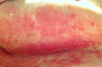
A 63-year-old woman presented with extremely painful plaques on the breasts, inframammary folds, and chest, and under the abdomen, for 3 years. The lesions had progressively increased in size with worsening pain over the past several months. She also reported painful raw areas on her genitals. Physical examination revealed multiple well-demarcated oval and geographic plaques with central white induration with areas of superficial erosion and pink peripheral borders on the breasts, inframammary folds, midchest, and abdomen that were exquisitely tender to light palpation. Genital examination revealed a vulvar white plaque with superficial erosions and an atrophic labia minora.
