User login
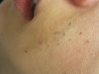
Diagnosis: Agminated blue nevus
Agminated blue nevi are aggregated clusters of nevi that appear blue due to their deep dermal location and the subsequent scattering of short wavelengths of light (Tyndall effect). The clusters are typically contained within a 10 cm area. The intervening background skin is usually without hyperpigmentation or skin discoloration, although speckled pigmentation can occur. Generally, all forms of agminated nevi likely result from a loss of heterozygosity (LOH) mutation in the somatic line of an embryo, inducing a localized predilection for the development of nevi.
The differential diagnosis for agminated blue nevi includes segmental melanocytic nevi, melanoma, and other agminated melanocytic nevi: benign melanocytic nevi, atypical or dysplastic nevi, and speckled lentiginous nevi (nevus spilus). The distribution of the nevi can help narrow the differential, as segmental melanocytic nevi are less clustered and spread over a larger area than agminated blue nevi. Congenital nevi are present at birth or within a few months and often have unique features, including perifollicular pigmentary changes, hypertrichosis, and focal hypopigmentation. Acquired nevi (blue nevi, benign melanocytic nevi, and atypical or dysplastic nevi) usually descend over time from the dermal-epidermal junction to the dermis and typically become a lighter color. Lastly, the presence or absence of background pigmentation can help distinguish between agminated blue nevi and nevus spilus. Nevus spilus is a subtype of congenital melanocytic nevi with a similar aggregated appearance but has a characteristic background hyperpigmentation between the discrete nevi.
Agminated blue nevi and nevus spilus can be differentiated in the clinic by using a Wood’s lamp or performing a biopsy, which is not indicated unless atypia is present. If a non-speckled background hyperpigmentation is present on Wood’s lamp illumination, the diagnosis of nevus spilus may be more appropriate, as agminated blue nevi typically show no background pigmentation. A biopsy of the skin would allow for distinguishing between nevus spilus and agminated nevi based on the pigment within the surrounding skin. The presence of melanocytic hyperplasia within the skin surrounding the discrete nevi would support the diagnosis of nevus spilus.
The main concern with any melanocytic proliferation is the potential for transformation into a melanoma. In general, agminated nevi carry an increased melanoma risk. Specifically, congenital melanocytic nevi result in a <1-5% risk of melanoma depending on the lesion size. The risk of melanoma associated with agminated blue nevi is currently unknown, but it should be noted that individual blue nevi rarely become malignant. Since more than 50% of cutaneous melanomas develop without a preceding nevus, removal of nevi to reduce melanoma risk is not typically indicated, except in the case of a large congenital melanocytic nevi (>20cm). However, long-term surveillance is crucial for agminated blue nevi, especially if the individual has dysplastic nevus syndrome. If the involved nevi develop any features of atypia, a biopsy should be performed to assess for the development of a melanoma.
This case and photo were submitted by Dr. Damon McClain of Three Rivers Dermatology in Coraopolis, Pa., and Mark Ash, a medical student at East Carolina University, Greenville, N.C.
Dr. Bilu Martin is in private practice at Premier Dermatology, MD, in Aventura, Fla. More diagnostic cases are available at edermatologynews.com. To submit your case for possible publication, send an email to dermnews@frontlinemedcom.com.
References:
- Ashfaq A. Marghoob, Robin Blum, Robert Nossa, Klaus J. Busam, Dana Sachs, Allan Halpern. Agminated Atypical (Dysplastic) Nevi Case Report and Review of the Literature. Arch Dermatol. 2001;137(7):917-920.
- Belinda Tan, Noah Craft, Lindy P. Fox, Lowell A. Goldsmith, Michael D. Tharp. Visual Dx. Blue Nevus. Accessed 2/11/16. Camila Roos Mariano da Rocha, Thaís Corsetti Grazziotin, Maria Carolina Widholzer Rey, Laura Luzzatto, Renan Rangel Bonamigo. Congenital agminated melanocytic nevus - Case report. An Bras Dermatol. 2013;88(6 Suppl 1):170-2.
- Chris G. Adigun, Jeffrey D. Bernhard, Sarah Stein, Karen Wiss, Sheila Galbraith, Craig N. Burkhart, Dean Morrell, Lynn Garfunkel, Nancy Esterly. Visual Dx. Agminated Nevus. Accessed 2/11/16.
- Julie V Schaffer and Jean L Bolognia. Acquired melanocytic nevi (moles). In: UpToDate, Post TW (Ed), UpToDate, Waltham, MA. (Accessed on February 11, 2016.)
- Julie V Schaffer and Jean L Bolognia. Congenital melanocytic nevi. In: UpToDate, Post TW (Ed), UpToDate, Waltham, MA. (Accessed on February 11, 2016.)
- Maria A. Pizzichetta, H. Peter Soyer, Cesare Massone, Lorenzo Cerroni. Clinical and Dermoscopic Features of Agminated Blue Nevus. Arch Dermatol. 2007;143(9):1209-1226.

A 23-year-old healthy female presented with a several-year history of asymptomatic dark spots on her left cheek. The lesions seemed to be darkening. Incidentally, she was in a four-wheeler accident in middle school and suffered facial trauma, which resulted in the replacement of two superior left front teeth, but she denied significant cutaneous injury at that time. On physical examination, there were several clustered 1-mm blue-gray macules in linear formation with a subtle underlying tan patch on her left cheek.