User login
ANSWER
The correct answer is vitiligo (choice “b”).
DISCUSSION
Vitiligo develops when pigment cells (melanocytes) fail or die. Although there appears to be a hereditary component in some cases, as well as a connection to autoimmune disease, environmental factors (eg, intense sun exposure, stress) may also play a role.
This patient has nonsegmental vitiligo (NSV), the most common form. It is usually symmetrically distributed on high-friction areas, such as hands, knees, and elbows, as well as around the eyes and mouth. Segmental vitiligo, which affects only 10% of all vitiligo patients, tends to manifest during adolescence and typically remains confined to one area.
Unfortunately, for the majority of those with NSV (including the case patient), the condition tends to be progressive—and it responds poorly to treatment with topical steroids, calcineurin inhibitors, or phototherapy. As seen in this case, it can cause pigment loss in or around lesions; in fact, if left alone, the lesion may completely lose color. And NSV can encompass wider areas of involvement—to the extent that some patients lose all the pigment in their bodies. The resulting psychiatric fallout is considerable, especially in darker-skinned patients.
This patient's lesion will likely double in size by adulthood, which will not only subject him to ridicule but also increase the risk for malignant transformation. For this reason, he was advised to have the lesion excised under general anesthesia. He was also started on a regimen of a topical steroid cream and a calcineurin inhibitor on alternating days, but his prognosis is, in all honesty, poor.
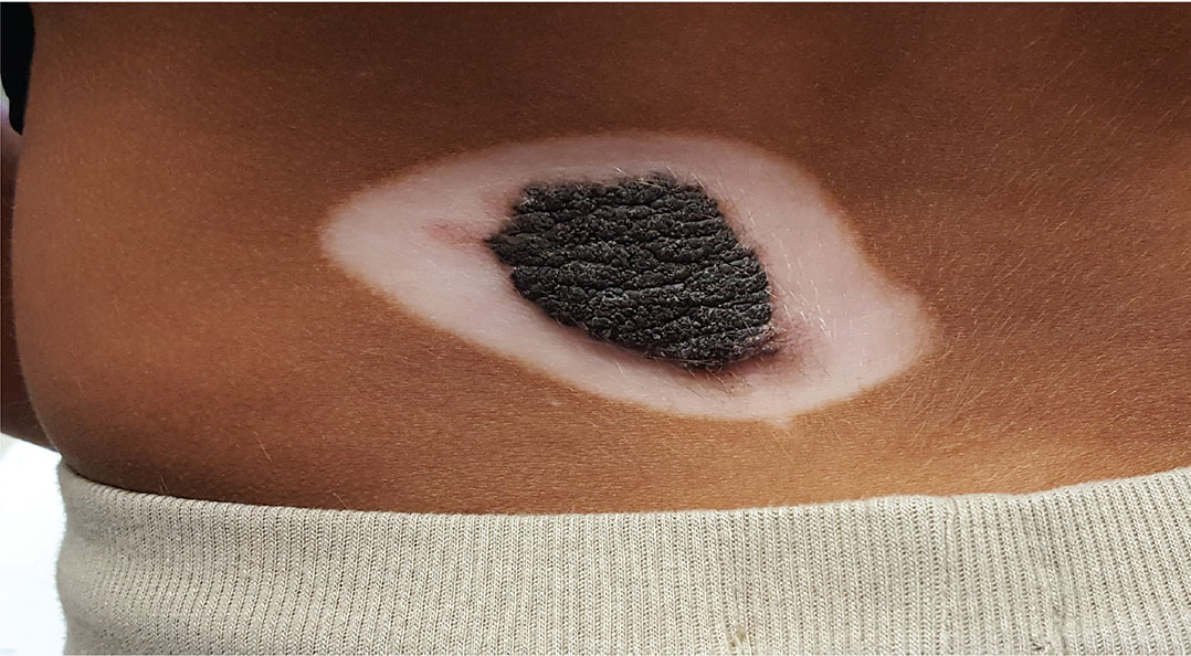
A 5-year-old boy is referred to dermatology for evaluation of recent color changes to the skin around a congenital lesion. Located on his mid low back, the polygonal lesion measures 8 x 5 cm and is uniformly dark brown with a mammillated, hair-bearing surface. The plaque has grown proportionately with the child but otherwise remained stable.
A year ago, however, the patient’s family noticed that the normal brown skin around the lesion was turning white. This “halo” became noticeably larger over the span of the year—effectively doubling the size of the lesion.
The child’s type V skin is in sharp contrast to the porcelain white band that parallels the margins of his lesion. The surface of the depigmented skin is completely smooth, with no epidermal changes. Faint but definite depigmentation is noted on the periocular skin of both eyes, in addition to well-defined depigmentation on his fingertips and the perionychial areas of all 10 fingers.
The family asserts that the boy is otherwise healthy and that there is no family history of similar phenomena. The rest of the examination is unremarkable.
