User login
A 47-YEAR-OLD WOMAN was admitted to our hospital for intravenous antibiotic treatment of recurrent cellulitis with ulceration of her left second and third toes. Previous outpatient management with trimethoprim-sulfamethoxazole followed by clindamycin had failed.
The patient had been treated repeatedly over the previous 10 years for similar episodes of methicillin-resistant Staphylococcus aureus cellulitis with ulceration of the same toes. These episodes began after the patient had been in multiple car accidents and had sustained lower extremity trauma.
When the patient was admitted, she was afebrile and had normal vital signs. The ulcerations on her left second and third toes (FIGURE) were painful. The distal dorsal foot was warm, erythematous, and indurated without fluctuance or crepitus. There were diffuse spider veins on the lower extremities and the peripheral pulses were 2+ symmetrically. Electrolytes, including calcium, phosphate, and alkaline phosphatase, were within normal limits, the white blood cell count was 4.9 × 10³/mm3 and C-reactive protein was 1.0 mg/dL. There was an elevated erythrocyte sedimentation rate of 46 mm/h, mild transaminitis (ALT>AST), and a finding of chronic hepatitis C infection (a few months prior).
FIGURE
Ulcerated toes in a nondiabetic patient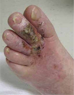
Wound and blood cultures were negative for infection. Radiologic examination of the left foot showed no signs of osteomyelitis or other bony abnormality. We sent punch biopsies out for pathologic assessment.
WHAT IS YOUR DIAGNOSIS?
HOW WOULD YOU TREAT THIS PATIENT?
Diagnosis: Osteoma cutis
Lesional biopsies revealed that the patient had osteoma cutis, a skin condition in which bone ossification (including lamellae, trabecular bone formation, osteocytes, and sometimes marrow) occurs within the dermis.
A rare condition
Osteoma cutis has an incidence of 1.2-1.7 cases per 1000 skin lesion biopsies.1 The primary form of osteoma cutis occurs in about 25% of cases and is associated with certain genetic disorders such as Gardner’s syndrome and Albright’s hereditary osteodystrophy; it arises without a preexisting lesion.1 The secondary type of osteoma cutis—which our patient had—often arises within a cancerous lesion (especially melanocytic nevi and basal cell carcinoma) or chronic inflammation (as is found in traumatic scars, acne vulgaris, chronic venous stasis, vasculitis, and other nonspecific inflammatory conditions).1-3
Sixty-eight percent of osteoma cutis cases are benign, and most patients are white females.1 Most lesions arise on the head, neck, and digits.1,4 Although foot lesions are much less common, a few cases of secondary osteoma cutis on the foot have been reported.4,5
Osteoma cutis is believed to arise via mesenchymal ossification (in contrast to endochondral bone formation from a cartilaginous precursor). Proposed mechanisms include aberrant embryological migration of mesenchymal cells into the dermis and metaplastic transformation of fibroblasts into osteoid-producing osteoblasts.4
Is it osteoma cutis or calcinosis cutis?
The differential includes calcinosis cutis, or deposition of insoluble calcium compounds in the skin without true bone formation. Lesions of both osteoma cutis and calcinosis cutis are sometimes palpable and seen on x-ray,2 but more often, plain radiographs are normal.3
Making the diagnosis requires a high suspicion for the condition—especially in patients without peripheral vascular disease or neuropathy who have nonhealing or slow-healing ulcers. A good patient history is also important to help rule out possible uncommon causes, such as cutaneous tuberculosis.6
Other diagnoses to consider for nonhealing or treatment-resistant ulcers include infection (eg, bacterial, mycobacterial, fungal, or underlying osteomyelitis), vasculopathy (arterial insufficiency, venous stasis, atheroembolism, or diabetes mellitus), pyoderma gangrenosum, and malnutrition.
Because the diagnosis of osteoma cutis is made primarily by pathology, suspicious lesions should be biopsied.
Conservative Tx is a good approach
Case reports suggest that bone removal speeds healing, but the benefits of surgical intervention are unclear because healing is observed in patients who receive only conservative management.2,3,5 With lesions arising on the head and neck, treatment goals are usually aesthetic. Surgical techniques of resection, curettage, or dermabrasion are most often used, and topical retinoic acid has proven to be a helpful adjuvant therapy.7
No surgery for our patient
Given our patient’s history of trauma followed by recurrent ulceration (which may not have completely resolved), we suspected that chronic inflammation was the cause of the osteoma cutis.
We prescribed minocycline 100 mg BID for 14 days for superficial wound infection, with plans to extend treatment as needed based on wound healing. She also received care at a local wound clinic for incomplete resolution of the ulceration and biopsy sites. The patient was lost to follow-up.
CORRESPONDENCE
Sally P. Weaver, MD, PhD, McLennan County Medical Education and Research Foundation, 1600 Providence Drive, Waco, TX 76707; sallyweaver@me.com
1. Conlin PA, Jimenez-Quintero LP, Rapini RP. Osteomas of the skin revisited. Am J Dermatopathol. 2002;24:479-483.
2. Sarkany I, Kreel L. Subcutaneous ossification of the legs in chronic venous stasis. Br Med J. 1966;2:27-28.
3. Duarte IG. Multiple injuries of osteoma skin in the face. An Bras Dermatol. 2010;85:695-698.
4. Burgdorf W, Nasemann T. Cutaneous osteomas. Arch Dermatol Res. 1977;260:121-135.
5. Titchener AG, Ramoutar DN, Al-Rufaie H, et al. Osteoma cutis masquerading as an ingrowing toenail. Cases J. 2009;2:7176.-
6. Ghosh SK, Bandyopadhyay D, Ghosh A, et al. Non-healing perianal ulcer. Dermatol Online J. 2009;15:9.-
7. Ayaviri NA, Nahas FX, Barbosa MV, et al. Isolated primary osteoma cutis of the head. Can J Plast Surg. 2006;14:33-36.
A 47-YEAR-OLD WOMAN was admitted to our hospital for intravenous antibiotic treatment of recurrent cellulitis with ulceration of her left second and third toes. Previous outpatient management with trimethoprim-sulfamethoxazole followed by clindamycin had failed.
The patient had been treated repeatedly over the previous 10 years for similar episodes of methicillin-resistant Staphylococcus aureus cellulitis with ulceration of the same toes. These episodes began after the patient had been in multiple car accidents and had sustained lower extremity trauma.
When the patient was admitted, she was afebrile and had normal vital signs. The ulcerations on her left second and third toes (FIGURE) were painful. The distal dorsal foot was warm, erythematous, and indurated without fluctuance or crepitus. There were diffuse spider veins on the lower extremities and the peripheral pulses were 2+ symmetrically. Electrolytes, including calcium, phosphate, and alkaline phosphatase, were within normal limits, the white blood cell count was 4.9 × 10³/mm3 and C-reactive protein was 1.0 mg/dL. There was an elevated erythrocyte sedimentation rate of 46 mm/h, mild transaminitis (ALT>AST), and a finding of chronic hepatitis C infection (a few months prior).
FIGURE
Ulcerated toes in a nondiabetic patient
Wound and blood cultures were negative for infection. Radiologic examination of the left foot showed no signs of osteomyelitis or other bony abnormality. We sent punch biopsies out for pathologic assessment.
WHAT IS YOUR DIAGNOSIS?
HOW WOULD YOU TREAT THIS PATIENT?
Diagnosis: Osteoma cutis
Lesional biopsies revealed that the patient had osteoma cutis, a skin condition in which bone ossification (including lamellae, trabecular bone formation, osteocytes, and sometimes marrow) occurs within the dermis.
A rare condition
Osteoma cutis has an incidence of 1.2-1.7 cases per 1000 skin lesion biopsies.1 The primary form of osteoma cutis occurs in about 25% of cases and is associated with certain genetic disorders such as Gardner’s syndrome and Albright’s hereditary osteodystrophy; it arises without a preexisting lesion.1 The secondary type of osteoma cutis—which our patient had—often arises within a cancerous lesion (especially melanocytic nevi and basal cell carcinoma) or chronic inflammation (as is found in traumatic scars, acne vulgaris, chronic venous stasis, vasculitis, and other nonspecific inflammatory conditions).1-3
Sixty-eight percent of osteoma cutis cases are benign, and most patients are white females.1 Most lesions arise on the head, neck, and digits.1,4 Although foot lesions are much less common, a few cases of secondary osteoma cutis on the foot have been reported.4,5
Osteoma cutis is believed to arise via mesenchymal ossification (in contrast to endochondral bone formation from a cartilaginous precursor). Proposed mechanisms include aberrant embryological migration of mesenchymal cells into the dermis and metaplastic transformation of fibroblasts into osteoid-producing osteoblasts.4
Is it osteoma cutis or calcinosis cutis?
The differential includes calcinosis cutis, or deposition of insoluble calcium compounds in the skin without true bone formation. Lesions of both osteoma cutis and calcinosis cutis are sometimes palpable and seen on x-ray,2 but more often, plain radiographs are normal.3
Making the diagnosis requires a high suspicion for the condition—especially in patients without peripheral vascular disease or neuropathy who have nonhealing or slow-healing ulcers. A good patient history is also important to help rule out possible uncommon causes, such as cutaneous tuberculosis.6
Other diagnoses to consider for nonhealing or treatment-resistant ulcers include infection (eg, bacterial, mycobacterial, fungal, or underlying osteomyelitis), vasculopathy (arterial insufficiency, venous stasis, atheroembolism, or diabetes mellitus), pyoderma gangrenosum, and malnutrition.
Because the diagnosis of osteoma cutis is made primarily by pathology, suspicious lesions should be biopsied.
Conservative Tx is a good approach
Case reports suggest that bone removal speeds healing, but the benefits of surgical intervention are unclear because healing is observed in patients who receive only conservative management.2,3,5 With lesions arising on the head and neck, treatment goals are usually aesthetic. Surgical techniques of resection, curettage, or dermabrasion are most often used, and topical retinoic acid has proven to be a helpful adjuvant therapy.7
No surgery for our patient
Given our patient’s history of trauma followed by recurrent ulceration (which may not have completely resolved), we suspected that chronic inflammation was the cause of the osteoma cutis.
We prescribed minocycline 100 mg BID for 14 days for superficial wound infection, with plans to extend treatment as needed based on wound healing. She also received care at a local wound clinic for incomplete resolution of the ulceration and biopsy sites. The patient was lost to follow-up.
CORRESPONDENCE
Sally P. Weaver, MD, PhD, McLennan County Medical Education and Research Foundation, 1600 Providence Drive, Waco, TX 76707; sallyweaver@me.com
1. Conlin PA, Jimenez-Quintero LP, Rapini RP. Osteomas of the skin revisited. Am J Dermatopathol. 2002;24:479-483.
2. Sarkany I, Kreel L. Subcutaneous ossification of the legs in chronic venous stasis. Br Med J. 1966;2:27-28.
3. Duarte IG. Multiple injuries of osteoma skin in the face. An Bras Dermatol. 2010;85:695-698.
4. Burgdorf W, Nasemann T. Cutaneous osteomas. Arch Dermatol Res. 1977;260:121-135.
5. Titchener AG, Ramoutar DN, Al-Rufaie H, et al. Osteoma cutis masquerading as an ingrowing toenail. Cases J. 2009;2:7176.-
6. Ghosh SK, Bandyopadhyay D, Ghosh A, et al. Non-healing perianal ulcer. Dermatol Online J. 2009;15:9.-
7. Ayaviri NA, Nahas FX, Barbosa MV, et al. Isolated primary osteoma cutis of the head. Can J Plast Surg. 2006;14:33-36.