User login
ANSWER
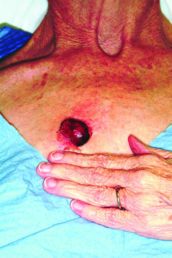
The correct answer is all of the above (choice “d”). All are reasonable diagnoses and important because they drive the decision as to how to proceed.
DISCUSSION
An incisional biopsy would also have been acceptable, but collection of the entire lesion, with modest margins where possible, is almost always the gold standard in establishing a diagnosis in cases such as this one. There are several reasons, one being that the pathologist will then have adequate tissue to examine and will have a good chance of determining whether the lesion began locally or represents spread from a distant site. The only argument against such an approach would be the potential for scarring.
Moreover, in the majority of cases, this procedure can be curative—as in this case, in which the pathology report revealed the lesion to be a well-differentiated squamous cell carcinoma that had been traumatized, resulting in the formation of reparative inappropriate granulation tissue. It was the latter that accounted for the friability and rapid growth. Fortunately, the margins were clear, so no additional surgery or radiation was necessary.
A complete differential diagnosis would also include other types of cancer, including colon cancer and melanoma. Considering the possibilities, the patient was all too happy to have the problem taken care of in one step.
ANSWER
The correct answer is all of the above (choice “d”). All are reasonable diagnoses and important because they drive the decision as to how to proceed.
DISCUSSION
An incisional biopsy would also have been acceptable, but collection of the entire lesion, with modest margins where possible, is almost always the gold standard in establishing a diagnosis in cases such as this one. There are several reasons, one being that the pathologist will then have adequate tissue to examine and will have a good chance of determining whether the lesion began locally or represents spread from a distant site. The only argument against such an approach would be the potential for scarring.
Moreover, in the majority of cases, this procedure can be curative—as in this case, in which the pathology report revealed the lesion to be a well-differentiated squamous cell carcinoma that had been traumatized, resulting in the formation of reparative inappropriate granulation tissue. It was the latter that accounted for the friability and rapid growth. Fortunately, the margins were clear, so no additional surgery or radiation was necessary.
A complete differential diagnosis would also include other types of cancer, including colon cancer and melanoma. Considering the possibilities, the patient was all too happy to have the problem taken care of in one step.

Rapid growth and bleeding are two of the most common reasons that patients seek evaluation for lesions. Both are concerns for this 69-year-old woman who has had a chest lesion for almost two years. Recently, the lesion suddenly tripled in size and began to bleed with minor trauma. Although painless, the lesion is of particular concern since the patient was diagnosed with breast cancer about 18 years ago and underwent a right radical mastectomy at that time. The patient is a smoker with a more than 50–pack-year history. She denies any recent history of shortness of breath, cough, or weight loss. Examination reveals an impressive nodule on the right midchest, measuring 4 cm and demonstrating a smooth, vascular surface and lobular shape. The base of the lesion is pedunculated, and the lesion itself is quite mobile. After consultation with the patient, the decision to excise is quickly reached and the procedure carried out. The excised lesion is sent for pathologic exam.