User login
ANSWER
The correct answer is pseudoxanthoma elasticum (PXE; choice “d”), details of which are discussed below. Eruptive xanthomata (choice “a”) usually present with widespread eruptive papules and/or nodules and are associated with extreme hypertriglyceridemia. There are several different types of Ehlers-Danlos syndrome, an inherited defect of connective tissues, but the most common involve hypermobile joints (so-called double-jointedness) and/or cutis laxa, neither of which is present in our patient. Moreover, the skin changes seen in our patient are totally unlike those seen in Ehlers-Danlos syndrome.
Solar elastosis (choice “c”) represents sun-caused basophilic degeneration of the deep dermis and presents with a whitish “chicken skin” look. Most commonly seen on the upper forehead of older men with an extensive history of overexposure to ultraviolet light, it almost never appears in intertriginous or other areas unexposed to sunlight.
DISCUSSION
PXE is an autosomal recessive disease with prevalence estimates ranging from 1:25,000 to 1:100,000. It does not demonstrate racial or geographic predilections, although it appears to have a slight preponderance in females. It typically begins to manifest in the second to third decade of life. This genetic defect results in the improper function of elastin and elastic fibers in the deep dermis, the intima of midsized arteries, and the Bruch membrane in the eye, where characteristic clinical and histopathologic changes occur. Thus, three main organ systems are affected: the skin, the eyes, and the cardiovascular system. However, the manifestations can vary a great deal—even among siblings.
Almost 100% of patients older than 30 eventually show signs of the disease in the eyes, with angioid streaks often becoming symptomatic only after trauma to the eye. The end point is reparative neovascularization, leakage from which can eventuate in hemorrhage, scarring, and loss of vision.
PXE also affects midsized arteries, especially of the extremities, where pathologic changes in the vessel walls lead to the premature formation of atheromatous plaques and eventual claudication, loss of peripheral pulses, hypertension, angina, and myocardial infarction that occur at a much younger age than in the unaffected population. Cerebral ischemic attacks, with the exception of aneurysms, are common with PXE. Gastrointestinal bleeds are common, but PXE does not affect the liver, lung, or kidneys.
“Treatment” for the appearance of PXE-affected skin is nearly nonexistent, but there are steps that should be taken. For example: (1) genetic counseling to assess the risk for future generations, as well as presymptomatic testing; (2) regular, periodic eye examination, including funduscopic examination, as well as advice to avoid heavy lifting, trauma, smoking, or other counterproductive activities; and (3) emphasis on preventive lifestyle choices to avoid cardiovascular complications, with early detection a more attainable goal with continued surveillance.
PXE is only one of several so-called “genodermatoses,” that is, inheritable conditions with dermatologic manifestations. Other examples include neurofibromatosis type 1, the aforementioned Ehlers-Danlos syndrome, and epidermolysis bullosa—among many, many others.
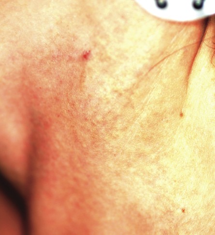
A 29-year-old woman presents for evaluation of changes in her skin, which she first noticed during her late teens. The changes have been asymptomatic and slowly progressive, and various providers have given different explanations for them at different times. However, she has never been seen by dermatology prior to this visit, which is ostensibly for treatment of hand warts and eyelid dermatitis. Aside from being somewhat atopic, the patient is in otherwise excellent health. Laboratory work done as part of a recent physical showed her lipid levels to be well within normal limits. A discussion of family history reveals she has a sister with the same type of skin changes; however, she too has never received a definitive diagnosis from any of the many providers who have evaluated her over the years. The patient points to changes in several intertriginous locations, including bilateral neck skin, both antecubital areas, axillae, and crural folds. The skin in those areas has an odd cobblestone-like papularity, appears atrophic, and is light yellow. The changes are barely palpable. The midline posterior neck is spared, but the same changes can be seen on oral mucosal surfaces. Elsewhere, there are no signs of hypermobile joints or of cutis laxa. No evidence of excessive sun exposure is observed. Several punch biopsies show distorted, fragmented elastic fibers in the mid to deep reticular dermis.
