User login
New medical and surgical treatments can minimize infantile hemangiomas in their proliferative phase and decrease residual scarring after they involute, according to Dr. Brandie J. Metz.
The first decision in treating a hemangioma is whether to use a surgical or medical approach, Dr. Metz said at a cosmetic dermatology seminar sponsored by Skin Disease Education Foundation in Santa Monica, Calif.
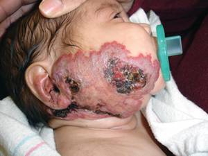
Medical treatment is usually a first-line choice, although surgery may be considered when the patient presents with severe refractory pain from ulceration or when residua after involution would likely require surgical treatment. "In that case, why wait?" said Dr. Metz, the director of pediatric dermatology at the University of California, Irvine.
In the past 2 years, propranolol has gained acceptance as an effective medical therapy. Its use was first noted in a case series published in 2008 (N. Engl. J. Med. 2008;358:2649-51). In this French report, 11 infants received propranolol for varying treatment periods. Within 24 hours, all of the hemangiomas responded with softening and a change in color from red to purple, indicating decreased vascularity.
The treatment is not without risks, Dr. Metz noted. "Possible side effects are bradycardia, hypotension, hypoglycemia, and bronchospasm. A gradual dose escalation with close monitoring is the best way to go."
She is an investigator for a large, double-blind, placebo-controlled trial of propranolol that is currently underway. Patients receive either placebo or propranolol at 3 mg/kg for 15 days, followed by another 15 days at 4 mg/kg. The primary outcome will be hemangioma thickness variation measured by ultrasonography from baseline to 1 month.
When a lesion is easily resectable and a patient has failed medical therapy, or if ulceration and pain are present, surgery can be considered. Generally, thin plaques with a gradual slope have a better surgical prognosis. A circular incision around the lesion, with purse string closure, is the usual technique. A 2002 study of 25 patients found that this method reduced hemangiomas by a mean of 73% in width and 45% in length (Plast. Reconstr. Surg. 2002;109:1544-54).
Lasers can treat superficial proliferating hemangiomas, as well as the residua left after involution, Dr. Metz said. The pulsed dye laser is one option. Short pulses of 0.45-1.5 millisecond are used at either a 585- or 595-nm wavelength. The concomitant use of a dynamic cooling device makes the treatment less painful and can allow the use of a higher fluence.
Several studies attest to the success of pulsed dye lasers for hemangiomas. A 2009 study of 90 patients with 105 hemangiomas reported complete or near-complete clearance of color for 81% and thickness for 64%. Although there was no scarring or atrophy, one patient did develop an ulceration that resolved. Hyperpigmentation occurred in 4% and hypopigmentation in 14% (Derm. Surg. 2009;35:1947-54).
A 2006 study noted complications related to pulsed dye laser treatment. Of 12 patients with segmental facial hemangiomas, there were 8 ulcerations and 4 with atrophic scarring (Lasers Surg. Med. 2006;38:116-23).
Two published reports detail the use of fractionated laser for residua. A 2008 report found that the laser effectively decreased fibrofatty tissue and redundant skin that resulted from an extensive mixed facial hemangioma (Derm. Surg. 2008;34:1112-4). A 2009 report found similar results on residua from another facial hemangioma (Arch. Dermatol. 2009;145:748-50).
Dr. Metz disclosed that she is an investigator on the current propranolol trial, sponsored by University Hospital, Bordeaux, France.
SDEF and this news organization are owned by Elsevier.
New medical and surgical treatments can minimize infantile hemangiomas in their proliferative phase and decrease residual scarring after they involute, according to Dr. Brandie J. Metz.
The first decision in treating a hemangioma is whether to use a surgical or medical approach, Dr. Metz said at a cosmetic dermatology seminar sponsored by Skin Disease Education Foundation in Santa Monica, Calif.
Medical treatment is usually a first-line choice, although surgery may be considered when the patient presents with severe refractory pain from ulceration or when residua after involution would likely require surgical treatment. "In that case, why wait?" said Dr. Metz, the director of pediatric dermatology at the University of California, Irvine.
In the past 2 years, propranolol has gained acceptance as an effective medical therapy. Its use was first noted in a case series published in 2008 (N. Engl. J. Med. 2008;358:2649-51). In this French report, 11 infants received propranolol for varying treatment periods. Within 24 hours, all of the hemangiomas responded with softening and a change in color from red to purple, indicating decreased vascularity.
The treatment is not without risks, Dr. Metz noted. "Possible side effects are bradycardia, hypotension, hypoglycemia, and bronchospasm. A gradual dose escalation with close monitoring is the best way to go."
She is an investigator for a large, double-blind, placebo-controlled trial of propranolol that is currently underway. Patients receive either placebo or propranolol at 3 mg/kg for 15 days, followed by another 15 days at 4 mg/kg. The primary outcome will be hemangioma thickness variation measured by ultrasonography from baseline to 1 month.
When a lesion is easily resectable and a patient has failed medical therapy, or if ulceration and pain are present, surgery can be considered. Generally, thin plaques with a gradual slope have a better surgical prognosis. A circular incision around the lesion, with purse string closure, is the usual technique. A 2002 study of 25 patients found that this method reduced hemangiomas by a mean of 73% in width and 45% in length (Plast. Reconstr. Surg. 2002;109:1544-54).
Lasers can treat superficial proliferating hemangiomas, as well as the residua left after involution, Dr. Metz said. The pulsed dye laser is one option. Short pulses of 0.45-1.5 millisecond are used at either a 585- or 595-nm wavelength. The concomitant use of a dynamic cooling device makes the treatment less painful and can allow the use of a higher fluence.
Several studies attest to the success of pulsed dye lasers for hemangiomas. A 2009 study of 90 patients with 105 hemangiomas reported complete or near-complete clearance of color for 81% and thickness for 64%. Although there was no scarring or atrophy, one patient did develop an ulceration that resolved. Hyperpigmentation occurred in 4% and hypopigmentation in 14% (Derm. Surg. 2009;35:1947-54).
A 2006 study noted complications related to pulsed dye laser treatment. Of 12 patients with segmental facial hemangiomas, there were 8 ulcerations and 4 with atrophic scarring (Lasers Surg. Med. 2006;38:116-23).
Two published reports detail the use of fractionated laser for residua. A 2008 report found that the laser effectively decreased fibrofatty tissue and redundant skin that resulted from an extensive mixed facial hemangioma (Derm. Surg. 2008;34:1112-4). A 2009 report found similar results on residua from another facial hemangioma (Arch. Dermatol. 2009;145:748-50).
Dr. Metz disclosed that she is an investigator on the current propranolol trial, sponsored by University Hospital, Bordeaux, France.
SDEF and this news organization are owned by Elsevier.