User login
The iPhone may be the latest “it” gadget, but a flurry of recent innovation has given portable ultrasound devices a healthy buzz within the biomedical community.
Beyond the gee-whiz factor, though, a growing number of studies demonstrate the everyday value of putting portable units in the hands of hospitalists.
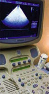
“The big news has been the tiny portable scanner,” says Stephen Smith, a biomedical engineer at Duke University, in North Carolina, and a pioneer in ultrasound technology. Siemens recently introduced a hand-held device called the Acuson P10, which weighs 1.6 pounds, retails for $9,499 and can fit within a hospitalist’s coat pocket. Not to be outdone, GE has announced plans to introduce an ultrasound unit no bigger than an iPod.
Smith and his collaborators have taken the technology one step farther. They incorporate electrocardial leads on the unit’s transducer face to permit electrocardiograms and a microphone to let hospitalists use the ultrasound like a stethoscope.
Eric Isaacs, MD, a clinical professor of medicine at San Francisco General Hospital, says he routinely uses ultrasound for vascular access “to ensure the safety of procedures that we previously performed either blind or by anatomical landmarks.” Beyond improving the accuracy of placing central and peripheral lines, he says, “the reason we are using ultrasound more now is that the machine is so portable. The radiologists are no longer in the hospital 24 hours a day, and so by necessity we are using the tools that were previously only accessible from 9 to 5.”
Range of Uses
Among the reports recognizing ultrasound’s value, he cited a 2003 study in the British Medical Journal affirming the technology’s superiority to relying on physical landmarks in gaining central venous access, resulting in a lower technical failure rate, reduced complications, and faster access.1 Dr. Issacs says ultrasound also has helped guide procedures such as thorancentesis and paracentesis, other applications once confined to radiology. “It’s something that’s allowing me to do at the bedside what I would otherwise have to wait several hours for,” he says.
For heart patients, he says, a hospitalist can bring ultrasound to the bedside during a cardiac arrest to inspect cardiac motion and fluid, and monitor the patient’s hydration status by examining the size of the inferior vena cava. Internists likewise could examine the size of a patient’s aorta to look for signs of an aneurism, especially for a patient experiencing abdominal pain in the middle of the night. “Quite frankly, it seems like the only limit to ultrasound use is imagination,” Dr. Isaacs says.
Robert Rodriguez, MD, a clinical professor of medicine and emergency medicine at San Francisco General Hospital, says he uses ultrasound on 25% of the patients he sees on an in-patient basis. His biggest use, he says, is for placing central lines—though that could soon change.
“I work with a population that has a very high percentage of injection drug abuse, in whom it’s very difficult to find even a peripheral vein,” he says. At least once a day, he uses ultrasound to locate the brachial vein for such peripheral lines, circumventing the need for a central line through the subclavian vein and the risk of a pneumothorax. “In the past, we would have to put in a central line for just about anything,” he says. “And now we can put in a peripheral line that saves them the risk.”
Another benefit, he says, is in breeding better patient interactions—for example, with gallstones. “You can say to the patient: ‘This is the gallbladder, these are the stones in the gallbladder, this is what’s causing the pain,’” he says. “I think patients appreciate being able to see that firsthand. I think they also appreciate that it’s going to lessen their likelihood of having a complication.”
At the University of Chicago Medical Center, cardiologist Kirk Spencer, MD, says ultrasound procedures still are performed mainly by sonographers and cardiologists. He hopes to change that with a slew of studies demonstrating the feasibility of putting portable ultrasound in the hands of internists.
In one study, hospitalized patients indicated for echocardiography received an echocardiogram, while all others were examined with ultrasound. “We found a significant number of cardio pathologies,” Dr. Spencer says. The findings, he says, were independent of specific medical complaints, such as endocrinology or orthopedic problems.2 “If you were sick enough to get in the hospital, there was a chance that you had a significant cardiac problem that needed to be addressed,” he says.
The study that most excites Dr. Spencer was presented at the 2007 IEEE International Ultrasonics Symposium in October.2 It looked at using ultrasound before releasing a cardiac patient. “One of the biggest problems, one of the most common diagnoses is congestive heart failure,” he says, with a six-month readmission rate of 30% to 40%. Giving ultrasound devices to internists allowed them to look at the amount of fluid around the heart of each cardiac patient.
“The patients who got readmitted all had more fluid detected by ultrasound,” Dr. Spencer says. “So we can do that and say, ‘Hey, you need to stay in the hospital two more days. But if that prevents you from coming back in six months, then that’s a good thing.’”
In patients diagnosed with congestive heart failure, he and his collaborators found, the mean fluid volume was higher for those who were later readmitted. Dr. Spencer plans to pick a reasonable cut-off value and prospectively test whether delaying the release of patients whose fluid levels exceed that value can cut readmission rates.
Most of the battery-operated units used by the medical center weigh between 6 to 10 pounds and cost between $12,000 and $20,000, he says. The devices, about twice the thickness of an iBook, can easily be carried on a shoulder strap. Echocardiogram machines, by contrast, weigh about 300 pounds, must be plugged in and retail for about $250,000.
Concerns, Obstacles
Dr. Spencer cautioned that ultrasound shouldn’t replace echocardiograms or other tools. “So no one is proposing that this would replace a full exam,” he says. “What we’re hoping is that this would detect things that have gone missing or would help ask very specific questions at the bedside.” His studies suggest the approach works well as long as the questions are simple: “Is there fluid or not? Is the heart good or bad?’ But not: ‘Is there an infection?’”
Beyond cardiology and the emergency room, Dr. Spencer says ultrasound has obvious imaging uses in the ICU. The dichotomy, he says, is that imaging intensive care patients can be especially difficult due to their edema, wounds, and lack of mobility. “That area has not blossomed as well as it could have,” he says.
Even so, the burgeoning number of applications for ultrasound “really has huge potential for good,” says Harvey Nisenbaum, MD, an associate professor of radiology at the University of Pennsylvania School of Medicine and president-elect of the American Institute of Ultrasound in Medicine (AIUM). “But the problem is that it’s an art form in the sense that it’s not automated.” No two ultrasound images will be identical, for example, because each depends upon the probe’s precise location. The key, Dr. Nisenbaum says, is proper training under agreed-upon guidelines, followed by continuing education and the maintenance of a hospitalist’s competency.
The AIUM, Nisenbaum says, is working to develop standard credentialing criteria for a range of ultrasound applications to help unify what has been a patchwork approach. Another limitation, he says, has been the lack of Food and Drug Administration (FDA) approval for ultrasound contrast agents Optison and Definity for noncardiac applications. Several deaths have been linked to the use of the intravenous agents in the sickest patients.
The institute is working with the FDA on trying to get the reagents approved for broader use, as they are in other countries. Nisenbaum cautioned the process likely will take a while. Once approved, getting a reimbursement code established for insurance purposes could take even longer.
A further obstacle, according to Dr. Spencer, is the lack of resolution surrounding medical legal issues. “Are we going to agree that this is like a physical examination?” he asks. “It’s unclear whether the medical legal community is going to accept that with ultrasound,” he says.
For cardiology applications, at least, he wonders if the push for reimbursement is such a good thing. “General internists are under incredible pressure [for billing],” he says. “They’re in a really tough spot, and so there would be enormous pressure to get reimbursed for every ultrasound.” As it is, he says, Medicare is targeting echocardiogram as an overutilized reimbursement item. “I hope the reason we’re using this is because we’re examining the patients anyway and this would allow us to find things that we might have missed,” he says. “It’s a better way of examining people, not a new technique for generating revenues. I think that would be a disaster.”
Jeffrey Wiese, MD, SHM board member and associate dean of graduate medical education at Tulane University School of Medicine’s Section of General Internal Medicine and Geriatrics in New Orleans, began putting ultrasound in the hands of his hospitalists and residents in 2007. It’s the “100% right thing to do,” he advises hospitalist groups. “It can be a meaningful way of improving safety. I hope that everyody would move that way.”
Dr. Wiese says residents began using ultrasound more and more for extra visualization during procedures.
“The reason we got into this was straightaway safety, independent of [Centers for Medicare and Medicaid Services] codes and billing—particularly regarding thoracentesis and internal lines,’’ he says. His hospitalists use SonaSite’s MicroMaxx system, “which was a key piece in the way of being able to bill. For all CMT just like endoscopy and bronchoscopy, you have to provide images of the procedure to prove you did it. With the MicroMaxx machine, it allows you to insert a USB and pull down images, take them to a print machine, print them out, and put them in a chart.”
Dr. Wiese touts the sheer amount of what hospitalists can use ultrasound for. “You can do echoes and abdominal ultrasound—not at the level of the radiology room or the cardiology lab, but you can get a quick look,” he says.
Should other hospitalist programs go in the same direction? “From a quality perspective there’s no question you go down that road,” Dr. Wiese asserts. “You do the math: How much does one pneumothorax cost? That’s especially true if [a] pneuothorax finds its way to CMS. One pneuomothorax that you prevent probably pays for your [$20,000-$30,000] machine. That’s even before you get into issues of billing for the use of it, which I think is a secondary way of funding the purchase.”
Forging Ahead
In the meantime, researchers are focusing on ever-diverse applications and smaller units.
At the Mayo Clinic in Jacksonville, Fla., director of regional anesthesia Steven Clendenen, MD, has pioneered the use of ultrasound for guiding nerve blocks.3 The imaging has “totally revolutionized” how the hospital manages pain, he says. As yet, the device still is cart-based, though he expects its size to shrink considerably. “You remember the first calculators, how big they were, and now look at them,” he says.
Beyond working toward miniaturized ultrasound units, Duke’s Smith has been developing real-time three-dimensional angiograms of blood vessels in the brain, a potential boon for stroke diagnoses.4 Another project may bring hospital-based ultrasound full circle: a device that produces a 3-D stereo-image, “like in the IMAX theater,” he says.5 Smith and his colleagues have modified a commercial scanner, “so the target comes out of the screen at you.” Among the many potential uses, expectant parents could see a 3-D stereo view of the developing fetus—something not even the iPhone can offer. TH
Bryn Nelson is a science journalist based in New York.
References
- Hind, D, Calvert, N, McWilliams, R, Davidson, A, Paisley, S, Beverley, C, Thomas, S. Ultrasonic locating devices for central venous cannulation: meta-analysis. Br Med J. 2003;327(7411):361.
- Fedson, S, Neithardt, G, Thomas, P, et al. Unsuspected clinically important findings detected with a small portable ultrasound device in patients admitted to a general medicine service. J Am Soc Echocardiogr. 2003;16(9):901-905.
- Feinglass NG, Clendenen SR, Torp KD, Wang RD, Castello R, Greengrass RA. Real-time three-dimensional ultrasound for continuous popliteal blockade: a case report and image description. Anesth Analg. 2007;105(1):272-274.
- Smith SW, Chu K, Idriss SF, Ivancevich NM, Light ED, Wolf PD. Feasibility Study: Real time 3D ultrasound imaging of the brain. Ultras Med Biol. 2004;30:1365-1371.
- Noble JR, Fronheiser MP, Smith SW. Real-time Stereo 3D Ultrasound. Ultrason Imaging. 2006;28:245-254.
The iPhone may be the latest “it” gadget, but a flurry of recent innovation has given portable ultrasound devices a healthy buzz within the biomedical community.
Beyond the gee-whiz factor, though, a growing number of studies demonstrate the everyday value of putting portable units in the hands of hospitalists.
“The big news has been the tiny portable scanner,” says Stephen Smith, a biomedical engineer at Duke University, in North Carolina, and a pioneer in ultrasound technology. Siemens recently introduced a hand-held device called the Acuson P10, which weighs 1.6 pounds, retails for $9,499 and can fit within a hospitalist’s coat pocket. Not to be outdone, GE has announced plans to introduce an ultrasound unit no bigger than an iPod.
Smith and his collaborators have taken the technology one step farther. They incorporate electrocardial leads on the unit’s transducer face to permit electrocardiograms and a microphone to let hospitalists use the ultrasound like a stethoscope.
Eric Isaacs, MD, a clinical professor of medicine at San Francisco General Hospital, says he routinely uses ultrasound for vascular access “to ensure the safety of procedures that we previously performed either blind or by anatomical landmarks.” Beyond improving the accuracy of placing central and peripheral lines, he says, “the reason we are using ultrasound more now is that the machine is so portable. The radiologists are no longer in the hospital 24 hours a day, and so by necessity we are using the tools that were previously only accessible from 9 to 5.”
Range of Uses
Among the reports recognizing ultrasound’s value, he cited a 2003 study in the British Medical Journal affirming the technology’s superiority to relying on physical landmarks in gaining central venous access, resulting in a lower technical failure rate, reduced complications, and faster access.1 Dr. Issacs says ultrasound also has helped guide procedures such as thorancentesis and paracentesis, other applications once confined to radiology. “It’s something that’s allowing me to do at the bedside what I would otherwise have to wait several hours for,” he says.
For heart patients, he says, a hospitalist can bring ultrasound to the bedside during a cardiac arrest to inspect cardiac motion and fluid, and monitor the patient’s hydration status by examining the size of the inferior vena cava. Internists likewise could examine the size of a patient’s aorta to look for signs of an aneurism, especially for a patient experiencing abdominal pain in the middle of the night. “Quite frankly, it seems like the only limit to ultrasound use is imagination,” Dr. Isaacs says.
Robert Rodriguez, MD, a clinical professor of medicine and emergency medicine at San Francisco General Hospital, says he uses ultrasound on 25% of the patients he sees on an in-patient basis. His biggest use, he says, is for placing central lines—though that could soon change.
“I work with a population that has a very high percentage of injection drug abuse, in whom it’s very difficult to find even a peripheral vein,” he says. At least once a day, he uses ultrasound to locate the brachial vein for such peripheral lines, circumventing the need for a central line through the subclavian vein and the risk of a pneumothorax. “In the past, we would have to put in a central line for just about anything,” he says. “And now we can put in a peripheral line that saves them the risk.”
Another benefit, he says, is in breeding better patient interactions—for example, with gallstones. “You can say to the patient: ‘This is the gallbladder, these are the stones in the gallbladder, this is what’s causing the pain,’” he says. “I think patients appreciate being able to see that firsthand. I think they also appreciate that it’s going to lessen their likelihood of having a complication.”
At the University of Chicago Medical Center, cardiologist Kirk Spencer, MD, says ultrasound procedures still are performed mainly by sonographers and cardiologists. He hopes to change that with a slew of studies demonstrating the feasibility of putting portable ultrasound in the hands of internists.
In one study, hospitalized patients indicated for echocardiography received an echocardiogram, while all others were examined with ultrasound. “We found a significant number of cardio pathologies,” Dr. Spencer says. The findings, he says, were independent of specific medical complaints, such as endocrinology or orthopedic problems.2 “If you were sick enough to get in the hospital, there was a chance that you had a significant cardiac problem that needed to be addressed,” he says.
The study that most excites Dr. Spencer was presented at the 2007 IEEE International Ultrasonics Symposium in October.2 It looked at using ultrasound before releasing a cardiac patient. “One of the biggest problems, one of the most common diagnoses is congestive heart failure,” he says, with a six-month readmission rate of 30% to 40%. Giving ultrasound devices to internists allowed them to look at the amount of fluid around the heart of each cardiac patient.
“The patients who got readmitted all had more fluid detected by ultrasound,” Dr. Spencer says. “So we can do that and say, ‘Hey, you need to stay in the hospital two more days. But if that prevents you from coming back in six months, then that’s a good thing.’”
In patients diagnosed with congestive heart failure, he and his collaborators found, the mean fluid volume was higher for those who were later readmitted. Dr. Spencer plans to pick a reasonable cut-off value and prospectively test whether delaying the release of patients whose fluid levels exceed that value can cut readmission rates.
Most of the battery-operated units used by the medical center weigh between 6 to 10 pounds and cost between $12,000 and $20,000, he says. The devices, about twice the thickness of an iBook, can easily be carried on a shoulder strap. Echocardiogram machines, by contrast, weigh about 300 pounds, must be plugged in and retail for about $250,000.
Concerns, Obstacles
Dr. Spencer cautioned that ultrasound shouldn’t replace echocardiograms or other tools. “So no one is proposing that this would replace a full exam,” he says. “What we’re hoping is that this would detect things that have gone missing or would help ask very specific questions at the bedside.” His studies suggest the approach works well as long as the questions are simple: “Is there fluid or not? Is the heart good or bad?’ But not: ‘Is there an infection?’”
Beyond cardiology and the emergency room, Dr. Spencer says ultrasound has obvious imaging uses in the ICU. The dichotomy, he says, is that imaging intensive care patients can be especially difficult due to their edema, wounds, and lack of mobility. “That area has not blossomed as well as it could have,” he says.
Even so, the burgeoning number of applications for ultrasound “really has huge potential for good,” says Harvey Nisenbaum, MD, an associate professor of radiology at the University of Pennsylvania School of Medicine and president-elect of the American Institute of Ultrasound in Medicine (AIUM). “But the problem is that it’s an art form in the sense that it’s not automated.” No two ultrasound images will be identical, for example, because each depends upon the probe’s precise location. The key, Dr. Nisenbaum says, is proper training under agreed-upon guidelines, followed by continuing education and the maintenance of a hospitalist’s competency.
The AIUM, Nisenbaum says, is working to develop standard credentialing criteria for a range of ultrasound applications to help unify what has been a patchwork approach. Another limitation, he says, has been the lack of Food and Drug Administration (FDA) approval for ultrasound contrast agents Optison and Definity for noncardiac applications. Several deaths have been linked to the use of the intravenous agents in the sickest patients.
The institute is working with the FDA on trying to get the reagents approved for broader use, as they are in other countries. Nisenbaum cautioned the process likely will take a while. Once approved, getting a reimbursement code established for insurance purposes could take even longer.
A further obstacle, according to Dr. Spencer, is the lack of resolution surrounding medical legal issues. “Are we going to agree that this is like a physical examination?” he asks. “It’s unclear whether the medical legal community is going to accept that with ultrasound,” he says.
For cardiology applications, at least, he wonders if the push for reimbursement is such a good thing. “General internists are under incredible pressure [for billing],” he says. “They’re in a really tough spot, and so there would be enormous pressure to get reimbursed for every ultrasound.” As it is, he says, Medicare is targeting echocardiogram as an overutilized reimbursement item. “I hope the reason we’re using this is because we’re examining the patients anyway and this would allow us to find things that we might have missed,” he says. “It’s a better way of examining people, not a new technique for generating revenues. I think that would be a disaster.”
Jeffrey Wiese, MD, SHM board member and associate dean of graduate medical education at Tulane University School of Medicine’s Section of General Internal Medicine and Geriatrics in New Orleans, began putting ultrasound in the hands of his hospitalists and residents in 2007. It’s the “100% right thing to do,” he advises hospitalist groups. “It can be a meaningful way of improving safety. I hope that everyody would move that way.”
Dr. Wiese says residents began using ultrasound more and more for extra visualization during procedures.
“The reason we got into this was straightaway safety, independent of [Centers for Medicare and Medicaid Services] codes and billing—particularly regarding thoracentesis and internal lines,’’ he says. His hospitalists use SonaSite’s MicroMaxx system, “which was a key piece in the way of being able to bill. For all CMT just like endoscopy and bronchoscopy, you have to provide images of the procedure to prove you did it. With the MicroMaxx machine, it allows you to insert a USB and pull down images, take them to a print machine, print them out, and put them in a chart.”
Dr. Wiese touts the sheer amount of what hospitalists can use ultrasound for. “You can do echoes and abdominal ultrasound—not at the level of the radiology room or the cardiology lab, but you can get a quick look,” he says.
Should other hospitalist programs go in the same direction? “From a quality perspective there’s no question you go down that road,” Dr. Wiese asserts. “You do the math: How much does one pneumothorax cost? That’s especially true if [a] pneuothorax finds its way to CMS. One pneuomothorax that you prevent probably pays for your [$20,000-$30,000] machine. That’s even before you get into issues of billing for the use of it, which I think is a secondary way of funding the purchase.”
Forging Ahead
In the meantime, researchers are focusing on ever-diverse applications and smaller units.
At the Mayo Clinic in Jacksonville, Fla., director of regional anesthesia Steven Clendenen, MD, has pioneered the use of ultrasound for guiding nerve blocks.3 The imaging has “totally revolutionized” how the hospital manages pain, he says. As yet, the device still is cart-based, though he expects its size to shrink considerably. “You remember the first calculators, how big they were, and now look at them,” he says.
Beyond working toward miniaturized ultrasound units, Duke’s Smith has been developing real-time three-dimensional angiograms of blood vessels in the brain, a potential boon for stroke diagnoses.4 Another project may bring hospital-based ultrasound full circle: a device that produces a 3-D stereo-image, “like in the IMAX theater,” he says.5 Smith and his colleagues have modified a commercial scanner, “so the target comes out of the screen at you.” Among the many potential uses, expectant parents could see a 3-D stereo view of the developing fetus—something not even the iPhone can offer. TH
Bryn Nelson is a science journalist based in New York.
References
- Hind, D, Calvert, N, McWilliams, R, Davidson, A, Paisley, S, Beverley, C, Thomas, S. Ultrasonic locating devices for central venous cannulation: meta-analysis. Br Med J. 2003;327(7411):361.
- Fedson, S, Neithardt, G, Thomas, P, et al. Unsuspected clinically important findings detected with a small portable ultrasound device in patients admitted to a general medicine service. J Am Soc Echocardiogr. 2003;16(9):901-905.
- Feinglass NG, Clendenen SR, Torp KD, Wang RD, Castello R, Greengrass RA. Real-time three-dimensional ultrasound for continuous popliteal blockade: a case report and image description. Anesth Analg. 2007;105(1):272-274.
- Smith SW, Chu K, Idriss SF, Ivancevich NM, Light ED, Wolf PD. Feasibility Study: Real time 3D ultrasound imaging of the brain. Ultras Med Biol. 2004;30:1365-1371.
- Noble JR, Fronheiser MP, Smith SW. Real-time Stereo 3D Ultrasound. Ultrason Imaging. 2006;28:245-254.