User login
American Society of Clinical Oncology (ASCO): Breast Cancer Symposium
ASCO: 80-gene profile pegs breast tumors resistant to trastuzumab
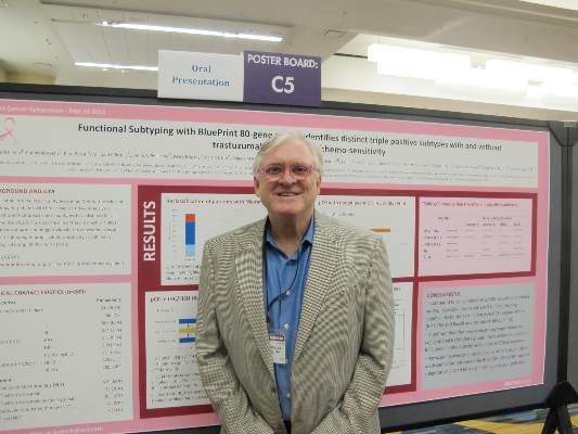
SAN FRANCISCO – Molecular subtyping of breast tumors with an 80-gene panel appears to be a better predictor of response or resistance to neoadjuvant therapy than do standard techniques and may identify patients who could benefit from dual HER-2 blockade, according to data presented by investigators.
In a prospective trial, molecular subtyping with the BluePrint 80-gene profile (Agendia) classified 23% of tumors into a different subgroup than did immunohistochemical staining and fluorescent in-situ hybridization (IHC/FISH) and identified a sub-group of so-called triple-positive tumors (positive for the HER-2, estrogen and progresterone receptors) with luminal features.
“This triple-positive, 80-gene luminal subtype, is relatively resistant to neoadjuvant chemotherapy with trastuzumab [Herceptin] alone. Pertuzumab [Perjeta] overcame resistance to chemotherapy with trastuzumab in a substantial proportion of the triple-positive 80-gene luminal subtype,” said Dr. Peter Beitsch from the Dallas (Texas) Surgical group, in an oral session at a breast cancer meeting sponsored jointly by the American Society of Clinical Oncology, American Society of Radiation Oncology, and Society of Surgical Oncology. The data were also presented in a poster session.
“This trial was about 4 years long, and over the course of the trial, we saw the triple-positive luminal group get more and more pathologically complete responses, and we finally figured it out. What had happened was that about midway through the trial, pertuzumab became the standard,” Dr. Beitsch said in an interview.
The investigators used the gene profile to subtype tumors from 889 women from the ages of 18 years through 90 years with histologically proven breast cancer who had not yet undergone excision biopsy, axillary dissection, or breast cancer therapy.
Patients were treated with neoadjuvant therapy at the discretion of their oncologists, provided that the regimen was peer-reviewed or accepted by the National Comprehensive Cancer Network.
As noted before, 23% of tumors were classified into a different subgroup than those assigned by ICH/FISH.
The gene profile re-classified 82 of 167 patients (49%) who were classified by IHC/FISH as triple-positive as BluePrint luminal instead. In this subgroup of patients, the pathologic complete response (pCR) rate was 17%. In contrast, of the 72 patients (43%) classified as triple-positive by IHC/FISH and as BluePrint HER2 by the gene profile, the pCR rate was 59%, a statistically significantly better response rate than patients with the BluePrint luminal designation (P = .0001).
The remaining 13 patients (8%) were reclassified as BluePrint basal.
“We looked at the patients who were triple-positive, luminal subtype, and they had an absymal [chance of achieving pathologic complete response] with just trastuzumab and chemotherapy, but, you add in pertuzumab, and it goes up 10-fold,” Dr. Beitsch said.
“Using this particular molecular platform may indeed allow us to better categorize patients over what ICH alone is able to do,” said Dr. William J. Gradishar from the Lurie Comprehensive Cancer Center at Northwestern University in Chicago, Illinois, the invited discussant.
He noted that therapy guided by intrinsic subtype could be even more potent and might result in superior outcomes.
“Of course, in the end, the only way we’re going to be able to definitively address this is by rigorously looking at this in a prospective manner,” he said.
AT THE ASCO BREAST CANCER SYMPOSIUM
Key clinical point: An 80-gene profile identified a subtype of breast cancer that may require dual HER2 blockade for effective treatment.
Major finding: A subgroup of patients with triple-positive breast cancer and luminal subtype had a pathologic complete response rate of 17%, compared with 59% of patients with tumors classified as HER2-driven.
Data source: Prospective study of 889 women enrolled in a gene profiling trial.
Disclosures: The study was sponsored by Agendia. Dr. Beitsch disclosed receiving research support and serving as a speaker for the company. Dr. Gradishar reported no relevant conflicts of interest.
MRI Improved Breast Cancer Detection in Average-Risk Women
SAN FRANCISCO – MRI-screening may improve the detection of biologically relevant breast cancer in women who are at average-risk, and reduce the interval-cancer rate down to 0%, at a low false-positive rate, say the authors of a new study presented at the 2015 Breast Cancer Symposium.
In this cohort of heavily pre-screened women at average risk, the additional cancer yield achieved through MRI was high, at 15.8 cases per 1,000 women screened, and the added cancers diagnosed by MRI tended to be of high nuclear grade.
“MRI improves the detection of small high grade cancers in women at average risk, to the extent that the interval cancer rate was zero,” said Dr. Christiane Kuhl, of the University of Aachen, Germany.
“Between 30% and 50% of cancers identified in women who undergo screening mammography are not detected by mammography,” she said during her presentation.
Breast cancer continues to be the main or second most important cause of cancer death in women, and is the main cause of life years lost in women. “Which tells you that the main problem with mammography screening is not overdiagnosis,” she said. “The main problem is actually underdiagnois of disease.”
Breast MRI is currently recommended for screening women who are at a high-risk of breast-cancer, but despite decades of mammographic screening, breast cancer continues to cause high mortality for women who are deemed to be at an average risk of the disease. These data suggest that there is a need for improved methods of early diagnosis in this population.
Dr. Kuhl and her colleagues investigated the utility of supplemental MRI-screening of women who carry an average-risk of breast-cancer, and they conducted a prospective observational cohort-study that assessed the use of MRI screening in this population. The participants were in the usual age range for screening-mammography (40-70 years).
The women underwent dynamic contrast-enhanced MRI of the breast in addition to mammography every 12, 24, or 36 months, plus follow-up of 2 years to establish a standard-of-reference. The cohort included 2,120 women who underwent a total 3,861 MRI screenings.
Breast-cancer was diagnosed in 61 women (DCIS: 20, invasive: 41), and the rate of interval cancers was 0%, irrespective of screening interval. Of these women, 48 were diagnosed at prevalence-screening by MRI alone (supplemental cancer detection rate: 22.6 cases per 1,000 women screened. In addition, 13 women were diagnosed with breast-cancer in 1,741 incidence-screening-rounds collected over 4,887 women-years. A total 12 of these 13 incident cancers were diagnosed by screening-MRI alone (supplemental cancer detection rate was 6.9 per 1,000), one by MRI and mammography, and none were by mammography alone.
The authors also found that the supplemental cancer detection rate was independent of mammographic breast density. Invasive cancers were small, with a mean size of 8mm. They were node-negative in 93.4%, ER/PR-negative in 32.8%, and de-differentiated in 41.7% at prevalence, and 46% at incidence-screening. The specificity of MRI-screening was 97.1%, while false positive rate was 2.9%.
In a discussion of the paper, Dr. A. Marilyn Leitch, of the University of Texas Southwestern Medical Center, Dallas, pointed out that this was a selected population with negative mammograms. But is it possible to “apply MRI at longer intervals, say every 3 years, without initial mammography as a screening tool, in average risk women?” she asked
One of her concerns, however, is that “we hear all the time about the high false positive rates with MRI,” and they are generally higher than was reported in this study. “MRI also detects lesions that might drive patients to avoid breast conserving surgery, and there can be unreasonable costs for screening and work up of false positives.”
Screening guidelines from the USPSTF say that evidence for replacing mammography with MRI is lacking and the balance of benefit versus harms cannot be determined. “Even more ‘liberal’ guidelines from the American Cancer Society reserve MRI for high risk patients.”
What is needed is a clinical trial, she summarized, that will compare screening MRI at 3 year intervals to tomosynthesis mammogram annually in average risk women.
2015 BREAST CANCER SYMPOSIUM
ASCO: MRI improved breast cancer detection in average risk women
SAN FRANCISCO – MRI-screening may improve the detection of biologically relevant breast cancer in women who are at average-risk, and reduce the interval-cancer rate down to 0%, at a low false-positive rate, say the authors of a new study presented at the 2015 Breast Cancer Symposium.
In this cohort of heavily pre-screened women at average risk, the additional cancer yield achieved through MRI was high, at 15.8 cases per 1,000 women screened, and the added cancers diagnosed by MRI tended to be of high nuclear grade.
“MRI improves the detection of small high grade cancers in women at average risk, to the extent that the interval cancer rate was zero,” said Dr. Christiane Kuhl, of the University of Aachen, Germany.
“Between 30% and 50% of cancers identified in women who undergo screening mammography are not detected by mammography,” she said during her presentation.
Breast cancer continues to be the main or second most important cause of cancer death in women, and is the main cause of life years lost in women. “Which tells you that the main problem with mammography screening is not overdiagnosis,” she said. “The main problem is actually underdiagnois of disease.”
Breast MRI is currently recommended for screening women who are at a high-risk of breast-cancer, but despite decades of mammographic screening, breast cancer continues to cause high mortality for women who are deemed to be at an average risk of the disease. These data suggest that there is a need for improved methods of early diagnosis in this population.
Dr. Kuhl and her colleagues investigated the utility of supplemental MRI-screening of women who carry an average-risk of breast-cancer, and they conducted a prospective observational cohort-study that assessed the use of MRI screening in this population. The participants were in the usual age range for screening-mammography (40-70 years).
The women underwent dynamic contrast-enhanced MRI of the breast in addition to mammography every 12, 24, or 36 months, plus follow-up of 2 years to establish a standard-of-reference. The cohort included 2,120 women who underwent a total 3,861 MRI screenings.
Breast-cancer was diagnosed in 61 women (DCIS: 20, invasive: 41), and the rate of interval cancers was 0%, irrespective of screening interval. Of these women, 48 were diagnosed at prevalence-screening by MRI alone (supplemental cancer detection rate: 22.6 cases per 1,000 women screened. In addition, 13 women were diagnosed with breast-cancer in 1,741 incidence-screening-rounds collected over 4,887 women-years. A total 12 of these 13 incident cancers were diagnosed by screening-MRI alone (supplemental cancer detection rate was 6.9 per 1,000), one by MRI and mammography, and none were by mammography alone.
The authors also found that the supplemental cancer detection rate was independent of mammographic breast density. Invasive cancers were small, with a mean size of 8mm. They were node-negative in 93.4%, ER/PR-negative in 32.8%, and de-differentiated in 41.7% at prevalence, and 46% at incidence-screening. The specificity of MRI-screening was 97.1%, while false positive rate was 2.9%.
In a discussion of the paper, Dr. A. Marilyn Leitch, of the University of Texas Southwestern Medical Center, Dallas, pointed out that this was a selected population with negative mammograms. But is it possible to “apply MRI at longer intervals, say every 3 years, without initial mammography as a screening tool, in average risk women?” she asked
One of her concerns, however, is that “we hear all the time about the high false positive rates with MRI,” and they are generally higher than was reported in this study. “MRI also detects lesions that might drive patients to avoid breast conserving surgery, and there can be unreasonable costs for screening and work up of false positives.”
Screening guidelines from the USPSTF say that evidence for replacing mammography with MRI is lacking and the balance of benefit versus harms cannot be determined. “Even more ‘liberal’ guidelines from the American Cancer Society reserve MRI for high risk patients.”
What is needed is a clinical trial, she summarized, that will compare screening MRI at 3 year intervals to tomosynthesis mammogram annually in average risk women.
2015 BREAST CANCER SYMPOSIUM
Key clinical point: MRI-screening improved the detection of biologically relevant breast-cancer in women at average risk.
Major finding: The additional cancer yield achieved through MRI was 15.8 cases per 1,000 women screened.
Data source: A prospective observational cohort-study that was conducted in two centers on 2,120 asymptomatic women, for a total of 3,861 MRI-studies, covering 7,007 women-years.
Disclosures: The authors have no disclosures.
ASCO: Radiotherapy not needed for all women post mastectomy
Postmastectomy radiotherapy should not be routinely recommended for breast cancer patients with microscopic nodal metastases (N1mic) and T1-2 tumors, according to new findings presented at the symposium.
In patients with T1-2, N1 disease who were treated with standard therapies, the study authors found that overall, there were low rates of locoregional failure. Dr. Lonika Majithia of Ohio State University, Columbus, and colleagues, reported that “patients with N1mic disease had no locoregional failure events and improved overall survival, compared with patients with macrometastases.”
The indications for postmastectomy radiotherapy have expanded to include patients with one to three axillary nodal metastases, note the authors, and with improvements in diagnostic evaluation, an increasing number of N1mic are being detected. Therefore, the challenge facing oncologists now is if the risk posed by N1mic warrants routine delivery of postmastectomy radiotherapy.
The authors identified 550 eligible patients from a prospectively maintained cancer registry, who had a 5-year median follow-up. All patients had pathologic T1-2N1 breast cancer and were treated with an initial mastectomy and adjuvant systemic therapy from 2000 to 2013.
The primary endpoint of the study was locoregional failure, defined as a recurrence in either the ipsilateral chest wall or regional lymphatics (axillary, internal mammary, or supraclavicular). Secondary endpoints included disease-free survival and overall survival.
The majority of patients in the cohort had received chemotherapy (78%; n = 428) and antiendocrine therapy (70%; n = 385), while 15% (n = 82) had received postmastectomy radiation. Among the patients with N1mic disease, 81 had 1+ node, 13 had 2+ nodes, and 1 had 3+ nodes.
The 5-year rate of locoregional failure was 0% for patients with N1mic disease, as compared with 4.6% for those with macrometastases (P = .84). For the entire patient cohort, the 5-year locoregional failure rate was 3.9%. For patients with 1+, 2+, and 3+ nodes, the 5-year rate was 2.6%, 4.7%, and 6.4%, respectively (P = .79).
The authors observed that patients with N1mic disease had a trend towards improved disease-free survival (91.6% vs. 82.3%; P = .07) as well as significantly improved overall survival (96.9% vs. 87.6%; P = .03), as compared with patients with macrometastases.
FROM THE 2015 ASCO BREAST CANCER SYMPOSIUM
Key clinical point: Radiotherapy following mastectomy should not be routinely recommended for breast cancer patients with microscopic nodal metastases (N1mic) and T1-2 tumors.
Major finding: The 5-year rate of locoregional failure was 0% for patients with N1mic disease vs. 4.6% in those with macrometastases (P = .84).
Data source: 550 eligible patients who met the inclusion criteria were identified from a prospectively maintained cancer registry.
Disclosures: No conflicts of interest were disclosed.
ASCO: Racial disparity in HER2+ breast cancer survival subsided after trastuzumab approval
Well-documented racial disparities in survival of patients with HER2+ breast cancer diminished after FDA approval of trastuzumab, according to research presented at the 2015 ASCO breast cancer symposium.
A retrospective study identified 32,597 cases of HER2+ breast cancer from the California Cancer Registry, and divided them into early (diagnosed 2000-2006) and late (diagnosed 2006-2012) cohorts. In the early cohort, diagnosed before trastuzumab was available, black patients had an increased risk of mortality (hazard ratio, 1.32; 95% confidence interval, 1.16-1.49), compared with whites. In the late cohort there were no survival differences based on race.
“Although we were unable to document use of anti-HER2 treatment, the era of adjuvant trastuzumab appears to have attenuated the black/white disparity in HER2-positive breast cancer,” wrote Dr. Vincent Caggiano, hematologist at Sutter Medical Center Sacramento, California, and colleagues in a meeting abstract.
The study analyzed risk of mortality, adjusted for stage, grade, age, and socioeconomic status, for African Americans, Hispanics, Asian/Pacific Islanders, and American Indians, compared with white patients. Considering estrogen-receptor and progesterone-receptor status, the combination of all HER2+ subtypes (ER+/PR+/HER2+, ER-/PR+/HER2+, ER+/PR-/HER2+, ER-/PR-/HER2+), showed increased risk for black patients before 2006. The ER-/PR-/HER2+ subtype showed no racial disparities for either cohort. The highest risk (HR, 1.43; 95% CI, 1.17-1.75) was observed for black patients of the ER+/PR+/HER2+ subtype diagnosed before 2006.
FROM THE 2015 ASCO BREAST CANCER SYMPOSIUM
Key clinical point: The well-documented disparity in survival between black and white patients with HER2+ breast cancer was not observed during a time period after 2006, when the FDA approved trastuzumab.
Major finding: Among patients diagnosed from 2000 to 2006, black patients had an increased risk of mortality, compared with white patients (HR, 1.32; 95% CI, 1.16-1.49); from 2006-2012 there were no survival differences based on race/ethnicity.
Data source: The retrospective study used the California Cancer Registry to identify 32,597 cases of HER2+ breast cancer diagnosed from 2000 to 2012, and divided them into early (2000-2006) and late (2006-2012) cohorts.
Disclosures: The authors reported having no disclosures.
ASCO: Overall survival similar regardless of distant relapse site in breast cancer
The initial distant relapse site had little influence on overall survival rates among women with metastatic breast cancer who underwent surgery and definitive radiotherapy, according to a study presented at the 2015 ASCO Breast Cancer Symposium.
In total, 3,417 patients with breast cancer treated with surgery and definitive radiotherapy in Tokyo between 1980 and 2014 were included in the study, with a median follow up of 113 months. Metastatic progression was a first relapse event in 370 patients, and median duration of OS after initial metastatic relapse was 69 months.
Tumors were classified as luminal A, luminal-human epidermal growth factor receptor 2 (HER2), luminal B, and triple negative. OS after distant relapse for patients whose initial tumor was luminal was significantly longer than triple negative and HER2 subtypes (P = .003).
OS was similar for all subtypes after initial bone or nonbone/brain relapse.
Despite the fact that bone metastasis as the initial distant relapse is commonly considered to have better prognosis than other sites in metastatic breast cancer, “OS rates of bone and non-bone/brain metastasis groups… were almost identical,” wrote Kenshiro Shiraishi of the University of Tokyo Hospital, Japan, and colleagues in a meeting abstract.
FROM THE 2015 ASCO BREAST CANCER SYMPOSIUM
Key clinical point: In patients with metastatic breast cancer, initial distant relapse site had little influence on overall survival rates.
Major finding: Overall survival (OS) rates for patients whose initial relapse site was bone were similar to those whose initial site was nonbone/brain. OS after distant relapse for patients whose initial tumor was luminal was significantly longer than triple negative and HER2 subtypes (P = .003).
Data source: In total, 3,417 patients with breast cancer treated with surgery and definitive radiotherapy in Tokyo between 1980 and 2014 were included in the study.
Disclosures: The authors reported having no disclosures.
Treating women with metastatic breast cancer takes toll
Medical oncologists empathize with their patients and feel responsible for providing them emotional/psychological support, but many experience the negative emotional impact of their work when treating women with metastatic breast cancer, according to survey results presented at the symposium.
Medical oncologists with less experience appeared to be more impacted by their emotions as compared to physicians with more experience, and the study authors noted that “acknowledging medical oncologists’ emotions is important and underscores their own need for psychological/emotional support.”
Dr. Adam M. Brufsky, associate chief, division of hematology/oncology and codirector, Comprehensive Breast Cancer Center at the University of Pittsburgh, and his colleagues surveyed medical oncologists who treat five or more women per month with metastatic breast cancer, and surveys were also completed by patients with the disease. The goal of the study was to evaluate the emotional impact on the oncologist. The surveys were conducted from June to August 2014.
A total of 359 patients (median age 53 years) and 252 medical oncologists (median age 49 years and a median of 15 years in practice) completed the survey.
At the initial diagnosis of metastatic breast cancer, a larger proportion of oncologists as compared with patients reported that showing care and compassion (81% vs. 72%) and helping patients cope with their diagnosis (63% vs. 51%) were very important. A smaller percentage of respondents felt that referring patients to support services (24% vs. 38%) was very important, but a greater number of oncologists who were practicing less than 15 years, as compared to those in practice longer, stated that referrals to support services were very important at first diagnosis (30% vs. 19%). Dr. Brufsky and his team also found that a slightly higher number of more experienced oncologists perceived emotions like anxiety, commitment, hopefulness, and determination in their patients at their initial diagnosis.
A large proportion of medical oncologists (42%) reported that treating women with this diagnosis generated a great deal of negative emotion, but a majority (81% strongly/somewhat) agreed that it is unprofessional to let emotions impact treatment recommendations. However, nearly a quarter (23%) reported that emotions kept them from providing some information to patients.
The vast majority of respondents (93%) said that they did not want to give their patients false hope, but yet 27% reported that in certain situations, they do not discuss the fact that metastatic breast cancer is incurable with their patients.
FROM THE ASCO BREAST CANCER SYMPOSIUM
Key clinical point: Many medical oncologists experience the negative emotional impact of their work when treating patients with metastatic breast cancer.
Major finding: Medical oncologists’ emotions are important and these practitioners should acknowledge their own need for psychological/emotional support.
Data source: A survey was conducted of 359 patients and 252 medical oncologists.
Disclosures: The investigators had no relevant disclosures.
NLR correlates with survival in advanced breast cancer
After treatment begins, an increased neutrophil lymphocyte ratio (NLR) can be correlated with poor disease-specific survival in stage IV breast cancer, according to research presented at the symposium. In addition, the change in NLR might be an index of response to systemic treatment.
Inflammatory response exacerbates mechanisms linked to tumor growth and dissemination, note the study authors, led by Dr. Hae-na Shin of the University of Ulsan, Seoul, South Korea. Used as an index of systemic inflammatory status, the NLR could be a predictive biomarker for both prognosis and treatment response.
To test their hypothesis, Dr. Shin and his colleagues evaluated the baseline NLR prior to beginning treatment, and then the change in posttreatment NLR, to assess if the initial NLR and its change after therapy would be predictive of disease outcome in stage IV breast cancer patients.
The study cohort included 250 women with stage IV breast cancer who were diagnosed at the Asan Medical Center between 1997 and 2012. The NLR was evaluated at their first visit to the center, and a posttreatment NLR was obtained during the first follow-up appointment after patients received their first treatment.
The authors divided the pretreatment NLR by quartile, and the change in NLR was calculated by dividing posttreatment NLR by pretreatment NLR. If the value was greater than or equal to 1.2, NLR change was increased, and if not, then it was considered to be unchanged or reduced. The prognostic value of NLR was then evaluated by comparison with Cancer Specific Survival (CSS).
When comparing pretreatment NLR and posttreatment NLR, the NLR was elevated in 85 patients (34%) but remained the same or decreased in 165 others (66%). There were no significant differences between these two groups in baseline characteristics. However, in CSS, there were differences between the two groups but they did not reach statistical significance (log rank P = 0.052). The 1-, 3-, 5-year CSS rate was 78.8%, 35.7%, 20.5% in the group with an increased NLR, and 87.1%, 49.3%, 26.9% in the other patient group.
Upon multivariate analysis, the results suggested that an increased NLR change (post/pre NLR greater than or equal to 1.2) had statistical significance as a prognostic factor of stage IV breast cancer patients after treatment (hazard ratio, 1.750; 95% confidence interval, 1.130-2.709; P = 0.012).
AT THE ASCO BREAST CANCER SYMPOSIUM
Key clinical point: Neutrophil lymphocyte ratio (NLR) can be correlated with poor disease-specific survival in stage IV breast cancer and the change in NLR might be an index of response to systemic treatment.
Major finding: The results suggest that an increased NLR change (post/pre NLR greater than or equal to 1.2) had statistical significance as a prognostic factor of stage IV breast cancer patients after treatment (HR, 1.750; 95% CI, 1.130-2.709; P = .012).
Data source: The cohort was comprised of 250 stage IV breast cancer patients diagnosed at a single center and pre and post treatment NLR were evaluated.
Disclosures: The investigators had no relevant disclosures.
Histologic information can’t replace recurrence score
Combining routine histologic information, including tumor size, grade, and cellular proliferation marker Ki67, failed to accurately reflect the recurrence score produced by the commercial diagnostic test, Oncotype Dx, according to researchers.
Although each variable individually was significantly correlated with Oncotype Dx, the combination of variables had a percentage of variance (R2) of 0.35 on the recurrence score and did not accurately predict the result generated by the 21-gene reverse-transcriptase polymerase chain reaction (RT-PCR) diagnostic test.
The single-institution study included 252 patients, (248 females and 4 males, median age 56 years) with early-stage, estrogen receptor–positive breast cancer. All patients had Oncotype Dx testing performed between 2007 and 2014. The median tumor size was 2.1 cm, and 49% were grade 2, 28% grade 3, and 23% grade 1.
Investigators examined baseline patient demographic data, such as age, race, and sex, and routine pathologic features, such as histologic grade, Ki-67, tumor size, and histologic type. The combination of three variables that individually were highly correlated to the Oncotype Dx score did not produce values that could substitute for the recurrence score.
Furthermore, the prediction capability of the three-variable combination decreased for tumors with high-recurrence scores.
“Therefore, we conclude the RS [recurrence score] can provide additional valuable information and should not be replaced by analysis of routine histologic variables alone,” wrote Dr. Kate Lathrop of the Cancer Therapy and Research Center at the University of Texas Health Science Center in San Antonio, and her colleagues.
FROM THE ASCO BREAST CANCER SYMPOSIUM
Key clinical point: A combination of three histologic variables (tumor size, grade, and cellular proliferation marker Ki67) failed to accurately predict the Oncotype Dx recurrence score.
Major finding: Based on the Oncotype Dx recurrence score, the score predicted by the three-variable combination had a percentage of variance (R2) of 0.35.
Data source: The retrospective, single-institution study evaluated data from 252 patients with early-stage estrogen receptor–positive breast cancer who had Oncotype Dx testing performed between 2007 and 2014.
Disclosures: The authors reported having no disclosures.
Radiation + lumpectomy tied to increased survival in older women with TNBC
Older women with triple-negative breast cancer appear to get an overall survival and disease-specific survival benefit with the addition of radiation to breast-conserving surgery, authors of a retrospective study said.
Among 974 women aged 70 and above with T1-2, N0, M0 triple-negative breast cancer (TNBC; lacking the Her2-neu, estrogen, and progesterone receptors), the addition of radiation to lumpectomy was associated at 23 months’ follow-up with an overall survival (OS) rate of 98.2%, compared with 85.6% for women who received lumpectomy alone (P less than .001). Respective rates of disease-specific survival (DSS) were 99% and 94% (P = .003).
“The use of adjuvant radiation therapy after lumpectomy for elderly women with early-stage TNBC was associated with improved OS and DSS. Noting the potential for selection bias in this study, future prospective study is required to define the management of early-stage triple-negative breast cancer,” wrote Dr. Sean Szjea and colleagues at the University of Texas, Galveston, in a meeting abstract. The study will be presented at a breast cancer symposium sponsored by the American Society of Clinical Oncology.
It’s known that older women with estrogen-receptor positive disease can have good clinical outcomes with lumpectomy and adjuvant therapy alone, but whether adding radiation to breast-conserving surgery in older women with TNBC offers clinical benefit is less certain, the investigators said, prompting them to dive for data into the Surveillance, Epidemiology, and End Results database.
They collected information on 974 women aged 70 or older who underwent lumpectomy for early-stage TNBC with no nodal invasion or metastatic disease from 2010 through 2011. Of this group, 662 (68%) also received radiation therapy.
In addition to determining the OS and DSS rates in the overall population, the investigators conducted multivariate regression modeling controlling for confounding variables, including the use of neoadjuvant chemotherapy, the number of lymph nodes sampled, age, laterality, grade, T stage, the extent of surgery, and the existence of other cancers. They found that the survival benefit for radiation held for both OS (hazard ratio [HR], 0.14; P less than .001) and DSS (HR, 0.14; P = .01).
The authors reported having no relevant financial disclosures.
FROM THE 2015 ASCO BREAST CANCER SYMPOSIUM
Key clinical point: Radiation added to breast-conserving surgery in women 70 and older with early-stage triple-negative breast cancer was associated with improved survival.
Major finding: Overall survival with radiation and lumpectomy was 98.2%, compared with 85.6% for women who received lumpectomy alone (P less than .001)
Data source: A prospective database review of 974 women aged 70 and older treated from 2010 through 2011.
Disclosures: The authors reported having no relevant financial disclosures.


