User login
European Society of Cardiology (ESC): EUROECHO 2014
Emergency cardiac echocardiography accepted by Europeans
VIENNA – Rapid echocardiographic assessment has become routine for many patients who arrive at an emergency department with suspected acute heart failure, and experts consider these examinations critical for quickly getting patients on the right treatment.
Growing use and the important role for emergency echo exams prompted the European echocardiography community to issue in 2014 both recommendations and a position statement on the practice.
With their actions, European echocardiographers joined their U.S. colleagues who had earlier endorsed rapid, focused echocardiography exams. The European position also highlighted the limitations and pitfalls of emergency echo and the need for proper training.

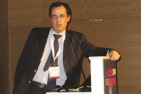
Use of limited, directed, ultrasound heart examinations on an emergency basis by physicians who are not cardiologists is “an irreversible process, but without appropriate training it may become dangerous,” Dr. Nuno Cardim said at the annual meeting of the European Association of Cardiovascular Imaging (EACVI).
A focused cardiac ultrasound (FoCUS) examination for patients with an emergency cardiac condition such as acute heart failure is not a new concept. In 2010, the American Society of Echocardiography and the American College of Emergency Physicians jointly issued a consensus statement on emergency FoCUS (J. Am. Soc. Echocardiogr. 2010;23:1225-30), and the American Society of Echocardiography followed with additional recommendations in 2013 that also dealt with nonemergency uses for FoCUS (J. Am. Soc. Echocardiogr. 2013;26:567-81).
In its 2014 position statement released last May, the EACVI directly addressed FoCUS for the first time (Eur. Heart J. Cardiovasc. Imaging 2014:15;956-60). The statement acknowledged the important role for a circumscribed, point-of-care ultrasound exam in patients undergoing cardiopulmonary resuscitation and in other critical cardiac conditions, but highlighted that a FoCUS exam does not substitute for a comprehensive echocardiographic exam, and that FoCUS should only be done by properly trained clinicians who appreciate the limits of a FoCUS exam.
The EASVI recommendations, which came out a few months later in collaboration with the Acute Cardiovascular Care Association, said that “echocardiography is now recommended (where appropriately trained practitioners are available) in the management of cardiac arrest. However, FoCUS should always be used and interpreted thoughtfully, since this fundamentally limited approach may lead to missing/misinterpretation of important findings unless the practitioner is aware of its (and their) limitations” (Eur. Heart J. Cardiovasc. Imaging 2014 [doi:10.1093/ehjci/jeu210]).
“Of course all patients with suspected acute heart failure in the emergency department should undergo an echo exam. The question is, who will do it? These are patients who are the most difficult to assess,” said Dr. Susanna Price, a member of the EACVI recommendations panel and a specialist in critical care cardiology at Royal Brompton Hospital in London.
“Without proper training, the person doing FoCUS could make a false positive diagnosis, or might miss something and make a false negative diagnosis,” said Dr. Cardim, professor and director of echocardiography and cardiac imaging at Hospital da Luz in Lisbon, and another member of the EACVI panel.
To avoid this, emergency-medicine physicians and others who often triage patients with acute heart disorders should be trained in echocardiography and especially the FoCUS exam, which aims to quickly evaluate several important abnormalities of cardiac function: pericardial effusion, cardiac tamponade, left and right ventricular size and function, and intravascular volume status. A FoCUS exam also screens for pulmonary embolism. FoCUS assesses each of these in a yes-or-no or present-or-absent way, information critical for guiding emergency management but lacking the quantitative and detailed information available with a comprehensive echocardiography exam.
“FoCUS must never substitute” for the comprehensive exam, which should always also be done, he said. FoCUS “should be used wisely and cautiously because of its limitations.”
The FoCUS exam also has equipment specifications. Ideally, clinicians should use a portable, hand-held ultrasound machine, which is larger than “pocket-sized” ultrasound devices and hence gives much better image quality compared with pocket-sized devices, Dr. Cardim said in an interview.
Dr. Cardim and Dr. Price had no disclosures.
On Twitter @mitchelzoler
VIENNA – Rapid echocardiographic assessment has become routine for many patients who arrive at an emergency department with suspected acute heart failure, and experts consider these examinations critical for quickly getting patients on the right treatment.
Growing use and the important role for emergency echo exams prompted the European echocardiography community to issue in 2014 both recommendations and a position statement on the practice.
With their actions, European echocardiographers joined their U.S. colleagues who had earlier endorsed rapid, focused echocardiography exams. The European position also highlighted the limitations and pitfalls of emergency echo and the need for proper training.
Use of limited, directed, ultrasound heart examinations on an emergency basis by physicians who are not cardiologists is “an irreversible process, but without appropriate training it may become dangerous,” Dr. Nuno Cardim said at the annual meeting of the European Association of Cardiovascular Imaging (EACVI).
A focused cardiac ultrasound (FoCUS) examination for patients with an emergency cardiac condition such as acute heart failure is not a new concept. In 2010, the American Society of Echocardiography and the American College of Emergency Physicians jointly issued a consensus statement on emergency FoCUS (J. Am. Soc. Echocardiogr. 2010;23:1225-30), and the American Society of Echocardiography followed with additional recommendations in 2013 that also dealt with nonemergency uses for FoCUS (J. Am. Soc. Echocardiogr. 2013;26:567-81).
In its 2014 position statement released last May, the EACVI directly addressed FoCUS for the first time (Eur. Heart J. Cardiovasc. Imaging 2014:15;956-60). The statement acknowledged the important role for a circumscribed, point-of-care ultrasound exam in patients undergoing cardiopulmonary resuscitation and in other critical cardiac conditions, but highlighted that a FoCUS exam does not substitute for a comprehensive echocardiographic exam, and that FoCUS should only be done by properly trained clinicians who appreciate the limits of a FoCUS exam.
The EASVI recommendations, which came out a few months later in collaboration with the Acute Cardiovascular Care Association, said that “echocardiography is now recommended (where appropriately trained practitioners are available) in the management of cardiac arrest. However, FoCUS should always be used and interpreted thoughtfully, since this fundamentally limited approach may lead to missing/misinterpretation of important findings unless the practitioner is aware of its (and their) limitations” (Eur. Heart J. Cardiovasc. Imaging 2014 [doi:10.1093/ehjci/jeu210]).
“Of course all patients with suspected acute heart failure in the emergency department should undergo an echo exam. The question is, who will do it? These are patients who are the most difficult to assess,” said Dr. Susanna Price, a member of the EACVI recommendations panel and a specialist in critical care cardiology at Royal Brompton Hospital in London.
“Without proper training, the person doing FoCUS could make a false positive diagnosis, or might miss something and make a false negative diagnosis,” said Dr. Cardim, professor and director of echocardiography and cardiac imaging at Hospital da Luz in Lisbon, and another member of the EACVI panel.
To avoid this, emergency-medicine physicians and others who often triage patients with acute heart disorders should be trained in echocardiography and especially the FoCUS exam, which aims to quickly evaluate several important abnormalities of cardiac function: pericardial effusion, cardiac tamponade, left and right ventricular size and function, and intravascular volume status. A FoCUS exam also screens for pulmonary embolism. FoCUS assesses each of these in a yes-or-no or present-or-absent way, information critical for guiding emergency management but lacking the quantitative and detailed information available with a comprehensive echocardiography exam.
“FoCUS must never substitute” for the comprehensive exam, which should always also be done, he said. FoCUS “should be used wisely and cautiously because of its limitations.”
The FoCUS exam also has equipment specifications. Ideally, clinicians should use a portable, hand-held ultrasound machine, which is larger than “pocket-sized” ultrasound devices and hence gives much better image quality compared with pocket-sized devices, Dr. Cardim said in an interview.
Dr. Cardim and Dr. Price had no disclosures.
On Twitter @mitchelzoler
EXPERT ANALYSIS FROM EUROECHO-IMAGING 2014
Early mitral-valve repair dampens tricuspid-valve regurgitation
VIENNA – One of the best ways to prevent advanced tricuspid-valve regurgitation and need for tricuspid-valve repair may be a more aggressive approach to mitral valve repair.
“If you operate on the mitral valve early, then tricuspid regurgitation does not tend to progress,” Dr. Sunil V. Mankad said at the annual meeting of the European Association of Cardiovascular Imaging. “If you wait until the mitral valve remodels and the atrium enlarges and remodels or there is pulmonary hypertension, then tricuspid regurgitation will progress,” said Dr. Mankad, a echocardiographer at the Mayo Clinic in Rochester, Minn.
Early intervention on mitral valve prolapse has other benefits as well, he said. Mitral disease causes atrial remodeling, which can then progress to atrial fibrillation, “and once that happens it’s a game changer for the patient, even if they later undergo valve repair,” because of atrial fibrillation’s long-term risks and consequences, Dr. Mankad said in an interview.
“We believe there is also subclinical left ventricular dysfunction” in patients with mitral-valve prolapse “even if their ejection fraction is normal.” Once that happens, even if the mitral valve is repaired “the heart is not normal anymore and there is subtle left ventricular dysfunction that is not captured by just looking at ejection fraction.”
To document the impact a more aggressive approach to mitral-valve repair can have on the tricuspid valve, Dr. Mankad cited a 2011 Mayo Clinic analysis of 699 patients who underwent mitral-valve repair at Mayo for severe mitral-valve prolapse and also had some amount of tricuspid regurgitation at the time of their surgery, including 115 patients (16%) with grade 3 or higher tricuspid regurgitation. One year after surgery, the severity of tricuspid regurgitation in these patients had decreased significantly overall, and throughout follow-up only one patient required surgery for tricuspid-valve repair, 4.5 years after that patient’s mitral-valve repair (J. Thoracic Cardiovasc. Surgery 2011;142:608-13).
Dr. Mankad also cited a recent editorial written by several of his Mayo Clinic colleagues that synthesized results from the 2011 report as well as from a second Mayo report published in 2014, and a third report from a different group also published in 2014. The authors of the editorial concluded that results from all three studies showed “the performance of early correction of mitral regurgitation is important not only for its own well known benefits (preservation of survival and minimization of late heart failure risk) but also to diminish the late occurrence of functional tricuspid regurgitation (J. Thoracic Cardiovasc. Surgery 2014;148:2810-2).
Because mitral-valve repair often improves tricuspid-valve function and durability, the editorialists suggested “strongly considering” tricuspid repair for a carefully defined, select subgroup of patients. Their list included patients with tricuspid regurgitation that is worse than moderate, right-heart dysfunction, symptoms of right-heart failure, pulmonary hypertension, reduced left ventricular systolic function, cardiomyopathy, or organic tricuspid pathology.
Existing evidence supports leaving the valve alone when patients have a tricuspid regurgitation that is less than moderate when they have also undergone effective correction of degenerative mitral regurgitation. Patients like these are “unlikely ever to have difficulty with the tricuspid valve or the right ventricle,” wrote the authors of the editorial.
Dr. Mankad offered his own suggestions for identifying patients with a tricuspid valve that requires repair at the time of mitral-valve surgery.
“The evidence supports tricuspid-valve repair at the time of mitral-valve surgery if there is tricuspid annular dilatation of more than 4.0 cm measured by three-dimensional echo or greater than moderate tricuspid regurgitation. This is based on observational data and not on results from randomized control trials, but it is what I recommend,” Dr. Mankad said. “I suggest measuring the tricuspid annulus; it is quite easy to do. Directly measuring the annulus size with three-dimensional echo is pretty basic, and I think it is ready for prime time.”
Dr. Mankad had no disclosures.
On Twitter @mitchelzoler
EXPERT ANALYSIS FROM EUROECHO-IMAGING 2014
3D echocardography underpins percutaneous mitral valve repair
VIENNA – Three-dimensional transesophageal echocardiography “may be considered the gatekeeper for assessing the feasibility of and planning the strategy for percutaneous mitral valve repair,” Dr. Giovanni La Canna said at the annual meeting of the European Association of Association of Cardiovascular Imaging.
“Real-time, three-dimensional transesophageal echo is a new way of looking at the mitral valve,” and it supplies a highly accurate and reproducible anatomic view. It adds important additional information when selecting patients for percutaneous mitral valve repair by giving a comprehensive picture of annular dimensions and shape, intercommissural extension of target-leaflet lesions, and the extent of calcification on the annulus and leaflets, said Dr. La Canna, an echocardiographer at San Raffaele Hospital in Milan.
Three-dimensional echo aids both forms of percutaneous mitral valve repair: annuloplasty and clipping with the MitraClip system.
When performing mitral-leaflet clipping, use of 3D echo adds incremental information beyond 2D echo that aids in the selection of the site for transseptal puncture; facilitates device alignment; optimizes grasping of the leaflet with the clip; and helps in assessment of mitral-valve area and residual regurgitation, the need for additional clips, and any residual defect in the interatrial septum. For especially challenging cases, data from 2D echo can be integrated with the 3D data for the most complete imaging guidance, Dr. La Canna said.
Despite its value, 3D echo still has limitations. Resolution is currently low, parts of the imaging can “drop out” or contain artifacts, the catheter can produce an image shadow, and the image can decay during the course of the procedure, he said.
Dr. La Canna has been a consultant to Abbott Vascular, which markets the MitraClip.
On Twitter @mitchelzoler
EXPERT ANALYSIS FROM EUROECHO-IMAGING 2014

CT shown most cost effective for chest-pain assessment
VIENNA – Coronary CT followed by some form of functional stress imaging when needed now stands as the most cost-effective way to screen patients with chest pain regardless of whether the pain is chronic or acute onset.
Results from a 2011 analysis established the cost efficacy of cardiac CT followed by stress imaging for patients with positive CT results for acute chest-pain patients at low or intermediate risk for coronary disease, and no data reported since then have changed that conclusion, Dr. Thomas H. Marwick said at the annual meeting of the European Association of Cardiovascular Imaging.
Results from a new analysis presented in a separate report at the meeting showed the superior cost effectiveness of the same approach for patients with stable chest pain who are at low or intermediate risk for having coronary disease, said Dr. Steffen E. Petersen, professor and head of the Centre for Advanced Cardiovascular Imaging at the William Harvey Research Institute in London.
“We did a cost-effectiveness analysis that took into account the cost of results that lead to additional testing and the cost when people return later with symptoms and need more testing. We compared about 15 different strategies, and found it was best to start with CT and if that was positive then do a functional test like stress echocardiography, stress single-photon emission CT (SPECT), or stress MRI. CT followed by stress echo seemed most cost effective, but using stress SPECT or MRI was not very different; they can all be used after CT,” Dr. Petersen said in an interview.
His analysis, which modeled the costs for a man or woman aged 60 years with a 30% probability of having coronary artery disease, showed that a testing strategy of coronary CT followed by stress echo for patients with a significant coronary stenosis cost roughly £32,000 (about $50,000) for each quality-adjusted life year gained.
Choice of the follow-up, functional test for patients with stable chest pain who show at least 50% stenosis in at least one segment of their coronary vessels on CT primarily depends on other considerations, such as local experience using the various functional-imaging methods as well as how long a patient might need to wait until the stress test is done, Dr. Petersen said. For higher-risk patients who likely have significant coronary artery disease, the cost effectiveness of an initial CT examination drops below that of the alternative approach of referring the patient directly for a functional test with no initial CT. He advised clinicians to assess a patient’s pretest probability of having coronary artery disease with a tool such as the modified Diamond-Forrester model (Eur. Heart J. 2011;32:1316-30).
Dr. Petersen and his associates used already-published data from the United States, United Kingdom, and The Netherlands that documented the positive and negative predictive values of various imaging methods used alone or in tandem as well as the impact of each method on additional testing, delays while testing proceeds, and eventual patient outcomes. “We basically all agreed on our model, and then we ran the model and looked at the results,” he said.
“I think our findings would change practice in a lot of places,” he added. For patients with stable disease access to various imaging methods is usually not an issue because stable patients can receive a referral to a facility with that method available.
Dr. Marwick asserted that cardiac CT followed by functional testing with SPECT for patients with an indeterminate CT result remains the most cost-effective way to screen a chest-pain patient who presents at an emergency department with a history and physical findings consistent with a low or intermediate risk of having coronary disease. He and his associates documented the consistent cost effectiveness of CT followed by SPECT in patients with a coronary artery disease prevalence of anywhere from 2% to 30% in a 2011 report (J. Am. Coll. Cardiol. Img. 2011;4:549-56). A big reason why CT first is so cost effective is because of its very high negative predictive value – its ability to reliably rule out patients who do not have significant coronary disease.
“It is very hard to overcome the cost effectiveness of CT for chest pain because of its very high predictive value, and because you can use it to study patients early,” said Dr. Marwick, professor and director of the Menzies Institute for Medical Research, University of Tasmania in Hobart, Australia. “Early CT can reduce the patient’s length of stay in the emergency department while avoiding the risk of sending someone home who is having a myocardial infarction,” Dr. Marwick said in an interview. Although his analysis showed that stress SPECT was the most cost-effective option for follow-up testing of a patient with an equivocal CT result, “it could be any functional test” because they all have nearly the same performance, he added. The other options are again stress echo or stress MRI.
“If a patient has chest pain and goes directly to CT the negative predictive value is so high that more than half the patients can safely go straight home. I’m not a big fan of CT, but this is what the numbers show,” he said
Dr. Marwick acknowledged that testing a patient’s blood level of troponin using a high-sensitivity assay might soon supplant CT, but for the time being high-sensitivity troponin remain too non specific, he said. A retrospective, Swedish study reported at the 2014 annual meeting of the American College of Cardiology documented that a high-sensitivity troponin assay could rule out myocardial infarction among patients presenting with chest pain at an emergency department with a negative predictive accuracy of nearly 100%. But at that time one expert commented that proof of the clinical utility of a high-sensitivity troponin assay for emergency chest pain patients needed validation in a well-designed, prospective trial.
Dr. Petersen and Dr. Marwick had no disclosures.
On Twitter @mitchelzoler
AT EUROECHO-IMAGING 2014
Key clinical point: For both stable and acute chest-pain patients initial assessment by coronary CT produced the most cost-effective results.
Major finding: Assessing stable chest-pain patients by coronary CT followed by stress echocardiography cost £32,000 per quality-adjusted life year.
Data source: Model that ran published data from the United States, the United Kingdom, and The Netherlands.
Disclosures: Dr. Petersen and Dr. Marwick had no disclosures.
VIDEO: Focused cardiac ultrasound aids acute heart failure patients
VIENNA – Bedside echocardiography has become a key part of quickly assessing patients with acute heart failure to decide the best management strategy.
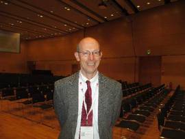
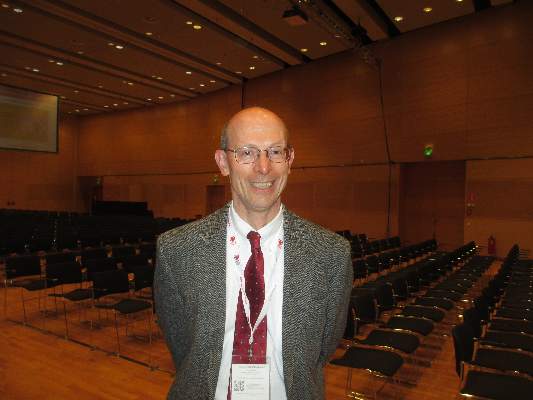
But bedside echo images often are challenging to interpret, so physicians performing an initial work-up of an acute heart failure patient need training in a basic echocardiography examination, Dr. Nuno Cardim said in an interview during the annual meeting of the European Association of Cardiovascular Imaging.
Known as Focused Cardiac Ultrasound (FoCUS), this triage-level exam is distinct from a comprehensive echocardiography assessment. Instead, FoCUS addresses some basic, yes-or-no, present-or-absent questions regarding systolic function, hypovolemia, tamponade, pleural effusion, and pulmonary embolus.
In a position paper and then in practice recommendations issued in 2014, the European Association of Cardiovascular Imaging acknowledged that use of FoCUS in acute heart failure patients was “irreversible,” said Dr. Cardim, professor and director of echocardiography and cardiac imaging at Hospital da Luz in Lisbon.
Cardiologists and echocardiography specialists need to make sure that FoCUS training is available to all physicians who might see suspected acute heart failure patients in emergency medicine settings, Dr. Cardim said.
Dr. Cardim had no disclosures.
The video associated with this article is no longer available on this site. Please view all of our videos on the MDedge YouTube channel
On Twitter @mitchelzoler
VIENNA – Bedside echocardiography has become a key part of quickly assessing patients with acute heart failure to decide the best management strategy.
But bedside echo images often are challenging to interpret, so physicians performing an initial work-up of an acute heart failure patient need training in a basic echocardiography examination, Dr. Nuno Cardim said in an interview during the annual meeting of the European Association of Cardiovascular Imaging.
Known as Focused Cardiac Ultrasound (FoCUS), this triage-level exam is distinct from a comprehensive echocardiography assessment. Instead, FoCUS addresses some basic, yes-or-no, present-or-absent questions regarding systolic function, hypovolemia, tamponade, pleural effusion, and pulmonary embolus.
In a position paper and then in practice recommendations issued in 2014, the European Association of Cardiovascular Imaging acknowledged that use of FoCUS in acute heart failure patients was “irreversible,” said Dr. Cardim, professor and director of echocardiography and cardiac imaging at Hospital da Luz in Lisbon.
Cardiologists and echocardiography specialists need to make sure that FoCUS training is available to all physicians who might see suspected acute heart failure patients in emergency medicine settings, Dr. Cardim said.
Dr. Cardim had no disclosures.
The video associated with this article is no longer available on this site. Please view all of our videos on the MDedge YouTube channel
On Twitter @mitchelzoler
AT EUROECHO-IMAGING 2014
Imaging helps cut TAVR’s paravalvular leaks
VIENNA – Routine use of CT or three-dimensional echocardiography before and during transcatheter aortic valve replacement has contributed to a dramatic drop in paravalvular leaks, the procedure’s Achilles heel, based on the experience at a single, high-volume U.S. center.
In recent months, during testing of the SAPIEN 3 transcatheter aortic valve replacement (TAVR) system under development by Edwards, no patients developed moderate or severe paravalvular regurgitation during follow-up, and mild paravalvular leaks occurred at a rate of 7%-9%, Dr. Rebecca T. Hahn said at the annual meeting of the European Association of Cardiovascular Imaging. She and her associates tallied these rates recently at their institution, Columbia University Medical Center in New York, during their participation in PARTNER II, a multicenter study of the SAPIEN 3 device.
Dr. Hahn attributed these unprecedentedly low rates to three factors: preprocedure imaging with CT or 3-D echocardiography to select the best-sized valve for each patient, routine use of intraprocedural 3-D echo to guide precise valve placement and monitor for complications, and the SAPIEN 3 valve.
To put these regurgitation rates in context, in results from the pivotal CoreValve trial reported last May, the 30-day rate of moderate or severe paravalvular regurgitation was 9%, and the rate of mild paravalvular regurgitation was 36% (N. Engl. J. Med. 2014;370:1790-8). The most recent data on paravalvular leak reported from a large trial using a SAPIEN valve currently on the U.S. market was from the PARTNER II trial of inoperable patients, which documented a roughly 20% rate of moderate or severe paravalvular regurgitation using either the SAPIEN XT or the original SAPIEN system.
Dr. Hahn acknowledged that one major factor contributing to the disappearance of moderate or severe leaks following TAVR was the design of the new SAPIEN 3 TAVR valve she is working with, which features a flexible layer around the outer perimeter of the valve to better seal it into the patient’s aortic valve annulus and prevent blood from leaking through gaps around the edge.
But she also credited intraprocedural 3-D echo as a major factor in the improved outcomes, as well as the use of CT or 3-D echo to accurately size the annulus before TAVR starts so that the patient’s annulus size can be matched with the most appropriately sized replacement valve.
“Each valve has a sweet spot” for the annulus size it will fit into, and CT imaging of the annulus to measure the size, or using three-dimensional echo when CT is not good enough, is the way that TAVR heart teams now make sure they use the valve size with a sweet spot for the patient receiving the valve, said Dr. Hahn, director of invasive and valvular echocardiography at Columbia. A key task for either imaging method is to measure annulus size on the “virtual” annular plane, a plane that is challenging to identify as it is defined by only the three hinge points of the aortic valve’s leaflets. “Three-dimensional echo allows us to quantify [annular size] in ways that we couldn’t before,” she said in an interview.
During the procedure, 3-D echocardiography is the only good imaging option. In addition to clarifying unusual morphologies of the annulus and surrounding tissues and blood vessels, and giving precise information on wire and valve placement, 3-D echo allows for rapid assessment of a hemodynamic emergency if one occurs during a TAVR procedure. Three-dimensional echo also affords the best way to assess the location and severity of paravalvular regurgitation. 3-D echo “allows us to make decisions during the procedure on what to do about paravalvular regurgitation,” Dr. Hahn said.
On Twitter @mitchelzoler
AT EUROECHO-IMAGING 2014
Key clinical point: Routine CT and three-dimensional echocardiography before and during transcatheter aortic valve replacement helped dramatically cut the incidence and severity of paravalvular leaks.
Major finding: Transcatheter aortic valve replacement with the SAPIEN 3 system produced no moderate or severe paravalvular leaks and a 7%-9% rate of mild leaks.
Data source: Recent, anecdotal single-center experience in the PARTNER II trial.
Disclosures: PARTNER II is sponsored by Edwards, the company developing the SAPIEN 3 TAVR system. Dr. Hahn is an investigator in the PARTNER II trial, and has received honoraria as a speaker on behalf of Boston Scientific, St. Jude, and Philips.
VIDEO: CT and 3D-echo imaging boost TAVR performance
VIENNA – Transcatheter aortic valve replacement has benefited enormously from the recent introduction of CT and three-dimensional echocardiography imaging into routine preprocedural planning and periprocedural guidance.
These two imaging methods have allowed clinicians to better select the right valve size for each patient, and 3D echo performed during transcatheter aortic valve replacement (TAVR) has also refined valve placement. The result has been TAVR procedures that go faster and have more accurate results while producing fewer complications, Dr. Rebecca Hahn said during an interview at the annual meeting of the European Association of Cardiovascular Imaging.
Perhaps the biggest impact of better valve-size selection and more detailed procedural guidance has been a reduced incidence and severity of paravalvular regurgitation once TAVR is complete and the replacement valve deployed. In recent months, Dr. Hahn and her associates at Columbia University Medical Center in New York have had no patients develop moderate or severe paravalvular regurgitation following TAVR, and the incidence of mild paravalvular leaks has dropped by about half, compared with when the procedure was first performed about 5 years ago, said Dr. Hahn, director of invasive and valvular echocardiography at Columbia.
The video associated with this article is no longer available on this site. Please view all of our videos on the MDedge YouTube channel
On Twitter@mitchelzoler
AT EUROECHO-IMAGING 2014