User login
World Congress on Treatment and Research in Multiple Sclerosis: 2014 Joint ACTRIMS-ECTRIMS Meeting
Lower frequency glatiramer acetate regimen appears safer
BOSTON– Delivering subcutaneous glatiramer acetate as a 40-mg/mL dose three times weekly rather than 20 mg/mL daily halved the annualized rate of injection-related adverse events in a randomized, open-label, phase IIIb trial of patients with relapsing-remitting multiple sclerosis.
The reduction in the annualized rate of moderate and severe injection-related adverse events in those with lower-frequency dosing was even greater, at about 60%, in the trial, called GLACIER (Glatiramer Acetate Low Frequency Safety and Patient Experience), Dr. Jerry S. Wolinsky reported at the joint meeting of the European and Americas committees for Treatment and Research in Multiple Sclerosis.

“As we all know, long-term, frequent injections are required for the first-line therapies for relapsing forms of multiple sclerosis, and this form of drug delivery presents a challenge for patients. Modification of the treatment regimen, such as using alternative dosages and lower-frequency administration schedules for these drugs with long-term proven efficacy, has the potential to improve the patient experience by reducing, perhaps, the number of adverse experiences that they have, improving tolerability, and in particular, potentially increasing adherence to treatments, especially in the longer-term,” Dr. Wolinsky said, explaining the basis for the GLACIER trial.
Glatiramer acetate 20 mg/mL administered by daily subcutaneous injection is a well-characterized treatment with a good long-term safety profile. It has been used for more than 20 years with more than 2 million patient-years of overall exposures, and it has established efficacy for the treatment of relapsing-remitting MS (RRMS). In January, the Food and Drug Administration approved a supplemental New Drug Application for the 40-mg/mL three times weekly regimen after it was shown to have an efficacy and safety profile similar to that of the 20-mg/mL regimen.
The GLACIER findings suggest that the glatiramer acetate 40-mg/mL three times per week dosing regimen is, for some patients, a favorable treatment option, particularly in those who want to be on glatiramer acetate, but who prefer fewer injections and more convenient dosing, concluded Dr. Wolinsky, director of the multiple sclerosis research group at the University of Texas Health Science Center at Houston.
The annualized rate of injection-related adverse events (IRAEs) in 108 adult patients with RRMS who were randomized to receive the thrice weekly injections was 35.3, compared with 70.4 in 101 patients who received daily injections (relative risk, 0.50). The annualized rate of moderate and severe IRAEs was 0.88 vs. 2.2 in the groups, respectively (relative risk, 0.40), he said.
The most common injection site reactions were pain, erythema, mass, swelling, and pruritus, and all of these were substantially reduced with lower-frequency dosing, compared with daily dosing. For example, the annualized rate of pain events was 24.8 vs. 55.3 with lower- vs. higher-frequency dosing, and the annualized rate of erythema events was 21.4 vs. 43.5.
GLACIER trial patients were aged 18 years or older (mean of 51 years) with a diagnosis of RRMS and an Expanded Disability Status Scale score of 0-5.5. Most (82%) were women, and 94% were white.
All were treated continuously with the 20-mg/mL daily dosing regimen for at least 6 months and were stable on that regimen for at least 2 months prior to screening for the GLACIER trial.
At least 75% treatment compliance was achieved by 99% of the GLACIER trial patients, and only 10 withdrew before completing the 4-month treatment phase, including 3 in the daily dosing group, and 7 in the lower-frequency dosing group. Only one patient withdrew because of an adverse event, Dr. Wolinsky said.
The GLACIER trial was funded by Teva Pharmaceutical Industries. Dr. Wolinsky disclosed ties to Teva and other companies marketing MS drugs.
BOSTON– Delivering subcutaneous glatiramer acetate as a 40-mg/mL dose three times weekly rather than 20 mg/mL daily halved the annualized rate of injection-related adverse events in a randomized, open-label, phase IIIb trial of patients with relapsing-remitting multiple sclerosis.
The reduction in the annualized rate of moderate and severe injection-related adverse events in those with lower-frequency dosing was even greater, at about 60%, in the trial, called GLACIER (Glatiramer Acetate Low Frequency Safety and Patient Experience), Dr. Jerry S. Wolinsky reported at the joint meeting of the European and Americas committees for Treatment and Research in Multiple Sclerosis.
“As we all know, long-term, frequent injections are required for the first-line therapies for relapsing forms of multiple sclerosis, and this form of drug delivery presents a challenge for patients. Modification of the treatment regimen, such as using alternative dosages and lower-frequency administration schedules for these drugs with long-term proven efficacy, has the potential to improve the patient experience by reducing, perhaps, the number of adverse experiences that they have, improving tolerability, and in particular, potentially increasing adherence to treatments, especially in the longer-term,” Dr. Wolinsky said, explaining the basis for the GLACIER trial.
Glatiramer acetate 20 mg/mL administered by daily subcutaneous injection is a well-characterized treatment with a good long-term safety profile. It has been used for more than 20 years with more than 2 million patient-years of overall exposures, and it has established efficacy for the treatment of relapsing-remitting MS (RRMS). In January, the Food and Drug Administration approved a supplemental New Drug Application for the 40-mg/mL three times weekly regimen after it was shown to have an efficacy and safety profile similar to that of the 20-mg/mL regimen.
The GLACIER findings suggest that the glatiramer acetate 40-mg/mL three times per week dosing regimen is, for some patients, a favorable treatment option, particularly in those who want to be on glatiramer acetate, but who prefer fewer injections and more convenient dosing, concluded Dr. Wolinsky, director of the multiple sclerosis research group at the University of Texas Health Science Center at Houston.
The annualized rate of injection-related adverse events (IRAEs) in 108 adult patients with RRMS who were randomized to receive the thrice weekly injections was 35.3, compared with 70.4 in 101 patients who received daily injections (relative risk, 0.50). The annualized rate of moderate and severe IRAEs was 0.88 vs. 2.2 in the groups, respectively (relative risk, 0.40), he said.
The most common injection site reactions were pain, erythema, mass, swelling, and pruritus, and all of these were substantially reduced with lower-frequency dosing, compared with daily dosing. For example, the annualized rate of pain events was 24.8 vs. 55.3 with lower- vs. higher-frequency dosing, and the annualized rate of erythema events was 21.4 vs. 43.5.
GLACIER trial patients were aged 18 years or older (mean of 51 years) with a diagnosis of RRMS and an Expanded Disability Status Scale score of 0-5.5. Most (82%) were women, and 94% were white.
All were treated continuously with the 20-mg/mL daily dosing regimen for at least 6 months and were stable on that regimen for at least 2 months prior to screening for the GLACIER trial.
At least 75% treatment compliance was achieved by 99% of the GLACIER trial patients, and only 10 withdrew before completing the 4-month treatment phase, including 3 in the daily dosing group, and 7 in the lower-frequency dosing group. Only one patient withdrew because of an adverse event, Dr. Wolinsky said.
The GLACIER trial was funded by Teva Pharmaceutical Industries. Dr. Wolinsky disclosed ties to Teva and other companies marketing MS drugs.
AT MSBOSTON 2014
Key clinical point: The glatiramer acetate 40-mg/mL three times per week dosing regimen may be a good option for patients who prefer fewer injections and more convenient dosing.
Major finding: The relative risk of IRAEs for lower- vs. higher-frequency dosing was 0.50.
Data source: The randomized, open-label GLACIER trial involving 209 patients.
Disclosures: Dr. Wolinsky disclosed ties to Teva Pharmaceutical Industries, which funded the trial, and other companies marketing MS drugs.

Watch for PML after switching from natalizumab to fingolimod
BOSTON– Cases of progressive multifocal leukoencephalopathy in multiple sclerosis patients who switch from natalizumab to fingolimod treatment are more likely a result of the natalizumab exposure than the fingolimod exposure, according to an analysis of cases in the Novartis safety reporting database for fingolimod.
About 15%-20% of fingolimod-treated patients have prior exposure to natalizumab, and of 11 patients from the safety reporting database who have developed progressive multifocal leukoencephalopathy (PML) to date, all had prior natalizumab exposure, according to Dr. Norman Putzki.
The 11 patients with PML had been treated with natalizumab for a median of 4 years, Dr. Putzki, an employee of Novartis Pharma AG, Basel, Switzerland, said at the joint meeting of the European and Americas Committees for Treatment and Research in Multiple Sclerosis.
The onset of PML, based on a retrospective review of MRI scans, was prior to the first fingolimod dose in 5 of the 11 patients, and between 3 weeks and 6 months after the first dose in 4 of the patients. Onset was unknown in the remaining 2 patients.
One additional case of possible PML was reported in a patient without prior natalizumab exposure. The patient had MRI features and the presence of AQP4 antibodies consistent with a diagnosis of neuromyelitis optica spectrum disorder, and clinical features that were consistent with a PML diagnosis, but the timing of onset in this patient could not be determined, Dr. Putzki said.
With more than 135,800 total patient-years of exposure, including during clinical trials and in the postmarketing period, no evidence of an association between fingolimod and PML has been found, he said.
Treatment with natalizumab, however, is an identified predisposing factor for PML development, and the risk is known to persist after treatment discontinuation. In fact, it is recommended that patients be monitored for any new signs or symptoms suggestive of PML for at least 6 months following discontinuation of natalizumab, he said, noting that this is particularly true for patients with John Cunningham virus (JCV) seropositivity, prior exposure to immunosuppressants, or long-term natalizumab exposure, as these patients may be at increased risk of developing PML.
With exposure to natalizumab for 25-48 months, the rate of PML in JCV-positive patients is 3/1,000 patients in those with no prior exposure to immunosuppressant therapy, and 13/1,000 in those with prior exposure to immunosuppressant therapy, whereas with exposure for 49-72 months, the respective rates are 7/1,000 and 9/1,000, he said.
Although definitive conclusions about PML risk and exposure to natalizumab and fingolimod cannot be made based on the current analysis, an excess risk due to an overlapping pharmacodynamic effect when switching from natalizumab to fingolimod was not established, and a risk of PML cannot be confidently attributed to fingolimod, Dr. Putzki said.
A baseline neurological examination and MRI scan prior to initiating fingolimod treatment may help – at least retrospectively – in attributing PML causality to natalizumab vs. fingolimod, he added.
“Vigilance for PML is warranted following natalizumab discontinuation irrespective of consecutive treatment approaches,” he concluded.
Key clinical point: Natalizumab-related PML risk may persist after switch to fingolimod.
Major finding: All PML cases were in patients with prior natalizumab exposure.
Data source: An analysis of 11 PML cases from the Novartis safety reporting database for fingolimod.
Disclosures: Dr. Putzki is employed by Novartis.
Nurses’ health studies show no link between diet, multiple sclerosis
BOSTON – Diet does not appear to contribute to the development of multiple sclerosis, according to findings from more than 185,000 prospectively followed women from the Nurses’ Health Study and the Nurses’ Health Study II.
None of three dietary indices measured at baseline – the Alternative Healthy Eating Index-2010 (AHEI-2010), the Alternate Mediterranean Diet index (aMED), and the Dietary Approaches to Stop Hypertension index (DASH) – were significantly associated with the risk of developing multiple sclerosis in the two longitudinal cohort studies, Dr. Dalia Rotstein reported at the a joint meeting of the European and Americas Committees for Treatment and Research in Multiple Sclerosis.

After adjustment for known confounders of multiple sclerosis (MS) risk, including age, latitude of residence at age 15 years, body mass index at age 18 years, pack-years of smoking, total energy intake, supplemental vitamin D intake, and ethnicity, the pooled relative risk of MS for the highest vs. the lowest quintile of scores for each index was 0.89 for the AHEI-2010, 1.10 for aMED, and 1.30 for DASH, said Dr. Rotstein of Brigham and Women’s Hospital, Boston.
A principal-components analysis identified two general dietary patterns among the subjects. One was a “Western” dietary pattern with high intake of red and processed meats, refined grains, and sweets, and the other was a “prudent” dietary pattern with high intake of vegetables, fruit, legumes, fish, poultry, and whole grains. Neither was associated with MS risk (relative risk for the highest vs. lowest quintile of scores, 0.71 and 1.09, respectively).
The results were similar when looking at mean cumulative dietary scores and when looking at the two cohorts separately, she said.
An exception in the analysis using mean cumulative dietary score was for the Western dietary pattern, which was shown to have a significant inverse association with MS risk (relative risk, 0.66). “This just met statistical significance, and we believe that this result is an artifact,” Dr. Rotstein said.
Study participants completed semiquantitative food frequency questionnaires every 4 years, beginning in 1984 for the Nurses’ Health Study (NHS) and in 1991 for NHS II. The MS cases were documented as of 2004 for NHS (130 cases) and as of 2009 for NHS II (350 cases).
With the exception of studies on vitamin D intake, prior studies have yielded null or inconsistent results with respect to the role of diet in MS development, and most have been limited by a focus on individual dietary elements and by small sample size.
The current study is the first large, prospective, population-based study to investigate the relationship, and it shows no evidence of an association between overall dietary quality and MS, she said.
The study is limited by the inherent subjectivity in dietary scores and by a basis in population norms rather than ideal consumption patterns. Also, the study is conceived around recommendations for cardiovascular health, but other dietary patterns may be more relevant for immunological health, she said, noting that additional study is needed to determine if dietary quality and individual dietary elements in the early years play a role in MS development.
Dr. Rotstein reported having no disclosures.
BOSTON – Diet does not appear to contribute to the development of multiple sclerosis, according to findings from more than 185,000 prospectively followed women from the Nurses’ Health Study and the Nurses’ Health Study II.
None of three dietary indices measured at baseline – the Alternative Healthy Eating Index-2010 (AHEI-2010), the Alternate Mediterranean Diet index (aMED), and the Dietary Approaches to Stop Hypertension index (DASH) – were significantly associated with the risk of developing multiple sclerosis in the two longitudinal cohort studies, Dr. Dalia Rotstein reported at the a joint meeting of the European and Americas Committees for Treatment and Research in Multiple Sclerosis.
After adjustment for known confounders of multiple sclerosis (MS) risk, including age, latitude of residence at age 15 years, body mass index at age 18 years, pack-years of smoking, total energy intake, supplemental vitamin D intake, and ethnicity, the pooled relative risk of MS for the highest vs. the lowest quintile of scores for each index was 0.89 for the AHEI-2010, 1.10 for aMED, and 1.30 for DASH, said Dr. Rotstein of Brigham and Women’s Hospital, Boston.
A principal-components analysis identified two general dietary patterns among the subjects. One was a “Western” dietary pattern with high intake of red and processed meats, refined grains, and sweets, and the other was a “prudent” dietary pattern with high intake of vegetables, fruit, legumes, fish, poultry, and whole grains. Neither was associated with MS risk (relative risk for the highest vs. lowest quintile of scores, 0.71 and 1.09, respectively).
The results were similar when looking at mean cumulative dietary scores and when looking at the two cohorts separately, she said.
An exception in the analysis using mean cumulative dietary score was for the Western dietary pattern, which was shown to have a significant inverse association with MS risk (relative risk, 0.66). “This just met statistical significance, and we believe that this result is an artifact,” Dr. Rotstein said.
Study participants completed semiquantitative food frequency questionnaires every 4 years, beginning in 1984 for the Nurses’ Health Study (NHS) and in 1991 for NHS II. The MS cases were documented as of 2004 for NHS (130 cases) and as of 2009 for NHS II (350 cases).
With the exception of studies on vitamin D intake, prior studies have yielded null or inconsistent results with respect to the role of diet in MS development, and most have been limited by a focus on individual dietary elements and by small sample size.
The current study is the first large, prospective, population-based study to investigate the relationship, and it shows no evidence of an association between overall dietary quality and MS, she said.
The study is limited by the inherent subjectivity in dietary scores and by a basis in population norms rather than ideal consumption patterns. Also, the study is conceived around recommendations for cardiovascular health, but other dietary patterns may be more relevant for immunological health, she said, noting that additional study is needed to determine if dietary quality and individual dietary elements in the early years play a role in MS development.
Dr. Rotstein reported having no disclosures.
AT MSBOSTON 2014
Key clinical point: Evidence suggests that dietary quality does not influence risk for MS.
Major finding: The top vs. bottom quintile of scores for a “Western” diet and a “prudent” diet were not associated with relative risk values that reached statistical significance (0.71 and 1.09, respectively).
Data source: The Nurses’ Health Study and the Nurses’ Health Study II, involving more than 185,000 subjects.
Disclosures: Dr. Rotstein reported having no disclosures.

Trial probes mechanism of THC-CBD improvement of multiple sclerosis spasticity
BOSTON – A tetrahydrocannabinol and cannabidiol (THC-CBD) oromucosal spray significantly improved spasticity symptoms in a randomized, placebo-controlled study of 43 multiple sclerosis patients.
The spray, which goes by the trade name Sativex, has been shown to reduce symptoms associated with spasticity in MS, and is approved in several European countries and commercially licensed in many others, including the United States, where it received fast-track status from the Food and Drug Administration earlier this year. But little has been known about the correlates of these effects on objective measures of spasticity or corticospinal excitability, Dr. Letizia Leocani said at the joint meeting of the Americas and European Committees for Treatment and Research in Multiple Sclerosis.
The findings of the study suggest, however, that mechanisms other than corticospinal excitability and the monosynaptic component of the stretch reflex may play a role in spasticity, Dr. Leocani reported.
The treatment effect, as measured by the modified Ashworth Scale (MAS), was significantly greater in those who were randomized to receive active treatment, compared with those who received placebo (change in MAS score, –1.51 vs. 0.16, respectively). Patients in the treatment group improved by about 18%; those in the placebo group improved by less than 7%, said Dr. Leocani, clinical group leader of the experimental neurophysiology unit of San Raffaele Hospital, Milan.
A 20% or better response on the MAS occurred in 44% of patients on active treatment, compared with about 15% of those who received placebo.
Patients in the study were adults with progressive MS and clinical evidence of spasticity. They were randomized in double-blind fashion to a 2-week titration period plus 2 weeks of stable dose treatment with either Sativex or a placebo formulation. A second double-blind, crossover cycle followed after another 2-week washout.
The MAS and several other clinical neurophysiological measures – including spasticity and pain numeric rating scales (NRS) - were obtained before and after each cycle. MAS improvement and improvement in NRS spasticity were significantly correlated (r = 0.38), but other neurophysiological measures did not differ in the treatment and placebo groups, and were not correlated with clinical parameters, with the exception of a trend between percent change in the MAS and in the bilateral soleus ratio of maximum H reflex to maximum M response.
While the current findings confirm the clinical benefit of treatment for spasticity, there was a lack of corresponding changes on measures of corticospinal excitability and on the monosynaptic component of the stretch reflex; these findings point to a role for – and a need for exploration of – other spinal and supraspinal mechanisms in spasticity physiopathology, she concluded.
Dr. Leocani reported having no relevant disclosures.
AT MSBOSTON 2014
Key clinical point: Sativex reduces symptoms of MS spasticity but does not induce corresponding changes on measures of corticospinal excitability and on the monosynaptic component of the stretch reflex.
Major finding: A 20% or better response on the MAS occurred in 44% of patients on active treatment, compared with about 15% of those who received placebo.
Data source: A randomized, placebo-controlled study of 43 patients.
Disclosures: Dr. Leocani reported having no relevant disclosures.
Optical coherence tomography appears valuable for tracking MS patients over time
BOSTON – Retinal changes over time mirror global central nervous system processes in patients with multiple sclerosis, according to findings from a longitudinal study comparing optical coherence tomography and magnetic resonance imaging findings.
The results validate optical coherence tomography (OCT) measures for both clinical monitoring of patients, and as an outcome measure in clinical trials, and could have implications that reach beyond MS, Dr. Shiv Saidha said at the joint meeting of the European and Americas committees for Treatment and Research in Multiple Sclerosis.
In 107 multiple sclerosis (MS) patients with a mean age of 44 years and median disease duration of 10 years who underwent biannual high-definition spectral-domain OCT with automated intraretinal segmentation for a mean of 46 months, and baseline and annual 3T magnetic resonance imaging including substructure volumetrics for a mean of 39 months, a significant association was seen over time between the rate of ganglion cell layer plus inner plexiform layer (GCIP) atrophy and the rate of whole brain atrophy (r = 0.45). The association was mainly driven by the relationship between GCIP thinning and cortical gray matter atrophy (r = 0.37), said Dr. Saidha of Johns Hopkins University, Baltimore.
"These results remained significant after correction for multiple comparisons, and they were verified by a number of additional statistical techniques," he said.
Adjustment was made for age, gender, MS subtype, disease duration, and history of optic neuritis.
With respect to subtype analyses, there also was a significant relationship between the rate of GCIP thinning and lesion accumulation (r = 0.30), and between the rate of inner nuclear layer (INL) atrophy and lesion accumulation (r = 0.28), in patients with relapsing-remitting MS. Thus, GCIP atrophy also mirrors lesion accumulation as well as brain atrophy (but to a lesser degree), and INL solely mirrors lesion accumulation and could also provide information regarding global inflammation, he said.
Additionally, the association between GCIP thinning and whole brain atrophy rate was stronger in patients with primary progressive MS (r = 0.54) – and particularly so in secondary progressive MS (r = 0.73) – than in those with relapsing-remitting MS (r = 0.33).
Optic neuritis in this study appeared to be relevant, he said, noting that with each of three sequential refinements for optic neuritis history (one model included all eyes with optic neuritis, one excluded all eyes with optic neuritis, and a third excluded all patients with any history of optic neuritis), a stepwise increase in the correlation between the rate of GCIP thinning and whole brain atrophy was seen.
"We did not observe a similar effect in secondary progressive MS. One possibility for this is that immediately following an optic neuritis event, you get this rapid disproportionate localized tissue injury which is sensitively captured with OCT, and the rate of brain change remains unchanged at that point. Then, as time goes on, the two rates (the rate of change in retinal and brain measures) may become realigned again in the future," he said.
The findings of this study are notable, because optic neuropathy is "virtually ubiquitous" as part of the MS disease process, with postmortem studies showing that up to 99% of patients have demyelinating plaques within their optic nerves at the time of death.
"The hypothesis is that demyelination within the optic nerve and/or transection of axons related to acute inflammation result in retrograde degeneration of the constituent fibers of the optic nerve," Dr. Saidha said, explaining that these fibers are derived from the innermost layer of the retina (the retinal fiber layer), that the axons are derived from the ganglion cell neurons from the ganglion cell layer, and that therefore, optical neuropathy results in atrophy of both the retinal nerve fiber and ganglion cell layers.
The majority of available studies looking at the relationship between OCT and brain substructure metrics have been cross-sectional, so a critically unanswered question is whether OCT really has utility in terms of tracking patients longitudinally.
"In other words, does atrophy or change within specific retinal layers parallel global concomitant neurodegeneration, which is really the principal correlate of disability in MS," he said, adding that the ways in which these relationships might vary by MS subtype has been "virtually unexplored."
Also, a history of optic neuritis has been shown on a cross-sectional level to potentially blunt or mask relationships between OCT measures and MRI, perhaps related to excessive localized tissue injury immediately following the optic neuritis; this raised the question of whether a history of optic neuritis is relevant to the interpretation of OCT measures over time for tracking patients.
"The results of this study suggest that GCIP atrophy ... really does mirror what’s happening globally as part of the MS disease process," he said, noting that it is among the first studies to show that over time, OCT probably does have utility in terms of tracking patients and providing information about both subclinical and clinical aspects of the disease process.
"This really cements and poises OCT as a potentially useful outcome measure in trials of putative neuroprotectants," he said, concluding that the results have far-reaching implications for the field of neurodegeneration in general, and that they could have profound implications if they are replicated in pure neurodegenerative disorders such as dementias.
Dr. Saidha reported that he has received consulting fees from Medical Logix for the development of CME programs in neurology, consulting fees from Axon Advisors, Educational Grant Support from Novartis and Teva Neurosciences, and speaking honoraria from the National Association of Managed Care Physicians.
AT MSBOSTON 2014
Key clinical point: Ganglion cell layer plus inner plexiform layer (GCIP) atrophy in multiple sclerosis patients can be tracked using optical coherence tomography to track disease progression.
Major finding: A significant association was seen over time between the rate of GCIP atrophy and the rate of whole brain atrophy (r = 0.45).
Data source: A 4-year longitudinal study involving 107 patients.
Disclosures: Dr. Saidha reported that he has received consulting fees from Medical Logix for the development of CME programs in neurology, consulting fees from Axon Advisors LLC, Educational Grant Support from Novartis and Teva Neurosciences, and speaking honoraria from the National Association of Managed Care Physicians.

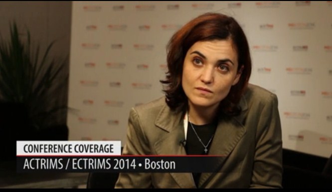
VIDEO: Study gathers insights on effects of cognitive rehab in MS
BOSTON – Brain-training with a video game appears to improve cognitive abilities in multiple sclerosis patients with cognitive impairment by increasing thalamo-cortical connectivity in the brain, according to Dr. Laura De Giglio.
She and her colleagues at Sapienza University of Rome conducted functional MRI scans of patients before and after 8 weeks of using "Dr. Kawashima's Brain Training" video game and compared them against patients who had been assigned to a wait-list group in a small, pilot, randomized study. They found improvements in processing speed and some aspects of working memory that correlated with increased functional connectivity between the thalamus and parts of the cortex.
In a video interview at the joint meeting of the European and Americas Committees for Treatment and Research in Multiple Sclerosis, Dr. De Giglio explained the rationale behind the study and how it might be applied further.
The video associated with this article is no longer available on this site. Please view all of our videos on the MDedge YouTube channel
AT MSBOSTON 2014
Video game improved cognition, brain connectivity in MS patients
BOSTON – Multiple sclerosis patients with cognitive impairment who played a brain-training video game on a regular basis had significant improvement in cognitive test results that correlated with an increase in functional connectivity between the thalamus and cortex in a small, randomized pilot study.
The results of the study provide some preliminary evidence that improvement in measures of the functional connectivity between thalamic and cortical areas serve as the functional substrate underlying clinical recovery, Dr. Laura De Giglio said at the joint meeting of the European and Americas Committees for Treatment and Research in Multiple Sclerosis.

Dr. De Giglio and her associates at Sapienza University of Rome based the premise of the current study on their research team’s recent functional MRI study in MS patients with cognitive impairment, which indicated that worse performance on cognitive testing is associated with increased functional thalamo-cortical connectivity in the thalamic resting state network (Radiology 2014 June [doi:http://dx.doi.org/10.1148/radiol.14131688]). That association could be interpreted to mean that "there are some neuroplastic changes in the thalamic resting state in MS that are not able to fully compensate for tissue damage to prevent cognitive dysfunction," Dr. De Giglio said.
Given the success of recent studies in cognitive rehabilitation of MS patients with face-to-face, computer-assisted, and home-based treatments, she and her colleagues sought to understand how this might work by randomizing 24 patients with cognitive impairment to 8 weeks of training with the video game, "Dr. Kawashimas Brain Training," or to a wait-list group that later received the same training after the study period. Patients used the Italian version of the video game at home for 30 minutes per day, 5 days per week during an 8-week period; their compliance with its use was recorded on the console. The patients had a similar mean level of education (14 years), disease duration (13 years), and Expanded Disability Status score (2).
The investigators assessed attention, processing speed, and working memory before and after the video game training with the Paced Auditory Serial Addition Test (PASAT) with a 3-second pace (0-60, higher scores better), the Symbol Digit Modalities Test (SDMT; 0-110, higher scores better), and the Stroop Test (higher scores better) while patients underwent 3-Tesla functional MRI.
The investigators defined cognitive impairment as failure on at least one of the three neuropsychological tests, based on a score below the 10th percentile of normative data in the Italian population.
Mean scores improved significantly in the intervention group, compared with the wait-list group, on the PASAT (from 35.5 to 46.2 vs. from 32.64 to 36.60) and the Stroop Test (from 22.83 to 28.83 vs. from 24.43 to 25.36). The intervention led to numerically greater mean improvement on the SDMT, but the results were not statistically significant.
Improvement on the PASAT correlated with patterns of increased thalamic connectivity to the left superior temporal gyrus and bilateral locations in the lingual, fusiform, and pre-central gyri. On the Stroop Test, improvement was correlated with improved connectivity between the right angular and supramarginal gyri and the thalamus. Improvement on the SDMT was associated with increased thalamic connectivity to bilateral locations on the temporal, occipital, and lateral parietal cortex.
The investigators observed no relapses or worsening of Expanded Disability Status Scale scores in the patients.
Dr. De Giglio had nothing to disclose. Two coauthors had consulting or lecture fees from pharmaceutical companies marketing MS therapies.
BOSTON – Multiple sclerosis patients with cognitive impairment who played a brain-training video game on a regular basis had significant improvement in cognitive test results that correlated with an increase in functional connectivity between the thalamus and cortex in a small, randomized pilot study.
The results of the study provide some preliminary evidence that improvement in measures of the functional connectivity between thalamic and cortical areas serve as the functional substrate underlying clinical recovery, Dr. Laura De Giglio said at the joint meeting of the European and Americas Committees for Treatment and Research in Multiple Sclerosis.
Dr. De Giglio and her associates at Sapienza University of Rome based the premise of the current study on their research team’s recent functional MRI study in MS patients with cognitive impairment, which indicated that worse performance on cognitive testing is associated with increased functional thalamo-cortical connectivity in the thalamic resting state network (Radiology 2014 June [doi:http://dx.doi.org/10.1148/radiol.14131688]). That association could be interpreted to mean that "there are some neuroplastic changes in the thalamic resting state in MS that are not able to fully compensate for tissue damage to prevent cognitive dysfunction," Dr. De Giglio said.
Given the success of recent studies in cognitive rehabilitation of MS patients with face-to-face, computer-assisted, and home-based treatments, she and her colleagues sought to understand how this might work by randomizing 24 patients with cognitive impairment to 8 weeks of training with the video game, "Dr. Kawashimas Brain Training," or to a wait-list group that later received the same training after the study period. Patients used the Italian version of the video game at home for 30 minutes per day, 5 days per week during an 8-week period; their compliance with its use was recorded on the console. The patients had a similar mean level of education (14 years), disease duration (13 years), and Expanded Disability Status score (2).
The investigators assessed attention, processing speed, and working memory before and after the video game training with the Paced Auditory Serial Addition Test (PASAT) with a 3-second pace (0-60, higher scores better), the Symbol Digit Modalities Test (SDMT; 0-110, higher scores better), and the Stroop Test (higher scores better) while patients underwent 3-Tesla functional MRI.
The investigators defined cognitive impairment as failure on at least one of the three neuropsychological tests, based on a score below the 10th percentile of normative data in the Italian population.
Mean scores improved significantly in the intervention group, compared with the wait-list group, on the PASAT (from 35.5 to 46.2 vs. from 32.64 to 36.60) and the Stroop Test (from 22.83 to 28.83 vs. from 24.43 to 25.36). The intervention led to numerically greater mean improvement on the SDMT, but the results were not statistically significant.
Improvement on the PASAT correlated with patterns of increased thalamic connectivity to the left superior temporal gyrus and bilateral locations in the lingual, fusiform, and pre-central gyri. On the Stroop Test, improvement was correlated with improved connectivity between the right angular and supramarginal gyri and the thalamus. Improvement on the SDMT was associated with increased thalamic connectivity to bilateral locations on the temporal, occipital, and lateral parietal cortex.
The investigators observed no relapses or worsening of Expanded Disability Status Scale scores in the patients.
Dr. De Giglio had nothing to disclose. Two coauthors had consulting or lecture fees from pharmaceutical companies marketing MS therapies.
AT MSBOSTON 2014
Key clinical point: Regular use of a brain training video game focusing on attention, processing speed, and working memory led to improvement on cognitive tests that correlated with increased connections between the thalamus and the cortex.
Major finding: Mean scores improved significantly in the intervention group, compared with the wait-list group, on the PASAT (from 35.5 to 46.2 vs. from 32.64 to 36.60) and the Stroop Test (from 22.83 to 28.83 vs. from 24.43 to 25.36).
Data source: A randomized, wait-list controlled, pilot study of 24 MS patients with cognitive impairment.
Disclosures: Dr. De Giglio had nothing to disclose. Two coauthors had consulting or lecture fees from pharmaceutical companies marketing MS therapies.

New insights gained in PML risk stratification, detection, pathogenesis
As powerful new drugs enter the market for the treatment of multiple sclerosis, methods are emerging for stratifying patients’ risk of progressive multifocal leukoencephalopathy.
Researchers are looking to immunosuppressive history and specific biomarkers to detect progressive multifocal leukoencephalopathy (PML) in its early stages. Also, they are gaining a better understanding of PML’s pathogenesis. At the joint meeting of the European and Americas Committees for Treatment and Research in Multiple Sclerosis in Boston, researchers will discuss each of these areas.
PML risk stratification
Two recently developed methods for determining a patient’s risk for PML while taking natalizumab – an anti–JC virus antibody index greater than 0.9 and a finding of less than 21.6% of CD4-positive T cells with l-selectin (CD62L) – may be best used for risk stratification when patients’ previous immunosuppressive drug use is known, according to research that Dr. Heinz Wiendl of the University of Münster in Germany will present Sept. 11. He and his colleagues’ analysis of nearly 2,000 natalizumab-treated MS patients found a statistical correlation between the anti-JCV antibody index and CD62L that existed only in patients who had previously been on immunosuppression. In addition, there was a stronger correlation between CD62L values and treatment duration in patients with previous immunosuppression, "suggesting CD62L as a first biological marker explaining why [previously immunosuppressed] patients are at higher risk to develop PML over time," the investigators wrote in their abstract.
Diagnosing PML regardless of etiology
A hypointense T2* band at the gray-white junction with variable thickness that at least partially encompasses the characteristic PML lesion sign of asymmetric hyperintensities on T2*-weight imaging may be helpful in detecting PML irrespective of the underlying etiology, according to new research from the National Institute of Neurological Disorders and Stroke that will be presented during the same session on Sept. 11. Dr. Varun Sethi and his colleagues at NINDS used a 3-T scanner to evaluate nine PML patients, including four with natalizumab-treated MS, two with lymphoma, one with idiopathic low CD4 T-cell count, and two with HIV infection. The band was never seen in association with preexisting MS lesions and had a tendency to spread over time in the five patients who had an additional scan 1-7 months after the first scan.
Insight into JC virus reactivation
On Sept. 11, Dr. Spyridon Chalkias of Beth Israel Deaconess Medical Center, Boston, will describe a study he led that provides further insight into how JC virus is reactivated in patients taking natalizumab, which has a mechanism of action that prevents trafficking of leukocytes out of the bloodstream. Dr. Chalkias and his colleagues found that JCV-seropositive patients taking natalizumab could be clinically asymptomatic and free of radiographic findings of PML despite evidence of JCV reactivation in the cerebrospinal fluid or in specific leukocyte populations. The researchers enrolled 43 JCV-seropositive MS patients, including 32 on natalizumab monotherapy for greater than 18 months, 6 on interferon beta-1a monotherapy for greater than 36 months, and 5 untreated controls. Of 27 natalizumab-treated patients who were asymptomatic and MRI-negative for PML, 2 had JCV DNA detected in their cerebrospinal fluid. Overall, the investigators found JCV DNA in the blood of 12 (28%) of the 43 patients. The two natalizumab-treated patients with JCV DNA in CSF did not have JCV DNA in blood or urine; however, the investigators detected JCV-specific CD4-positive T cells ex vivo more frequently in natalizumab-treated patients who were viremic, and in vitro stimulation led to more frequent detection of JCV-specific CD4-positive T cells than of CD8-positive T cells, indicating that "viremia might trigger a JCV-specific CD4-positive mediated response."
PML after natalizumab switchover to fingolimod
An analysis of a Novartis safety reporting database for fingolimod points to no new risk for PML that can be attributed to the drug, although 11 new cases have been reported in patients who switched from natalizumab to fingolimod. The study, to be presented by Dr. Norman Putzki of Novartis on Sept. 12, is based on 135,800 patient-years of fingolimod exposure in MS patients through February 2014. About 15%-20% of patients in the database had previously been on natalizumab. The 11 natalizumab-associated PML cases have been reported following 4 years’ mean duration of natalizumab use, and in all cases the onset of PML was either confirmed prior to the switch (in retrospect) or occurred within 6 months after the switch to fingolimod. In one additional case, a patient who had not been exposed to natalizumab was reported to have PML after 7 months of fingolimod treatment based on the results of a low JCV copy number in cerebrospinal fluid, but it was later determined that the patient’s clinical features and MRI evolution were consistent with neuromyelitis optica.
FROM MSBOSTON 2014
Generic glatiramer acetate has similar safety, efficacy to Copaxone
Generic glatiramer acetate provided reductions in gadolinium-enhancing lesions and annualized relapse rate that were similar to the brand-name version of the drug, Copaxone, in patients with relapsing-remitting multiple sclerosis without any additional safety concerns in a phase III trial.
The combined number of gadolinium-enhancing lesions over months 7, 8, and 9 of the 9-month, double-blind, placebo-controlled, randomized GATE trial, sponsored by Synthon BV, served as the primary endpoint and indicated equivalent efficacy based on the predefined equivalence margin for the ratio of new lesions between generic glatiramer acetate and Copaxone in the study of 794 total patients. Both drugs reduced the number of new lesions, compared with placebo. The annualized relapse rates were comparable across all the groups: 0.31 (generic glatiramer acetate), 0.41 (Copaxone), and 0.39 (placebo).

The active treatment groups had similar incidence, spectrum, and severity of reported adverse events, including injection site reactions. Dr. Jeffrey A. Cohen, director of the experimental therapeutics program at the Cleveland Clinic’s Mellen Center for Multiple Sclerosis Treatment and Research, will report the results on Sept. 12 at the joint meeting of the European and Americas Committees for Treatment and Research in Multiple Sclerosis in Boston.
On Sept. 11, Dr. Cohen also will speak about the need for generic equivalents to drugs that will soon be coming off patent protection. Although there is great potential for "cost savings to patients and third-party payers based on the more limited testing required for approval and resultant reduced development costs," Dr. Cohen wrote in his abstract that there are many difficulties in the process of developing these generics. As is the case for the highly complex peptide mixture that constitutes glatiramer acetate, interferons or monoclonal antibodies are biologics with highly complex chemical structures whose properties are vulnerable to changes on many levels that are difficult to predict without thorough testing. However, it will probably be necessary for developers to use more sensitive biologic markers of drug response, such as MRI, that have been previously well characterized for the original innovator product, as well as trials that use new designs and statistical analyses, he wrote.
Generic glatiramer acetate provided reductions in gadolinium-enhancing lesions and annualized relapse rate that were similar to the brand-name version of the drug, Copaxone, in patients with relapsing-remitting multiple sclerosis without any additional safety concerns in a phase III trial.
The combined number of gadolinium-enhancing lesions over months 7, 8, and 9 of the 9-month, double-blind, placebo-controlled, randomized GATE trial, sponsored by Synthon BV, served as the primary endpoint and indicated equivalent efficacy based on the predefined equivalence margin for the ratio of new lesions between generic glatiramer acetate and Copaxone in the study of 794 total patients. Both drugs reduced the number of new lesions, compared with placebo. The annualized relapse rates were comparable across all the groups: 0.31 (generic glatiramer acetate), 0.41 (Copaxone), and 0.39 (placebo).
The active treatment groups had similar incidence, spectrum, and severity of reported adverse events, including injection site reactions. Dr. Jeffrey A. Cohen, director of the experimental therapeutics program at the Cleveland Clinic’s Mellen Center for Multiple Sclerosis Treatment and Research, will report the results on Sept. 12 at the joint meeting of the European and Americas Committees for Treatment and Research in Multiple Sclerosis in Boston.
On Sept. 11, Dr. Cohen also will speak about the need for generic equivalents to drugs that will soon be coming off patent protection. Although there is great potential for "cost savings to patients and third-party payers based on the more limited testing required for approval and resultant reduced development costs," Dr. Cohen wrote in his abstract that there are many difficulties in the process of developing these generics. As is the case for the highly complex peptide mixture that constitutes glatiramer acetate, interferons or monoclonal antibodies are biologics with highly complex chemical structures whose properties are vulnerable to changes on many levels that are difficult to predict without thorough testing. However, it will probably be necessary for developers to use more sensitive biologic markers of drug response, such as MRI, that have been previously well characterized for the original innovator product, as well as trials that use new designs and statistical analyses, he wrote.
FROM MSBOSTON 2014

Thalamic abnormalities lie at heart of cognitive impairment in MS
Cognitive impairment continues to be a growing area of research in patients with multiple sclerosis, and the scientific program of the joint meeting of the European and Americas committees for Treatment and Research in Multiple Sclerosis has plenty to offer.
In the first young investigators session on Sept. 10, a multicenter study will provide evidence in support of the view that regional thalamic structural connectivity abnormalities play a significant role in cognitive impairment in relapsing-remitting MS patients. Dr. Alvino Bisecco of Vita-Salute San Raffaele University in Milan, Italy, and his colleagues will show that particular patterns of damage to gray matter (viewed as increased fractional anisotropy on diffusion tensor MR imaging) and to white matter (decreased fractional anisotropy) in thalamic subregions contributes to cognitive impairment in MS, compared with patterns observed in healthy control patients.
Another group, led by Dr. Prejaas Tewarie of VU University Medical Center in Amsterdam also will report on Sept. 11 evidence linking thalamic atrophy with disrupted cortical function in MS patients with cognitive impairment. Dr. Tewarie will present data indicating that in MS patients, reduced thalamic volume on MRI is positively associated with a shift in functional network topology between the thalamus and cortex on magnetoencephalography and increased thalamocortical functional connectivity, compared with healthy controls, that proved to be associated with worse cognition and functional status on the Kurtzke Expanded Disability Status Scale.
However, these abnormalities in thalamic functional connectivity could be treated with a brain-training video game, according to a study also to be presented at the first young investigators session on Sept. 10. An 8-week training period in patients who had failing results on at least one cognitive test led to improved test results and a significantly changed pattern of thalamocortical resting-state functional connectivity on MRI, when compared with a wait-list control group. Lead investigator Dr. Laura De Giglio and her associates at Sapienza University of Rome had previously shown that thalamocortical resting-state functional connectivity is disrupted in MS.
During the Sept. 11 hot topics session on symptomatic and rehabilitation strategies, Dr. Maria Pia Amato of the University of Florence (Italy) will review promising results coming from well-designed studies involving behavioral treatment and computerized training, as well as findings from other interventions on cognition, including modest improvement with interferon treatments in patients with relapsing-remitting disease and negative or inconsistent results seen with symptomatic therapies.
Finally, in the second young investigators session on Sept. 10, a unique study will reveal how patients with pediatric-onset and adult-onset relapsing-remitting MS appear to have similar cognitive outcomes at the same age in young adulthood. At mean ages of about 26-27 years in patients who were matched for level of education and physical disability, there were similar mean scores on neuropsychological tests, similar numbers of failed tests, and no difference in the percentage of patients classified as cognitively impaired (14% among pediatric-onset and 27% for adult-onset disease), according to data that will be presented by Dr. Bahia Hakiki of the University of Florence. Because "disease onset in a period of active brain growth and maturation may render pediatric-onset subjects more vulnerable to cognitive issues, our findings suggest good compensatory/recovery abilities in these subjects, possibly related with enhanced brain plasticity in early life," Dr. Hakiki and colleagues wrote.
FROM MSBOSTON 2014