User login
Not on My Watch!
The itchy patch of skin on this 60-year-old man’s forearm, beneath his watch, has troubled him intermittently for a year. It is unresponsive to topical medications, including tolnaftate cream and 1% hydrocortisone cream; the latter seemed to improve the condition initially, but with time, the patch grew larger and itchier.
The patient’s primary care provider believed the rash might be precancerous and prescribed 5-fluorouracil cream, which showed no benefit. The patient was then referred to a dermatologist, who performed a KOH examination. No fungal elements were found.
At that point, the patient decided to simply ignore the problem. That resolve ended when the itchiness increased—leading to his presentation today. He is in decent health but has had a kidney transplant and is taking the standard immunosuppressant (anti-rejection) medications.
EXAMINATION
A faintly pink, scaly rash is located on the patient’s forearm. It is unimpressive in appearance but very bothersome to the patient.

Despite the patient’s history, fungal origin remains a possibility, as does psoriasis. A punch biopsy is performed.
What is the diagnosis?
DISCUSSION
The biopsy results showed numerous fungal elements crowded around deeper aspects of hair follicles and a granulomatous reaction in the surrounding skin.
Normally, dermatophytes (the organisms that cause superficial fungal infections) are incapable of invading deeper tissues, making KOH exam a simple diagnostic method. But with immune suppression, the infection is able to invade deeper structures, making it more difficult to diagnose and treat. In this patient’s case, the use of steroid creams under occlusion (ie, the watch) had suppressed the body’s immune response to the infection.
The terminology used to describe this phenomenon varies according to the severity of infection. Tinea incognito might be used to describe this patient’s case, in which the steroid rendered the condition almost impossible to diagnose visually. With continued use of stronger topical steroids, the degree of inflammation might have been worse, involving deeper, larger nodules instead of faint pink scaling. In such cases, the term Majocchi fungal granuloma is used.
This patient moved his watch to the other arm temporarily and was successfully treated with twice-daily application of topical econazole cream and a two-week course of oral terbinafine (250 mg/d).
TAKE-HOME LEARNING POINTS
- Immunosuppression can make fungal infections more difficult to diagnose and treat.
- Dermatophytes don’t normally invade deeper structures and can usually be detected with a KOH exam of surface skin cells.
- If the patient is immunocompromised, a fungal diagnosis is best confirmed with a punch biopsy, and treatment achieved with a combination of oral and topical antifungal products.
- Mild cases of this kind are termed tinea incognito, while more severe cases are called Majocchi fungal granuloma.
The itchy patch of skin on this 60-year-old man’s forearm, beneath his watch, has troubled him intermittently for a year. It is unresponsive to topical medications, including tolnaftate cream and 1% hydrocortisone cream; the latter seemed to improve the condition initially, but with time, the patch grew larger and itchier.
The patient’s primary care provider believed the rash might be precancerous and prescribed 5-fluorouracil cream, which showed no benefit. The patient was then referred to a dermatologist, who performed a KOH examination. No fungal elements were found.
At that point, the patient decided to simply ignore the problem. That resolve ended when the itchiness increased—leading to his presentation today. He is in decent health but has had a kidney transplant and is taking the standard immunosuppressant (anti-rejection) medications.
EXAMINATION
A faintly pink, scaly rash is located on the patient’s forearm. It is unimpressive in appearance but very bothersome to the patient.
Despite the patient’s history, fungal origin remains a possibility, as does psoriasis. A punch biopsy is performed.
What is the diagnosis?
DISCUSSION
The biopsy results showed numerous fungal elements crowded around deeper aspects of hair follicles and a granulomatous reaction in the surrounding skin.
Normally, dermatophytes (the organisms that cause superficial fungal infections) are incapable of invading deeper tissues, making KOH exam a simple diagnostic method. But with immune suppression, the infection is able to invade deeper structures, making it more difficult to diagnose and treat. In this patient’s case, the use of steroid creams under occlusion (ie, the watch) had suppressed the body’s immune response to the infection.
The terminology used to describe this phenomenon varies according to the severity of infection. Tinea incognito might be used to describe this patient’s case, in which the steroid rendered the condition almost impossible to diagnose visually. With continued use of stronger topical steroids, the degree of inflammation might have been worse, involving deeper, larger nodules instead of faint pink scaling. In such cases, the term Majocchi fungal granuloma is used.
This patient moved his watch to the other arm temporarily and was successfully treated with twice-daily application of topical econazole cream and a two-week course of oral terbinafine (250 mg/d).
TAKE-HOME LEARNING POINTS
- Immunosuppression can make fungal infections more difficult to diagnose and treat.
- Dermatophytes don’t normally invade deeper structures and can usually be detected with a KOH exam of surface skin cells.
- If the patient is immunocompromised, a fungal diagnosis is best confirmed with a punch biopsy, and treatment achieved with a combination of oral and topical antifungal products.
- Mild cases of this kind are termed tinea incognito, while more severe cases are called Majocchi fungal granuloma.
This Baby's Got Flare
At birth, this child had a lesion on his shoulder that now—a year later—has doubled in size. His parents report no systemic symptoms or medication use for their son. They say that the child exhibits no distress; he does not attempt to scratch at the affected patch of skin. However, they observe that if the lesion is touched, it swells and then (within minutes) returns to normal.
There is no family history of similar problems. However, both the patient and his mother are highly atopic.
EXAMINATION
The lesion—a low, orange, oval plaque—measures about 3.5 x 2 cm. Barely palpable, it urticates when stroked with a fingernail edge but does not appear to cause any discomfort.

No other lesions of note are found. The child appears quite healthy and is in no distress.
What is the diagnosis?
DISCUSSION
Mastocytosis is caused by a localized accumulation of mast cells (a type of white blood cell) and CD34-positive mast cell precursors, which are normally present but widely scattered and sparse. This child has the most common form of cutaneous mastocytosis, which can manifest with solitary lesions or with dozens or hundreds of scattered lesions (the latter known as urticaria pigmentosa). Both types are typically benign and self-limited.
When stroked, mast cell lesions degranulate portions of the cell, releasing histamine precursors and leukotrienes (eg, IL 1 and IL 31). In most cases, stroking merely leads to short-lived urtication. But if the problem is more widespread (eg, urticaria pigmentosa) and the lesions are sufficiently traumatized, the release of these substances can lead to problems such as hypotension, malaise, fever, and abdominal pain.
Fortunately, this is rare, as is systemic mastocytosis—a condition in which mast cells infiltrate internal organs and bone marrow, interrupting normal function and, in the extreme, leading to mast cell leukemia. Our patient is not at risk for these complications; his lesion should resolve completely by age 3.
The differential for this patient’s lesion includes congenital nevus, lichen aureus, and café au lait spot.
TAKE-HOME LEARNING POINTS
- Cutaneous mastocytosis manifests as a reddish orange maculopapular patch, which urticates upon forceful stroking.
- Stroking the lesion degranulates the mast cells comprising it, leading to the release of histamine precursors.
- Mast cells can infiltrate internal organs and bone marrow, leading, in the extreme, to mast cell leukemia.
- Urticaria pigmentosa is a variation of mastocytosis in which hundreds of such lesions develop all over the body.
At birth, this child had a lesion on his shoulder that now—a year later—has doubled in size. His parents report no systemic symptoms or medication use for their son. They say that the child exhibits no distress; he does not attempt to scratch at the affected patch of skin. However, they observe that if the lesion is touched, it swells and then (within minutes) returns to normal.
There is no family history of similar problems. However, both the patient and his mother are highly atopic.
EXAMINATION
The lesion—a low, orange, oval plaque—measures about 3.5 x 2 cm. Barely palpable, it urticates when stroked with a fingernail edge but does not appear to cause any discomfort.
No other lesions of note are found. The child appears quite healthy and is in no distress.
What is the diagnosis?
DISCUSSION
Mastocytosis is caused by a localized accumulation of mast cells (a type of white blood cell) and CD34-positive mast cell precursors, which are normally present but widely scattered and sparse. This child has the most common form of cutaneous mastocytosis, which can manifest with solitary lesions or with dozens or hundreds of scattered lesions (the latter known as urticaria pigmentosa). Both types are typically benign and self-limited.
When stroked, mast cell lesions degranulate portions of the cell, releasing histamine precursors and leukotrienes (eg, IL 1 and IL 31). In most cases, stroking merely leads to short-lived urtication. But if the problem is more widespread (eg, urticaria pigmentosa) and the lesions are sufficiently traumatized, the release of these substances can lead to problems such as hypotension, malaise, fever, and abdominal pain.
Fortunately, this is rare, as is systemic mastocytosis—a condition in which mast cells infiltrate internal organs and bone marrow, interrupting normal function and, in the extreme, leading to mast cell leukemia. Our patient is not at risk for these complications; his lesion should resolve completely by age 3.
The differential for this patient’s lesion includes congenital nevus, lichen aureus, and café au lait spot.
TAKE-HOME LEARNING POINTS
- Cutaneous mastocytosis manifests as a reddish orange maculopapular patch, which urticates upon forceful stroking.
- Stroking the lesion degranulates the mast cells comprising it, leading to the release of histamine precursors.
- Mast cells can infiltrate internal organs and bone marrow, leading, in the extreme, to mast cell leukemia.
- Urticaria pigmentosa is a variation of mastocytosis in which hundreds of such lesions develop all over the body.
The Mother of All Skin Problems
Each time this 32-year-old woman has a baby—she’s had four to date—she notices that sections of her face darken. Early on, she observed a pattern in which the coming of winter coincided with a lightening of these affected areas—but now the effect lasts year-round, with progressive darkening. She has not tried any products (OTC or prescription) for this problem.
Growing up in the South, the patient and her family spent most summers boating, swimming, and fishing. Her use of sunscreen was sporadic, but she would tan easily regardless.
Her health is good, aside from a 15-year history of smoking.
EXAMINATION
There is excessive hyperpigmentation (brown) on the patient’s face. It follows a mask-like pattern, including her maxilla and the periphery of her face.

Elsewhere, there is abundant evidence of excessive sun exposure, with focal hyperpigmentation and telangiectasias on her arms. She has type IV skin, consistent with her Native American ancestry.
What is the diagnosis?
DISCUSSION
Melasma, also known as chloasma and dubbed the “mask of pregnancy,” is an extremely common problem that results from a combination of naturally dark skin, lots of sun exposure, and increased levels of estrogenic hormones. The latter can result from pregnancy or from oral contraceptive or estrogen replacement therapy use. Another precipitating factor is thyroid disease, certain types of which lead to an increase in melanocytic stimulating hormone.
Melasma, as one might expect, is seen almost exclusively in women, though a rare male is affected. It is especially common among Latina, Native American, and African-American women, whose melanocytes are especially able to produce pigment.
There are several treatments for melasma, none of them perfect, including tretinoin, azelaic acid, chemical peels, dermabrasion, and lasers. The most common treatment is hydroquinone cream, available in the US in both OTC (2%) and prescription (4%) strengths. However, hydroquinone is available OTC in stronger formulations (15% to 20%) in many Central and South American countries; unfortunately, many women who obtain and use these products experience exogenous ochronosis—a worsening or even precipitation of melasma, resulting from excessive production of tyrosinase.
Any treatment must be used in conjunction with rigorous sunscreen application. A full-spectrum product, with titanium dioxide and zinc oxide as the only active ingredients, must be used, because chemical-laden sunscreens don’t do as good a job covering UVA, UVB, and visible light. Convincing women who are unaccustomed to needing sunscreen to use it religiously is part of what makes treating melasma difficult.
The differential for melasma includes postinflammatory hyperpigmentation (eg, following an episode of contact dermatitis) and simple solar lentigines.
This patient was treated with hydroquinone 4% cream bid, plus sunscreen. She was also given information about other treatment options, such as laser and dermabrasion.
TAKE-HOME LEARNING POINTS
- Melasma, also known at chloasma, is quite common, especially among women with darker skin who live in sunny parts of the world.
- It results from a combination of dark skin, an increased level of estrogenic hormones (eg, with pregnancy, birth control pills, or estrogen replacement therapy), and excessive exposure to UV light.
- While hydroquinone cream can be an effective treatment, the maximum strength should be 4%; overuse of stronger concentrations (available in other countries, such as Mexico), can actually cause melasma to worsen.
Each time this 32-year-old woman has a baby—she’s had four to date—she notices that sections of her face darken. Early on, she observed a pattern in which the coming of winter coincided with a lightening of these affected areas—but now the effect lasts year-round, with progressive darkening. She has not tried any products (OTC or prescription) for this problem.
Growing up in the South, the patient and her family spent most summers boating, swimming, and fishing. Her use of sunscreen was sporadic, but she would tan easily regardless.
Her health is good, aside from a 15-year history of smoking.
EXAMINATION
There is excessive hyperpigmentation (brown) on the patient’s face. It follows a mask-like pattern, including her maxilla and the periphery of her face.
Elsewhere, there is abundant evidence of excessive sun exposure, with focal hyperpigmentation and telangiectasias on her arms. She has type IV skin, consistent with her Native American ancestry.
What is the diagnosis?
DISCUSSION
Melasma, also known as chloasma and dubbed the “mask of pregnancy,” is an extremely common problem that results from a combination of naturally dark skin, lots of sun exposure, and increased levels of estrogenic hormones. The latter can result from pregnancy or from oral contraceptive or estrogen replacement therapy use. Another precipitating factor is thyroid disease, certain types of which lead to an increase in melanocytic stimulating hormone.
Melasma, as one might expect, is seen almost exclusively in women, though a rare male is affected. It is especially common among Latina, Native American, and African-American women, whose melanocytes are especially able to produce pigment.
There are several treatments for melasma, none of them perfect, including tretinoin, azelaic acid, chemical peels, dermabrasion, and lasers. The most common treatment is hydroquinone cream, available in the US in both OTC (2%) and prescription (4%) strengths. However, hydroquinone is available OTC in stronger formulations (15% to 20%) in many Central and South American countries; unfortunately, many women who obtain and use these products experience exogenous ochronosis—a worsening or even precipitation of melasma, resulting from excessive production of tyrosinase.
Any treatment must be used in conjunction with rigorous sunscreen application. A full-spectrum product, with titanium dioxide and zinc oxide as the only active ingredients, must be used, because chemical-laden sunscreens don’t do as good a job covering UVA, UVB, and visible light. Convincing women who are unaccustomed to needing sunscreen to use it religiously is part of what makes treating melasma difficult.
The differential for melasma includes postinflammatory hyperpigmentation (eg, following an episode of contact dermatitis) and simple solar lentigines.
This patient was treated with hydroquinone 4% cream bid, plus sunscreen. She was also given information about other treatment options, such as laser and dermabrasion.
TAKE-HOME LEARNING POINTS
- Melasma, also known at chloasma, is quite common, especially among women with darker skin who live in sunny parts of the world.
- It results from a combination of dark skin, an increased level of estrogenic hormones (eg, with pregnancy, birth control pills, or estrogen replacement therapy), and excessive exposure to UV light.
- While hydroquinone cream can be an effective treatment, the maximum strength should be 4%; overuse of stronger concentrations (available in other countries, such as Mexico), can actually cause melasma to worsen.

Birthmark-ipelago
At birth, this now 18-year-old man had what he was told was “only a birthmark.” In the years since, the lesion has darkened and become increasingly well-defined and raised. It is occasionally irritated.
His parents deny any history of seizures, skeletal problems, vision problems, or developmental disabilities in the patient or the family. All parties want the lesion removed.
EXAMINATION
A contiguous linear collection of brown-to-tan papules and nodules runs entirely across the left upper chest, ending on the left shoulder. It measures about 1 cm in width. On closer examination, many of the marks appear to be a mix of milia and comedones.

No similar lesions are seen elsewhere, and the patient’s type IV skin appears normal in all other respects.
What is the diagnosis?
DISCUSSION
Linear epidermal nevi (LEN) are congenital hamartomatous lesions of embryonal ectodermal origin. They are classified based on their main components, which can be
- Sebaceous
- Apocrine
- Eccrine
- Follicular
- Keratinocytic.
The significance of the linear configuration is that in up to one-third of patients another organ system (eg, brain, eyes, bone) will be affected. Associated neurologic problems include seizures and intellectual disability, which are caused by a variety of neuropathologic lesions.
Other forms of epidermal nevi include nevus comedonicus (associated with cataracts) and inflammatory linear verrucous epidermal nevus (ILVEN). Most often seen in early childhood on the limbs, ILVEN accounts for about 5% of all epidermal nevi.
The case patient appeared to have a mixed form, composed of epidermal and comedonal elements. Fortunately, he did not have any other associated abnormalities.
Other items in the differential include lichen striatus, wart, and koebnerized psoriasis.
Treatment for LEN is problematic, due to not only pain and scarring but also the loss of normal pigment. Options include laser, dermabrasion, and surgery. Smaller lesions can simply be excised.
TAKE-HOME LEARNING POINTS
- Linear epidermal nevi (LEN) are benign, unusual, congenital, hamartomatous tumors of embryonal ectodermal origin, which mostly manifest above the waist (ie, shoulders, trunk) in a linear configuration.
- LEN are classified according to their predominant structural element (sebaceous, eccrine, apocrine, follicular, keratinocytic).
- About one-third of all cases involve other organs, including the brain, eyes, or skeleton.
- The differential for LEN includes wart, koebnerized psoriasis, and lichen striatus.
- Treatment can be problematic, but options include laser, dermabrasion, and surgery.
At birth, this now 18-year-old man had what he was told was “only a birthmark.” In the years since, the lesion has darkened and become increasingly well-defined and raised. It is occasionally irritated.
His parents deny any history of seizures, skeletal problems, vision problems, or developmental disabilities in the patient or the family. All parties want the lesion removed.
EXAMINATION
A contiguous linear collection of brown-to-tan papules and nodules runs entirely across the left upper chest, ending on the left shoulder. It measures about 1 cm in width. On closer examination, many of the marks appear to be a mix of milia and comedones.
No similar lesions are seen elsewhere, and the patient’s type IV skin appears normal in all other respects.
What is the diagnosis?
DISCUSSION
Linear epidermal nevi (LEN) are congenital hamartomatous lesions of embryonal ectodermal origin. They are classified based on their main components, which can be
- Sebaceous
- Apocrine
- Eccrine
- Follicular
- Keratinocytic.
The significance of the linear configuration is that in up to one-third of patients another organ system (eg, brain, eyes, bone) will be affected. Associated neurologic problems include seizures and intellectual disability, which are caused by a variety of neuropathologic lesions.
Other forms of epidermal nevi include nevus comedonicus (associated with cataracts) and inflammatory linear verrucous epidermal nevus (ILVEN). Most often seen in early childhood on the limbs, ILVEN accounts for about 5% of all epidermal nevi.
The case patient appeared to have a mixed form, composed of epidermal and comedonal elements. Fortunately, he did not have any other associated abnormalities.
Other items in the differential include lichen striatus, wart, and koebnerized psoriasis.
Treatment for LEN is problematic, due to not only pain and scarring but also the loss of normal pigment. Options include laser, dermabrasion, and surgery. Smaller lesions can simply be excised.
TAKE-HOME LEARNING POINTS
- Linear epidermal nevi (LEN) are benign, unusual, congenital, hamartomatous tumors of embryonal ectodermal origin, which mostly manifest above the waist (ie, shoulders, trunk) in a linear configuration.
- LEN are classified according to their predominant structural element (sebaceous, eccrine, apocrine, follicular, keratinocytic).
- About one-third of all cases involve other organs, including the brain, eyes, or skeleton.
- The differential for LEN includes wart, koebnerized psoriasis, and lichen striatus.
- Treatment can be problematic, but options include laser, dermabrasion, and surgery.
Under the Cover of Darkness
When he was about 12, a now 41-year-old man noticed that the skin on his left chest was darkening. For several years afterward, the darkness spread and deepened, and the area became hairy. In young adulthood, he experienced occasional outbreaks of what looked like acne on the lesion; this eventually cleared.
He now finds the hairiness increasingly bothersome, so he shaves the worst parts of it. Upon consulting his primary care provider, he was assured that the lesion is “a birthmark.” Unsatisfied with this answer, the patient took the advice of a friend and decided to consult dermatology.
EXAMINATION
A polygonal, hyperpigmented, hypertrichotic patch covers most of the patient’s left pectoral area. The lateral margin is irregular but well-defined. There is obvious partial regrowth of the shaved hair on the lateral margin, but it stops abruptly at that point.

The breast and surrounding tissue appear normal. No areas of hyperpigmentation or hypertrichosis are seen elsewhere.
What is the diagnosis?
DISCUSSION
First described by William Becker in 1948 (and subsequently named for him), the Becker nevus (BN) received little research attention until a French study of 20,000 young men showed a prevalence of 0.5%. Nearly half of the subjects had first noticed the lesion before the age of 10—a somewhat surprising finding, since abundant evidence implicates androgens in its genesis. (This is supported by the condition’s predominance in males, the increased numbers of androgen receptors and melanocytes in the affected skin, and the prevalence of hypertrichosis.)
The researchers were also surprised to find that only 30% of the reported lesions occurred above the nipple, because the first descriptions of BN gave the impression that the shoulder and chest were most commonly affected. We now know that BN can also be found on arms and legs.
Usually a benign condition, BN can be associated with skeletal or soft-tissue deformities in the affected area (eg, ipsilateral breast hypoplasia). Malignancies—most notably melanoma—have also been reported with BN but are especially uncommon.
The differential includes the café-au-lait macules of neurofibromatosis, Albright disease, and congenital melanocytic nevus. The history of BN (ie, presentation, hypertrichosis, gender of patient, and distribution) usually allow a clinical diagnosis.
Treatment is limited to laser hair removal or laser removal or reduction of pigment.
TAKE-HOME LEARNING POINTS
- Becker nevus (BN) is far more common in males than females.
- BN typically manifests during puberty, which aligns with the suspected androgenic etiology.
- Though the shoulders and chest are the most commonly affected areas, BNs can also appear on the flank, arms, or legs.
- The lesions are rarely associated with serious pathology; hypoplasia of the ipsilateral breast is the most common of these complications.
When he was about 12, a now 41-year-old man noticed that the skin on his left chest was darkening. For several years afterward, the darkness spread and deepened, and the area became hairy. In young adulthood, he experienced occasional outbreaks of what looked like acne on the lesion; this eventually cleared.
He now finds the hairiness increasingly bothersome, so he shaves the worst parts of it. Upon consulting his primary care provider, he was assured that the lesion is “a birthmark.” Unsatisfied with this answer, the patient took the advice of a friend and decided to consult dermatology.
EXAMINATION
A polygonal, hyperpigmented, hypertrichotic patch covers most of the patient’s left pectoral area. The lateral margin is irregular but well-defined. There is obvious partial regrowth of the shaved hair on the lateral margin, but it stops abruptly at that point.
The breast and surrounding tissue appear normal. No areas of hyperpigmentation or hypertrichosis are seen elsewhere.
What is the diagnosis?
DISCUSSION
First described by William Becker in 1948 (and subsequently named for him), the Becker nevus (BN) received little research attention until a French study of 20,000 young men showed a prevalence of 0.5%. Nearly half of the subjects had first noticed the lesion before the age of 10—a somewhat surprising finding, since abundant evidence implicates androgens in its genesis. (This is supported by the condition’s predominance in males, the increased numbers of androgen receptors and melanocytes in the affected skin, and the prevalence of hypertrichosis.)
The researchers were also surprised to find that only 30% of the reported lesions occurred above the nipple, because the first descriptions of BN gave the impression that the shoulder and chest were most commonly affected. We now know that BN can also be found on arms and legs.
Usually a benign condition, BN can be associated with skeletal or soft-tissue deformities in the affected area (eg, ipsilateral breast hypoplasia). Malignancies—most notably melanoma—have also been reported with BN but are especially uncommon.
The differential includes the café-au-lait macules of neurofibromatosis, Albright disease, and congenital melanocytic nevus. The history of BN (ie, presentation, hypertrichosis, gender of patient, and distribution) usually allow a clinical diagnosis.
Treatment is limited to laser hair removal or laser removal or reduction of pigment.
TAKE-HOME LEARNING POINTS
- Becker nevus (BN) is far more common in males than females.
- BN typically manifests during puberty, which aligns with the suspected androgenic etiology.
- Though the shoulders and chest are the most commonly affected areas, BNs can also appear on the flank, arms, or legs.
- The lesions are rarely associated with serious pathology; hypoplasia of the ipsilateral breast is the most common of these complications.

Mother Knows Best
About six months ago, an 8-year-old girl developed an asymptomatic rash near her ear. Her mother suspects it is psoriasis, which runs heavily in the family—but their primary care provider favors a fungal diagnosis. He prescribes a succession of topical and oral antifungal medications (including nystatin and terbinafine), which yield no discernable improvement. At this point, referral to dermatology is made.
The child’s mother denies any history of recent infections (eg, strep throat) on her daughter’s behalf. Furthermore, there are no reports of pain associated with the rash or elsewhere.
EXAMINATION
The rash, which is confined to the external right ear, is composed of uniformly smooth white scale on a faintly salmon base. The entire lesion measures about 3 cm at its widest point, and the margins are arciform and well-defined.

No such lesions are seen elsewhere, but tiny pits can be seen on one fingernail.
What is the diagnosis?
DISCUSSION
A punch biopsy could have confirmed the diagnosis, but with the family history, classic appearance, and lack of response to antifungal medication, there was little doubt that this was a case of psoriasis. This autoimmune disease affects nearly 3% of the white population in this country and has a genetic component about 30% of the time.
In psoriasis, keratinocytes matriculate upward from the basal layer to the skin surface at four times the normal rate—so quickly that they have no chance to lose their nuclei (as they normally would). They then pile up, creating plaques of micaceous white scale on a salmon-pink base. Histologically, the smoothly undulating dermoepidermal junction is jammed together, producing fused ridges with clumps of neutrophils on their tips.
While it favors extensor surfaces of extremities, psoriasis can show up anywhere on the body—on the genitals, mouth, and in the nails, where it can cause pits, dystrophy, discoloration, onycholysis, and onychorrhexis.
Unfortunately, this is probably just the beginning of this child’s psoriasis. The good news is that we’re in a golden age of psoriasis treatment, with more drugs than ever to choose from and even more in development. For this patient, we used a keratolytic agent (urea lotion) to thin out the surface scale, in order to allow a class 4 steroid cream to reach the pink inflammatory portion. Within a month, most of this patch had cleared, though we can be fairly sure it and others like it will be back. Education and ongoing follow-up will be needed, in case she is among the 20% to 25% of patients who will develop psoriatic arthropathy, a crippling form of arthritis.
It is certainly possible to develop a fungal infection on or in an ear, but for that to happen, there has to be a source (eg, animal, person, soil). Moreover, the scale would look entirely different, with clearing centers and advancing margins. The likely truth is that this was called “fungal” for lack of any other suspects.
TAKE-HOME LEARNING POINTS
- White scale on a salmon-pink base typifies psoriasis vulgaris, a very common diagnosis that is often mistaken for fungal infection; biopsy can be extremely helpful in establishing or ruling out this diagnosis.
- Psoriasis has a genetic basis, with many gene loci identified to date, but only about 30% of affected patients can attest to a family history.
- In addition to having unsightly, often itchy lesions, psoriasis patients are also at risk for psoriatic arthropathy, a potentially crippling condition.
- The best news is that we have many drugs with which to treat this disease, including a whole family of drugs termed “the biologics,” which directly (and successfully!) address the disease.
About six months ago, an 8-year-old girl developed an asymptomatic rash near her ear. Her mother suspects it is psoriasis, which runs heavily in the family—but their primary care provider favors a fungal diagnosis. He prescribes a succession of topical and oral antifungal medications (including nystatin and terbinafine), which yield no discernable improvement. At this point, referral to dermatology is made.
The child’s mother denies any history of recent infections (eg, strep throat) on her daughter’s behalf. Furthermore, there are no reports of pain associated with the rash or elsewhere.
EXAMINATION
The rash, which is confined to the external right ear, is composed of uniformly smooth white scale on a faintly salmon base. The entire lesion measures about 3 cm at its widest point, and the margins are arciform and well-defined.
No such lesions are seen elsewhere, but tiny pits can be seen on one fingernail.
What is the diagnosis?
DISCUSSION
A punch biopsy could have confirmed the diagnosis, but with the family history, classic appearance, and lack of response to antifungal medication, there was little doubt that this was a case of psoriasis. This autoimmune disease affects nearly 3% of the white population in this country and has a genetic component about 30% of the time.
In psoriasis, keratinocytes matriculate upward from the basal layer to the skin surface at four times the normal rate—so quickly that they have no chance to lose their nuclei (as they normally would). They then pile up, creating plaques of micaceous white scale on a salmon-pink base. Histologically, the smoothly undulating dermoepidermal junction is jammed together, producing fused ridges with clumps of neutrophils on their tips.
While it favors extensor surfaces of extremities, psoriasis can show up anywhere on the body—on the genitals, mouth, and in the nails, where it can cause pits, dystrophy, discoloration, onycholysis, and onychorrhexis.
Unfortunately, this is probably just the beginning of this child’s psoriasis. The good news is that we’re in a golden age of psoriasis treatment, with more drugs than ever to choose from and even more in development. For this patient, we used a keratolytic agent (urea lotion) to thin out the surface scale, in order to allow a class 4 steroid cream to reach the pink inflammatory portion. Within a month, most of this patch had cleared, though we can be fairly sure it and others like it will be back. Education and ongoing follow-up will be needed, in case she is among the 20% to 25% of patients who will develop psoriatic arthropathy, a crippling form of arthritis.
It is certainly possible to develop a fungal infection on or in an ear, but for that to happen, there has to be a source (eg, animal, person, soil). Moreover, the scale would look entirely different, with clearing centers and advancing margins. The likely truth is that this was called “fungal” for lack of any other suspects.
TAKE-HOME LEARNING POINTS
- White scale on a salmon-pink base typifies psoriasis vulgaris, a very common diagnosis that is often mistaken for fungal infection; biopsy can be extremely helpful in establishing or ruling out this diagnosis.
- Psoriasis has a genetic basis, with many gene loci identified to date, but only about 30% of affected patients can attest to a family history.
- In addition to having unsightly, often itchy lesions, psoriasis patients are also at risk for psoriatic arthropathy, a potentially crippling condition.
- The best news is that we have many drugs with which to treat this disease, including a whole family of drugs termed “the biologics,” which directly (and successfully!) address the disease.
Tiny Tot, Big Lesion
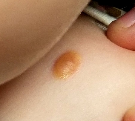
About six months ago, the parents of this 1-year-old boy first noticed the lesion on his shoulder. It started as a pinpoint papule but has grown to its current size—at which point, it caught their full attention. Although there are no associated symptoms, the parents request referral to dermatology to clear up the matter.
The child is reportedly healthy in all other respects, maintaining weight as expected, and normally active and reactive to verbal and visual stimuli.
EXAMINATION
A distinctive orangish brown, ovoid, 8 x 4–mm nodule is located on the child’s right superior shoulder. The lesion has a smooth, soft surface, and there is no tenderness on palpation. No additional lesions are seen elsewhere.
Eye examination reveals normal and symmetrical red reflexes.
What is the diagnosis?
Juvenile xanthogranuloma (JXG) is a rare, benign variant of non-Langerhans cell histiocytosis. This patient’s lesion is typical, but JXG can vary in appearance; some patients present with darker or larger lesions—or multiple lesions.
JXGs are essentially granulomatous tumors that, on histologic examination, display multinucleated giant cells called Touton giant cells. These macrophage-derived foam cells are seen in lesions with high lipid content.
JXG tends to favor the neck, face, and trunk but can appear around or (rarely) inside the eye, typically unilaterally in the iris. Benign in all other respects, ocular JXG lesions can cause spontaneous hyphema, glaucoma, or blindness; they must therefore be dealt with by a specialist. Fortunately, only about 10% of patients display ocular involvement.
JXGs can be confused with compound nevi, warts, or Spitz tumors. Therefore, biopsy is often necessary to establish the diagnosis.
TAKE-HOME LEARNING POINTS
- Juvenile xanthogranuloma (JXG) is a rare non-Langerhans cell tumor usually seen on the neck, face, or trunk of children younger than 2.
- The orangish brown, soft appearance of this patient’s papule was typical.
- Although atypical JXG lesions may require shave biopsy to confirm the diagnosis, they typically resolve on their own without treatment.
- When JXG lesions appear in the eye (most commonly in the iris), there is potential for serious complications, including heterochromia, glaucoma, spontaneous hyphema, or even blindness.
Time Won't Heal This Wound
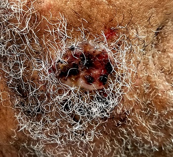
An 85-year-old black man presents with a nonhealing, asymptomatic lesion on his cheek. He says the problem began several months ago, during a fishing trip, when some fishing line got caught in his beard, pulling out a few hairs in the process.
An accompanying relative, however, is quite certain that the lesion predates the fishing incident (for which he was present). He believes the lesion has been there for two years. He also advises that the patient’s memory is “not what it used to be.”
The patient has a significant history of sun exposure from his job as a stonemason, which kept him outdoors most of the time. He has been seen by a variety of providers and diagnosed with several infections, including pyoderma—but antibiotics have had no effect on the lesion.
EXAMINATION
Located on the right lateral cheek is a 2.4-cm, full-thickness ulceration that penetrates well into adipose tissue. Little if any redness can be seen around the lesion, and no adjacent nodes are palpable. A shave biopsy of the lesion is obtained.
What is the diagnosis?
The pathology report showed evidence of a basosquamous cell carcinoma.
This case effectively illustrates a key message: Nonhealing lesions should be considered cancerous until proven otherwise (via biopsy). This remains true even in individuals with darker skin; they may have lower risk for skin cancer than do fair-skinned individuals, but they do not have no risk—especially if there is a lifetime history of sun exposure.
The depth and width of the lesion suggest it had been present for many years, slowing growing. This timeframe, along with the lack of response to antibiotics, made infection unlikely. Furthermore, an infection serious enough to cause ulceration would be red and painful.
The mixed picture on the pathology report is unusual but not at all unknown; it just means the lesion had features of both basal and squamous cell carcinoma. Unfortunately, the biopsy results, in conjunction with the lesion’s dimensions, indicate an increased risk for metastasis (or at least spread to local nodes). There could also be perineural involvement if the cancer cells spread to deeper structures through the penetrating nerves.
The entire clinical picture in this case made the patient a candidate for Mohs surgery, which would ensure two things: clear excision margins and optimal wound closure. Should the surgeon find perineural involvement, he or she might advise postoperative radiation therapy to guarantee complete eradication of the cancer.
TAKE-HOME LEARNING POINTS
- Nonhealing lesions should be considered cancerous until proven otherwise by biopsy.
- Even though dark-skinned individuals have far less risk for skin cancer than those with fair skin, a lifetime of sun exposure can overcome the odds.
- Size and depth of the lesion increases risk for metastasis or perineural involvement (spreading to deeper structures through the nerves).
- Mohs surgery, as well as postoperative radiation therapy, can be used to completely eradicate the cancer.
It's Just a Growth Spurt
A 38-year-old Latino man self-refers to dermatology for evaluation of a mass on his back that first appeared three years ago. Since then, it has grown steadily. There is no pain or discomfort associated with the lesion, and the patient claims to be quite healthy otherwise. There is no antecedent history for the affected area.
EXAMINATION
There is a subcutaneous, rubbery mass in the left infrascapular area. It measures 11 x 6 cm. Palpation reveals the lesion to be uniformly smooth and readily mobile. The overlying skin is free of abnormalities and increased warmth.

What is the diagnosis?
Lipomas are by far the most common soft-tissue tumor to affect humans and are totally benign. They typically measure 2 to 3 cm in diameter, but as this case demonstrates, they can grow much larger. While the rate of growth in this case was unusual, the location—and other features—are typical.
Lipomas are actual tumors, composed completely of adipose tissue contained in a thin, fragile, membranous capsule. Their tendency to develop can be hereditary, though most are spontaneous. They can manifest internally as well.
Superficial lipomas, which often manifest as multiple lesions on the arms and trunk, are usually easy to remove surgically. Lesions that are deeper and older or that appear on the face, however, often require considerable dissection to be freed from surrounding tissue. When excision is attempted, it is essential to remove the entire lesion to prevent recurrence. And, as always, the specimen must be sent for pathologic examination.
Patients often decide against surgery once they understand the issues. This is acceptable, but any deviation from the norm—such as pain, irregular surface texture, change in overlying skin, lack of mobility, or rapid growth—would constitute reasonable grounds for excision.
This man’s lesion likely extended down to the muscle fascia if not into the muscle itself. As a result, surgery would require general anesthesia and placement of a drain in the inferior portion of the wound, since such a large defect would likely invite a collection of blood and serum. For these reasons, he was referred to a general surgeon.
The differential for lipoma includes liposarcoma and angiolipoma. The latter are common and benign but become painful and are often more firm than normal. Histologically, they’re often indistinguishable from ordinary lipomas. Liposarcomas, when superficial, can imitate ordinary lipomas, but their surfaces tend to be more irregular and firm and the lesions themselves less mobile.
TAKE-HOME LEARNING POINTS
- Lipomas are the most common soft-tissue tumor encountered in outpatient practices.
- While the vast majority are benign and easy to remove surgically, most lipomas can be safely left alone.
- When excision is attempted, the entire lesion must be removed lest it regrow.
- Patients with larger, deeper lesions, or those in busy anatomical areas, should be referred to a general surgeon.
A 38-year-old Latino man self-refers to dermatology for evaluation of a mass on his back that first appeared three years ago. Since then, it has grown steadily. There is no pain or discomfort associated with the lesion, and the patient claims to be quite healthy otherwise. There is no antecedent history for the affected area.
EXAMINATION
There is a subcutaneous, rubbery mass in the left infrascapular area. It measures 11 x 6 cm. Palpation reveals the lesion to be uniformly smooth and readily mobile. The overlying skin is free of abnormalities and increased warmth.
What is the diagnosis?
Lipomas are by far the most common soft-tissue tumor to affect humans and are totally benign. They typically measure 2 to 3 cm in diameter, but as this case demonstrates, they can grow much larger. While the rate of growth in this case was unusual, the location—and other features—are typical.
Lipomas are actual tumors, composed completely of adipose tissue contained in a thin, fragile, membranous capsule. Their tendency to develop can be hereditary, though most are spontaneous. They can manifest internally as well.
Superficial lipomas, which often manifest as multiple lesions on the arms and trunk, are usually easy to remove surgically. Lesions that are deeper and older or that appear on the face, however, often require considerable dissection to be freed from surrounding tissue. When excision is attempted, it is essential to remove the entire lesion to prevent recurrence. And, as always, the specimen must be sent for pathologic examination.
Patients often decide against surgery once they understand the issues. This is acceptable, but any deviation from the norm—such as pain, irregular surface texture, change in overlying skin, lack of mobility, or rapid growth—would constitute reasonable grounds for excision.
This man’s lesion likely extended down to the muscle fascia if not into the muscle itself. As a result, surgery would require general anesthesia and placement of a drain in the inferior portion of the wound, since such a large defect would likely invite a collection of blood and serum. For these reasons, he was referred to a general surgeon.
The differential for lipoma includes liposarcoma and angiolipoma. The latter are common and benign but become painful and are often more firm than normal. Histologically, they’re often indistinguishable from ordinary lipomas. Liposarcomas, when superficial, can imitate ordinary lipomas, but their surfaces tend to be more irregular and firm and the lesions themselves less mobile.
TAKE-HOME LEARNING POINTS
- Lipomas are the most common soft-tissue tumor encountered in outpatient practices.
- While the vast majority are benign and easy to remove surgically, most lipomas can be safely left alone.
- When excision is attempted, the entire lesion must be removed lest it regrow.
- Patients with larger, deeper lesions, or those in busy anatomical areas, should be referred to a general surgeon.
Bubble Trouble
A 13-year-old girl is referred to dermatology by her pediatrician for evaluation of “warts” that manifested several months ago. The asymptomatic lesions are simply a cosmetic concern—albeit a persistent one.
Treatments including liquid nitrogen, salicylic acid–based OTC products, and an electric device (purchased online) have been tried, but none have helped. The topical products caused pain and blistering, and although they did eliminate several of the lesions, more soon appeared to take their place.
Further history-taking reveals that the child (like her family) is highly atopic, with seasonal allergies and a history of eczema, hives, and asthma. Two of her siblings have had similar lesions, which cleared fairly quickly without treatment.
EXAMINATION
Approximately 20 papules are randomly arranged on the patient’s anterior neck. They are pink and round, each measuring 2 to 3 mm. Closer inspection reveals that most display a central umbilication. The lesions are firm on palpation.
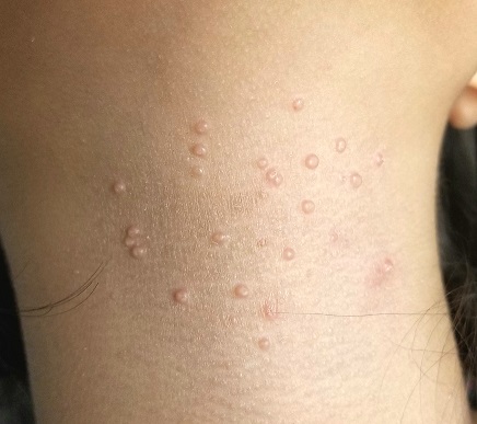
The child is multiracial; her type IV skin is quite dry but otherwise free of lesions.
What is the diagnosis?
One of the most frequently encountered skin lesions in primary care, molluscum contagiosum (MC) is seen mostly in children. It is strongly associated with immune suppression, especially atopy, which affects around 20% of newborns. Commonly called “water warts,” MC lesions have no connection to human papillomavirus and are instead caused by the molluscum contagiosum virus—the largest virus to infect humans.
This virus is transmitted through direct contact with an infected individual, which often occurs during the summertime when many children swim. Though the warts cause little if any harm, they can be a source of embarrassment for the child and can be concerning to parents, who are often given erroneous information about the diagnosis.
There’s also the unfortunate fact that the occasional MC lesion fills with pus, turning red and swollen—a fair imitation of bacterial infection. This is simply a sign that the lesion is dying and will soon disappear, but it’s understandably frightening to parents.
As this case illustrates so well, destroying a few MC lesions does nothing to keep a whole new crop from taking their place. And although the condition is self-limiting, it is common for the warts to take two or more years to go away.
The truth is, to date, there has been no proven, safe, painless, effective treatment for MC. Modalities include laser, electrodessication, and simple curettage.
A new treatment that combines dilute povidone-iodine with dimethyl sulfoxide in an OTC compounded liquid mixture (applied bid) has shown some promise in limited trials (Capriotti et al. J Clin Aesthet Dermatol. 2017;10[3]:41). This is what the case patient was treated with. I have given it to perhaps a dozen patients over the past several months, but to date, none have returned to report results. (This, as far as I know, is not a proprietary product and I have no financial interest in it.)
TAKE-HOME LEARNING POINTS
- Firm, 2- to 3-mm, umbilicated papules on children are almost certainly molluscum contagiosum (MC), usually related to atopy.
- MC is acquired by direct contact but can be spread by scratching or picking.
- It often appears admixed with eczema, especially in the antecubital and popliteal areas.
- Although MC eventually resolves with or without treatment, the process can take a while, making patient/parent education important.
- The newest treatment (that I am aware of) is a mixture of povidone-iodine and dimethyl sulfoxide, to be applied bid.
A 13-year-old girl is referred to dermatology by her pediatrician for evaluation of “warts” that manifested several months ago. The asymptomatic lesions are simply a cosmetic concern—albeit a persistent one.
Treatments including liquid nitrogen, salicylic acid–based OTC products, and an electric device (purchased online) have been tried, but none have helped. The topical products caused pain and blistering, and although they did eliminate several of the lesions, more soon appeared to take their place.
Further history-taking reveals that the child (like her family) is highly atopic, with seasonal allergies and a history of eczema, hives, and asthma. Two of her siblings have had similar lesions, which cleared fairly quickly without treatment.
EXAMINATION
Approximately 20 papules are randomly arranged on the patient’s anterior neck. They are pink and round, each measuring 2 to 3 mm. Closer inspection reveals that most display a central umbilication. The lesions are firm on palpation.
The child is multiracial; her type IV skin is quite dry but otherwise free of lesions.
What is the diagnosis?
One of the most frequently encountered skin lesions in primary care, molluscum contagiosum (MC) is seen mostly in children. It is strongly associated with immune suppression, especially atopy, which affects around 20% of newborns. Commonly called “water warts,” MC lesions have no connection to human papillomavirus and are instead caused by the molluscum contagiosum virus—the largest virus to infect humans.
This virus is transmitted through direct contact with an infected individual, which often occurs during the summertime when many children swim. Though the warts cause little if any harm, they can be a source of embarrassment for the child and can be concerning to parents, who are often given erroneous information about the diagnosis.
There’s also the unfortunate fact that the occasional MC lesion fills with pus, turning red and swollen—a fair imitation of bacterial infection. This is simply a sign that the lesion is dying and will soon disappear, but it’s understandably frightening to parents.
As this case illustrates so well, destroying a few MC lesions does nothing to keep a whole new crop from taking their place. And although the condition is self-limiting, it is common for the warts to take two or more years to go away.
The truth is, to date, there has been no proven, safe, painless, effective treatment for MC. Modalities include laser, electrodessication, and simple curettage.
A new treatment that combines dilute povidone-iodine with dimethyl sulfoxide in an OTC compounded liquid mixture (applied bid) has shown some promise in limited trials (Capriotti et al. J Clin Aesthet Dermatol. 2017;10[3]:41). This is what the case patient was treated with. I have given it to perhaps a dozen patients over the past several months, but to date, none have returned to report results. (This, as far as I know, is not a proprietary product and I have no financial interest in it.)
TAKE-HOME LEARNING POINTS
- Firm, 2- to 3-mm, umbilicated papules on children are almost certainly molluscum contagiosum (MC), usually related to atopy.
- MC is acquired by direct contact but can be spread by scratching or picking.
- It often appears admixed with eczema, especially in the antecubital and popliteal areas.
- Although MC eventually resolves with or without treatment, the process can take a while, making patient/parent education important.
- The newest treatment (that I am aware of) is a mixture of povidone-iodine and dimethyl sulfoxide, to be applied bid.