User login
The Rash That Outlasts
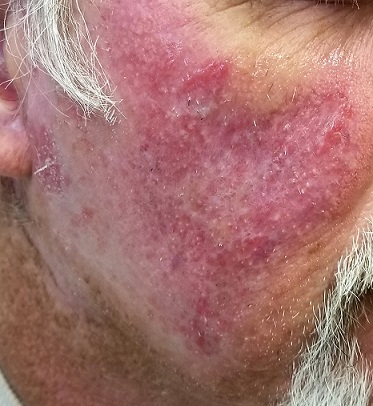
A 63-year-old man says the 20-year-old rash on his face first appeared one summer. Although it slackens a bit each winter, it flares up again when the weather warms—despite a bevy of OTC and prescription topical treatments.
The patient has consulted many providers, including several dermatologists, who have diagnosed the butterfly rash of lupus. But blood tests failed to bear out that theory, and no one has ever biopsied it.
The patient spent many years working in the sun with minimal to no protection. He denies fever, malaise, joint pain, or other illness. He denies having a similar rash elsewhere on his body.
EXAMINATION
A symmetrical, strikingly red, extensive rash covers most of both sides of the patient’s face. There is epidermal scaling and roughness and large areas of obvious follicular enlargement, atrophy, and telangiectasias.
Punch biopsy shows a multitude of changes: atrophic epithelium, basal vacuolar changes, an intense dermal lymphocytic infiltrate, liquefaction degeneration, and apoptotic keratinocytes. Compact orthokeratosis is noted on the surface, and increased mucin formation in the dermis.
What is the diagnosis?
These skin changes, in conjunction with the biopsy findings, are consistent with a diagnosis of discoid lupus erythematosus (DLE). The vast majority of affected patients do not have systemic lupus erythematosus (SLE), although nearly 17% eventually progress to it. This patient’s seronegative status had puzzled his previous providers—a common mistake that frequently delays diagnosis and treatment.
DLE is the most common type of chronic cutaneous lupus, occurring in about 17 to 48 of every 100,000 people in the general population. It affects far more women than men, mostly those ages 20 to 40. More blacks than whites are affected by DLE, which is thought to be a polygenic autoimmune disease linked to various human leukocyte antigen groups.
DLE affects sun-exposed areas, including the scalp (where it often goes undiagnosed, leading to scarring alopecia). It can also occur in the mouth, manifesting as annular eroded areas. In this patient’s case, the combination of UV exposure and apparent genetic predisposition caused his malady.
Following workup to rule out systemic disease, the patient was started on hydroxychloroquine HCL (200 mg bid) and given strict instructions to protect himself from the sun. This should yield considerable improvement, although the scarring on large sections of his face (eg, the posterior cheeks) will be permanent. He will also be monitored for possible progression to SLE.
TAKE-HOME LEARNING POINTS
- Discoid lupus erythematosus (DLE) is a form of chronic cutaneous lupus caused by overexposure to the sun in a genetically susceptible individual.
- Most DLE patients never develop systemic lupus, though it’s far from unknown (17%).
- DLE, an autoimmune disease, is far more common in women (ages 20 – 40) than in men.
- Diagnosis is often made clinically, but biopsy reveals characteristic findings that confirm the disease.
- Besides sun protection, DLE is treated with the oral antimalarial hydroxychloroquine HCL (200 mg bid) and topical steroids as needed.
Farmer Flummoxed by Finger Lesion
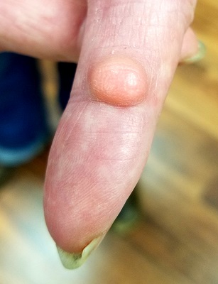
A 70-year-old man self-refers to dermatology for evaluation of a “risin’ in my finger,” which has existed for “at least 40 years.” While the lesion doesn’t really hurt, the patient wants it gone because he traumatizes it almost daily while working on his farm.
He has tried innumerable removal methods, including acids, blood root, and duct tape. Most recently, his primary care provider attempted treatment with cryotherapy.
The patient’s health is excellent in other respects, with no history of similar lesions elsewhere.
EXAMINATION
A dome-like pink nodule is seen on the palmar surface of the patient’s left index finger. The 1-cm lesion is smooth, moderately firm, and nontender. Although the majority of the lesion protrudes above the skin’s surface, there is an intradermal component. Normal skin lines on the surface are preserved, and there is no surface punctum. Palpation of the epitrochlear and axillary areas above the hand reveals no masses.
What is the diagnosis?
Traumatic puncture—especially of the palms and fingers—can invaginate the surface of skin, effectively burying the surface follicular infundibulum and associated sebaceous gland as the wound heals. These structures can continue to produce sebum and epidermal cells, which then accumulate in a space delineated by the lining of the infundibulum and form a sac called an epidermal inclusion cyst (EIC).
EICs differ from the more common epidermoid (or epidermal) cyst for several reasons. For one, epidermoid cysts are confined almost exclusively to oily skin (usually above the waist, with the back being the most common location). Unlike EICs, epidermoid cysts almost always have at least one central comedone, which acts as a plug, preventing the discharge of its contents.
The differential also includes pilar cysts—another common type, popularly known as “wens.” But these occur almost exclusively in the scalp and only rarely display a central punctum.
All of these cysts have an organized wall that contains their characteristic cheesy contents, and all can be removed surgically—preferably in one piece—under local anesthesia. Once excised, the lesion should always be submitted for pathologic examination, since cystic lesions can (albeit rarely) undergo malignant transformation.
In this case, excision was performed with anesthesia via digital block, supplemented with small amounts of lidocaine and epinephrine for hemostasis. The lesion was removed in one transverse elliptical piece and with special care to avoid trauma to underlying structures (visualized with the aid of a tourniquet). The wound was closed with interrupted sutures.
The pathology report confirmed the preoperative diagnosis, and the patient recovered uneventfully.
TAKE-HOME LEARNING POINTS
- Epidermal inclusion cysts (EICs) result from a puncture wound that buries surface follicular infundibula and their sebaceous glands under the skin, where they continue to produce material that collects in a sac.
- Unlike the more common epidermoid (epidermal) cysts, which affect the oily areas of the body (above the waist), EICs don’t have a central punctum.
- EICs can be left alone or excised, but removal must be done in one piece, lest they recur. The same is true for all aforementioned cysts.
Girl Faces Down Lesion
The lesion on this 8-year-old girl’s face was first noted about a year ago. Recently, however, it has started to grow, prompting her parents to consult the child’s primary care provider (PCP). The lesion is completely asymptomatic but nonetheless concerning to the parents and to the PCP, who has no idea what it could be. He therefore refers them to dermatology.
The child is otherwise healthy. There is no family history of chronic or inheritable disease.
EXAMINATION
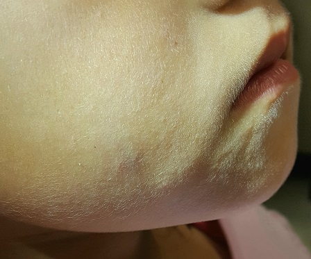
The lesion is a 2-cm subcutaneous firm round mass located along the superior aspect of the right jawline. The only overlying skin change is a bluish discoloration. No surface punctum is seen. No other lesions are seen or felt on examination of the rest of the head and neck.
What is the diagnosis?
These findings are typical of pilomatricoma, a rather unusual lesion with multiple alternate names (among them: calcifying epithelioma of Malherbe and pilomatrixoma). These benign tumors arising from hair matrix cells are typically seen on the head, face, neck, and upper extremities, most often on children.
This patient’s lesion was typical in size, although they can vary from 3 mm (the smallest I’ve seen) to more than 20 cm. The firm feel, lack of a punctum (which would suggest an epidermal cyst), and bluish discoloration are all typical features.
These lesions generally merit little or no concern. However, in the rare instance when the patient has multiple lesions, the possibility of at least two conditions should be considered: Gardner disease and myotonic seizure.
In terms of treatment, pilomatricoma can be safely left alone; understandably, though, most parents will only be satisfied by excision. It is important to note that, unlike most true cysts, pilomatricomas have a very poorly defined cyst wall with contents that are equally odd—watery and full of tiny white flecks that represent calcified cells. All of this must be totally removed to prevent recurrence. In most cases, defects must be closed in two layers, to minimize “dead” space that might otherwise fill with blood.
One could argue that this child’s lesion should have been removed by a plastic surgeon—but the family had no insurance. Excision was therefore the treatment of choice; the most difficult aspect was persuading the patient to cooperate. (Sometimes, you have to wait years for the child to mature before you attempt it.) The outcome in this case proved to be quite acceptable. Of course, the lesion was sent to pathology, which confirmed the pre-op diagnosis.
The differential includes epidermal cysts (which are almost unknown in prepubertal children), and sweat gland cysts.
TAKE-HOME LEARNING POINTS
- Pilomatricomas are benign cysts that originate from hair matrix cells, usually appearing on the head, neck, face, and upper extremities of children.
- The cysts are typically firm and round and often display a faintly bluish tone (as seen in this case).
- As solitary lesions, they are of little or no significance; when seen in multiples, however, pilomatricomas suggest the possibility of Gardner syndrome or myotonic seizure.
- Excision is the best treatment, though these can be left alone long enough to allow the patient to mature sufficiently to cooperate with the surgical process.
The lesion on this 8-year-old girl’s face was first noted about a year ago. Recently, however, it has started to grow, prompting her parents to consult the child’s primary care provider (PCP). The lesion is completely asymptomatic but nonetheless concerning to the parents and to the PCP, who has no idea what it could be. He therefore refers them to dermatology.
The child is otherwise healthy. There is no family history of chronic or inheritable disease.
EXAMINATION
The lesion is a 2-cm subcutaneous firm round mass located along the superior aspect of the right jawline. The only overlying skin change is a bluish discoloration. No surface punctum is seen. No other lesions are seen or felt on examination of the rest of the head and neck.
What is the diagnosis?
These findings are typical of pilomatricoma, a rather unusual lesion with multiple alternate names (among them: calcifying epithelioma of Malherbe and pilomatrixoma). These benign tumors arising from hair matrix cells are typically seen on the head, face, neck, and upper extremities, most often on children.
This patient’s lesion was typical in size, although they can vary from 3 mm (the smallest I’ve seen) to more than 20 cm. The firm feel, lack of a punctum (which would suggest an epidermal cyst), and bluish discoloration are all typical features.
These lesions generally merit little or no concern. However, in the rare instance when the patient has multiple lesions, the possibility of at least two conditions should be considered: Gardner disease and myotonic seizure.
In terms of treatment, pilomatricoma can be safely left alone; understandably, though, most parents will only be satisfied by excision. It is important to note that, unlike most true cysts, pilomatricomas have a very poorly defined cyst wall with contents that are equally odd—watery and full of tiny white flecks that represent calcified cells. All of this must be totally removed to prevent recurrence. In most cases, defects must be closed in two layers, to minimize “dead” space that might otherwise fill with blood.
One could argue that this child’s lesion should have been removed by a plastic surgeon—but the family had no insurance. Excision was therefore the treatment of choice; the most difficult aspect was persuading the patient to cooperate. (Sometimes, you have to wait years for the child to mature before you attempt it.) The outcome in this case proved to be quite acceptable. Of course, the lesion was sent to pathology, which confirmed the pre-op diagnosis.
The differential includes epidermal cysts (which are almost unknown in prepubertal children), and sweat gland cysts.
TAKE-HOME LEARNING POINTS
- Pilomatricomas are benign cysts that originate from hair matrix cells, usually appearing on the head, neck, face, and upper extremities of children.
- The cysts are typically firm and round and often display a faintly bluish tone (as seen in this case).
- As solitary lesions, they are of little or no significance; when seen in multiples, however, pilomatricomas suggest the possibility of Gardner syndrome or myotonic seizure.
- Excision is the best treatment, though these can be left alone long enough to allow the patient to mature sufficiently to cooperate with the surgical process.
Digging for the Diagnosis
The lesions on this 41-year-old African-American woman’s skin have waxed and waned over the years—but they’re always on her mind. They are most prominent on her arms and trunk but crop up almost anywhere on her body.
When they manifest—for no apparent reason—they itch, creating an irresistible urge for the patient to pick at them. This provides some relief, both from the itching and from her feeling that there is “something in there” that she needs to remove. Fairly often, her digging (with fingernails) results in “finding” white bumps at the ends of tiny hairs.
As these small excoriations heal, the wounds itch—compelling her to pick the site open again. She knows she is caught in a vicious cycle but doesn’t know how to stop. Selective serotonin reuptake inhibitors have been tried, with little to no effect.
EXAMINATION
The patient’s type IV skin is covered with dark brown, maculopapular lesions that are so numerous and large (average diameter, 2 to 3 cm) that they are impossible to ignore. Her palms, soles, face, and midback are spared.

Only a few of the newer lesions are palpable, showing faint signs of central excoriation. Previous biopsies failed to show significant pathology.
The patient appears ill at ease during history-taking. She admits to picking her skin for many years but doesn’t believe she is inhabited by any kind of bug. The skin on her wrists and between her fingers is clear. Her 3-year-old daughter’s skin is free of notable changes.
What is the diagnosis?
Known in the DSM-5 and ICD-10 as skin-picking disorder, this condition has also been called dermatillomania. For unknown reasons, its incidence is far greater among women than men.
While it has been posited as a form of obsessive compulsive disorder (OCD), dermatillomania responds poorly, if at all, to standard OCD treatments. It is considered by others to more closely resemble addiction because, despite knowing its harm, patients persistently pick at the skin and often report a subsequent sense of relief.
This patient’s type IV skin lent itself to postinflammatory hyperpigmentation upon injury. Although she knew this, she still felt that she could somehow pick the darkness away.
Bloodwork was done to rule out other conditions, such as porphyria, hematologic disease, and renal or liver disease. Had a recent biopsy not been performed, this would have been included to rule out systemic disease.
The patient was given a topical steroid cream to put on any itchy lesions and counseled to avoid picking or scratching them, since this was the only way her skin could ever clear.
TAKE-HOME LEARNING POINTS
- One common term for this patient’s disorder is dermatillomania, though the DSM-5 and ICD-10 refer to it as skin-picking disorder.
- This patient experienced postinflammatory hyperpigmentation, which caused her considerable embarrassment.
- Many affected patients have unresolved underlying psychologic issues that contribute to their problem.
- The solution (which may require extensive counseling): Stop picking, and the dark lesions will eventually resolve.
The lesions on this 41-year-old African-American woman’s skin have waxed and waned over the years—but they’re always on her mind. They are most prominent on her arms and trunk but crop up almost anywhere on her body.
When they manifest—for no apparent reason—they itch, creating an irresistible urge for the patient to pick at them. This provides some relief, both from the itching and from her feeling that there is “something in there” that she needs to remove. Fairly often, her digging (with fingernails) results in “finding” white bumps at the ends of tiny hairs.
As these small excoriations heal, the wounds itch—compelling her to pick the site open again. She knows she is caught in a vicious cycle but doesn’t know how to stop. Selective serotonin reuptake inhibitors have been tried, with little to no effect.
EXAMINATION
The patient’s type IV skin is covered with dark brown, maculopapular lesions that are so numerous and large (average diameter, 2 to 3 cm) that they are impossible to ignore. Her palms, soles, face, and midback are spared.
Only a few of the newer lesions are palpable, showing faint signs of central excoriation. Previous biopsies failed to show significant pathology.
The patient appears ill at ease during history-taking. She admits to picking her skin for many years but doesn’t believe she is inhabited by any kind of bug. The skin on her wrists and between her fingers is clear. Her 3-year-old daughter’s skin is free of notable changes.
What is the diagnosis?
Known in the DSM-5 and ICD-10 as skin-picking disorder, this condition has also been called dermatillomania. For unknown reasons, its incidence is far greater among women than men.
While it has been posited as a form of obsessive compulsive disorder (OCD), dermatillomania responds poorly, if at all, to standard OCD treatments. It is considered by others to more closely resemble addiction because, despite knowing its harm, patients persistently pick at the skin and often report a subsequent sense of relief.
This patient’s type IV skin lent itself to postinflammatory hyperpigmentation upon injury. Although she knew this, she still felt that she could somehow pick the darkness away.
Bloodwork was done to rule out other conditions, such as porphyria, hematologic disease, and renal or liver disease. Had a recent biopsy not been performed, this would have been included to rule out systemic disease.
The patient was given a topical steroid cream to put on any itchy lesions and counseled to avoid picking or scratching them, since this was the only way her skin could ever clear.
TAKE-HOME LEARNING POINTS
- One common term for this patient’s disorder is dermatillomania, though the DSM-5 and ICD-10 refer to it as skin-picking disorder.
- This patient experienced postinflammatory hyperpigmentation, which caused her considerable embarrassment.
- Many affected patients have unresolved underlying psychologic issues that contribute to their problem.
- The solution (which may require extensive counseling): Stop picking, and the dark lesions will eventually resolve.
Man's Condition Gets Out of Hand
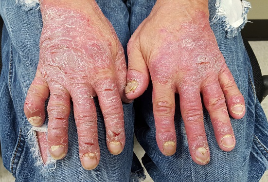
This 46-year-old man’s skin disease has gotten so serious that he is essentially disabled. The problem started about six months ago, with joint pain that particularly affected his left ankle. Now, his hands are fissured and swollen to the point that he is unable to button his shirt or hold a fork. He is referred to dermatology by his attorney, who is helping him pursue possible disability benefits, for evaluation and treatment.
He has been seen by a variety of primary care providers, who have collectively prescribed topical triamcinolone 0.1% and several antifungal medications, including a two-month course of oral terbinafine. When those failed, he was treated with prednisone; at the start of the three-week course, there were signs of improvement but by the end, his hands were worse than ever.
EXAMINATION
The dorsal and palmar surfaces of the patient’s hands are covered with thick, white scales atop salmon-colored erythematous bases. Multiple fissures and marked edema can be seen. Seven of 10 fingernails are dystrophic, yellowed, and thickened.
The patient’s elbows, knees, and upper intergluteal area show less impressive involvement.
There is marked tenderness on palpation of the left Achilles insertion, made worse by dorsiflexion.
What is the diagnosis?
Psoriasis can affect one or more areas, typically the hands, scalp, genitals, or feet. When it’s focused in one area, as in this case, it can be baffling to diagnose; sometimes it’s hard to see the forest for the trees. But because the condition affects almost 3% of white Americans, you’ll see it regularly—if you’re looking for it.
Nearly 25% of patients with psoriasis eventually develop psoriatic arthritis (PsA), which not only affects the joints but also can cause complications such as enthesitis, or inflammation of the entheses (the sites of insertion of the tendon into bone; eg, the Achilles). This can be confused with plantar fasciitis, which this patient had been previously diagnosed with.
This diagnosis could have been proved or disproved by a KOH prep (which would have shown evidence of fungal disease) or a biopsy (which would have shown fused rete ridges, microabscesses at the papillary tips, hyperkeratosis, and parakeratosis). Providers should first establish a firm diagnosis to dictate effective treatment. In this case, a visual diagnosis was possible.
Given the severity of the problem, the patient was started on a biologic; he showed vast improvement within a week. He was referred to rheumatology for evaluation and management of PsA, and the severity of his disease was communicated to his attorney.
TAKE-HOME LEARNING POINTS
- In some cases, psoriasis can be isolated to the hands, feet, genitals, or scalp, complicating detection of the condition.
- Almost 25% of patients with psoriasis develop psoriatic arthritis (PsA), which can manifest with dactylitis, arthritis, or enthesitis.
- Left untreated, PsA is potentially debilitating.
- Establishing a firm diagnosis with KOH prep or biopsy will dictate effective treatment.
A Sore Subject
For several years, a 60-year-old woman has had a nonhealing, asymptomatic “sore” on her upper right cheek. The lesion causes no pain or discomfort; it bothers her simply because it will not go away. It does occasionally bleed.
Several attempts at treatment—including antibiotic ointment, peroxide, and topical alcohol— have failed. A dermatologist once treated the lesion with cryotherapy; this initially reduced its size, but the effect didn’t last.
The patient admits to “worshipping the sun” as a youngster, tanning at every opportunity. Several family members, including her sister and mother, have had skin cancer.
EXAMINATION
A 6.5-mm, round, red nodule is located on the mid-upper right cheek, below the eye. On closer inspection, it appears glassy and translucent, with several obvious telangiectasias. It is surprisingly firm on palpation, though not at all tender to touch.
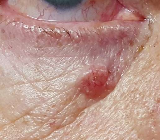
Elsewhere, the patient’s fair skin has abundant evidence of sun damage, including wrinkles, discoloration, and focal telangiectasias. No other lesions are seen. No nodes are palpable in her head or neck.
With the patient’s permission, and after discussion of the indications, procedure, alternatives, and risks, the lesion is removed by curettement. It is quite friable and shallow, allowing complete removal. The area is left to heal by secondary intention, and the specimen is submitted to pathology.
What is the diagnosis?
The pathology report confirmed the suspected diagnosis: noduloulcerative basal cell carcinoma (BCC). By far the most common type of skin cancer in the world, noduloulcerative BCCs are caused by overexposure to the sun.
As obvious as this diagnosis seemed to be, biopsy was warranted to confirm it and to rule out several other items in the differential. The latter include squamous cell carcinoma (which has far more harmful potential than BCC), sebaceous carcinoma, Merkel cell carcinoma, and melanoma.
Not all BCCs are created equal—certain clinical and histologic characteristics predict a more aggressive course. While most BCCs require surgical excision, other treatment options do exist. In this patient’s case, the size and location of the lesion lent it to curettement and healing by secondary intention. Other methods could have included immunotherapy with imiquimod, antitumor creams (eg, 5-fluorouracil), or even radiation therapy. Her lesion was small and well-defined enough that Mohs surgery was not indicated—though some might disagree with that assessment.
TAKE-HOME LEARNING POINTS
- Like all basal cell carcinomas (BCCs), ulcerative BCC—the most common type—is caused by overexposure to the sun.
- BCCs are often mistaken for infection or other sore, which delays the diagnosis.
- Nonhealing lesions should be considered cancerous until proven otherwise.
- Though this diagnosis was fairly obvious, biopsy is necessary to rule out other items in the differential—some of which (ie, Merkel cell carcinoma, melanoma) are potentially fatal.
For several years, a 60-year-old woman has had a nonhealing, asymptomatic “sore” on her upper right cheek. The lesion causes no pain or discomfort; it bothers her simply because it will not go away. It does occasionally bleed.
Several attempts at treatment—including antibiotic ointment, peroxide, and topical alcohol— have failed. A dermatologist once treated the lesion with cryotherapy; this initially reduced its size, but the effect didn’t last.
The patient admits to “worshipping the sun” as a youngster, tanning at every opportunity. Several family members, including her sister and mother, have had skin cancer.
EXAMINATION
A 6.5-mm, round, red nodule is located on the mid-upper right cheek, below the eye. On closer inspection, it appears glassy and translucent, with several obvious telangiectasias. It is surprisingly firm on palpation, though not at all tender to touch.
Elsewhere, the patient’s fair skin has abundant evidence of sun damage, including wrinkles, discoloration, and focal telangiectasias. No other lesions are seen. No nodes are palpable in her head or neck.
With the patient’s permission, and after discussion of the indications, procedure, alternatives, and risks, the lesion is removed by curettement. It is quite friable and shallow, allowing complete removal. The area is left to heal by secondary intention, and the specimen is submitted to pathology.
What is the diagnosis?
The pathology report confirmed the suspected diagnosis: noduloulcerative basal cell carcinoma (BCC). By far the most common type of skin cancer in the world, noduloulcerative BCCs are caused by overexposure to the sun.
As obvious as this diagnosis seemed to be, biopsy was warranted to confirm it and to rule out several other items in the differential. The latter include squamous cell carcinoma (which has far more harmful potential than BCC), sebaceous carcinoma, Merkel cell carcinoma, and melanoma.
Not all BCCs are created equal—certain clinical and histologic characteristics predict a more aggressive course. While most BCCs require surgical excision, other treatment options do exist. In this patient’s case, the size and location of the lesion lent it to curettement and healing by secondary intention. Other methods could have included immunotherapy with imiquimod, antitumor creams (eg, 5-fluorouracil), or even radiation therapy. Her lesion was small and well-defined enough that Mohs surgery was not indicated—though some might disagree with that assessment.
TAKE-HOME LEARNING POINTS
- Like all basal cell carcinomas (BCCs), ulcerative BCC—the most common type—is caused by overexposure to the sun.
- BCCs are often mistaken for infection or other sore, which delays the diagnosis.
- Nonhealing lesions should be considered cancerous until proven otherwise.
- Though this diagnosis was fairly obvious, biopsy is necessary to rule out other items in the differential—some of which (ie, Merkel cell carcinoma, melanoma) are potentially fatal.
It's A Slow Grow
For more than two years, this 40-year-old African-American woman has had a lesion on her left maxilla. While it does not cause pain or discomfort, its presence is alarming to the patient. The lesion has grown steadily without responding to various topical OTC medications (tolnaftate, clotrimazole, miconazole, 1% hydrocortisone cream), oral anti-yeast medication, and antibiotics (fluconazole, erythromycin).
The patient claims to be in excellent health otherwise. She reports a strong family history of autoimmune disease, including rheumatoid arthritis and lupus.
EXAMINATION
There is an oval, scaly, atrophic patch on the upper left maxilla, just below the left nostril. The lesion measures 1.2 cm, has well-defined margins, and appears darker than the patient’s type V skin.
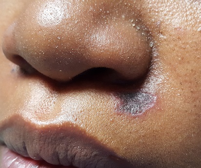
No redness or edema are seen, and no nodes are palpable in the area. Examination of the adjacent oral mucosal surface shows nothing amiss.
Under sterile conditions, and after local anesthesia (1% lidocaine with epinephrine) is administered, a 3-mm punch biopsy is obtained from the center of the lesion. The defect is closed with two interrupted nylon sutures.
What is the diagnosis?
The results showed follicular hyperkeratosis, epidermal atrophy, acanthosis with basal-layer degeneration, periadnexal lymphocytic infiltrate, and increased mucin in the dermis. These findings—along with the clinical picture and family history—are consistent with discoid lupus erythematosus (DLE).
This manifestation of an autoimmune process is especially common in younger women of color. DLE primarily affects sun-exposed areas (eg, face, ears, neck) and involves scaly, round-to-oval patches and plaques with atrophic centers and follicular accentuation.
These lesions are frequently misdiagnosed as “fungal.” The differential also includes lichen planus and Jessner lymphocytic infiltrate.
Of the three types of cutaneous lupus erythematosus (discoid, acute, subacute), DLE is the most common. The acute form is defined by a “butterfly rash” across the face, while the subacute form involves multiple round-to-oval scaly lesions in wide photodistribution.
The chronic cutaneous lupus category includes DLE, tumid (characterized by deep, painful, indurated nodules), and panniculitis, which affects large areas of adipose tissue. DLE is, once again, the most common.
DLE can be an entity unto itself or part of a larger diagnosis of systemic lupus erythematosus (SLE). The good news: Only about 15% of patients with DLE progress to SLE. It is debatable whether patients with DLE need a full workup for SLE, since corroborative findings are rarely found.
Emphasis is placed on effective treatment of DLE, which includes the use of class 3 or 4 topical steroids and oral antimalarials (eg, hydroxychloroquine). Even with treatment, DLE can take weeks or months to resolve and often leaves permanent scarring. Ongoing sun protection is necessary to prevent recurrence.
TAKE-HOME LEARNING POINTS
- Discoid lupus erythematosus (DLE) is a common form of chronic cutaneous lupus, which has an autoimmune origin but is triggered by sun exposure.
- DLE typically presents with scaly round-to-oval patches and plaques with atrophic centers and follicular accentuation, often on sun-exposed areas.
- Women of color are at increased risk for lupus, and biopsy is often needed to confirm the diagnosis.
- Only about 15% of DLE patients ever progress to systemic lupus erythematosus (SLE), but DLE can be part of a larger SLE diagnosis.
- Treatment includes topical steroids and oral antimalarials (eg, hydroxychloroquine)—and sun protection is crucial.
For more than two years, this 40-year-old African-American woman has had a lesion on her left maxilla. While it does not cause pain or discomfort, its presence is alarming to the patient. The lesion has grown steadily without responding to various topical OTC medications (tolnaftate, clotrimazole, miconazole, 1% hydrocortisone cream), oral anti-yeast medication, and antibiotics (fluconazole, erythromycin).
The patient claims to be in excellent health otherwise. She reports a strong family history of autoimmune disease, including rheumatoid arthritis and lupus.
EXAMINATION
There is an oval, scaly, atrophic patch on the upper left maxilla, just below the left nostril. The lesion measures 1.2 cm, has well-defined margins, and appears darker than the patient’s type V skin.
No redness or edema are seen, and no nodes are palpable in the area. Examination of the adjacent oral mucosal surface shows nothing amiss.
Under sterile conditions, and after local anesthesia (1% lidocaine with epinephrine) is administered, a 3-mm punch biopsy is obtained from the center of the lesion. The defect is closed with two interrupted nylon sutures.
What is the diagnosis?
The results showed follicular hyperkeratosis, epidermal atrophy, acanthosis with basal-layer degeneration, periadnexal lymphocytic infiltrate, and increased mucin in the dermis. These findings—along with the clinical picture and family history—are consistent with discoid lupus erythematosus (DLE).
This manifestation of an autoimmune process is especially common in younger women of color. DLE primarily affects sun-exposed areas (eg, face, ears, neck) and involves scaly, round-to-oval patches and plaques with atrophic centers and follicular accentuation.
These lesions are frequently misdiagnosed as “fungal.” The differential also includes lichen planus and Jessner lymphocytic infiltrate.
Of the three types of cutaneous lupus erythematosus (discoid, acute, subacute), DLE is the most common. The acute form is defined by a “butterfly rash” across the face, while the subacute form involves multiple round-to-oval scaly lesions in wide photodistribution.
The chronic cutaneous lupus category includes DLE, tumid (characterized by deep, painful, indurated nodules), and panniculitis, which affects large areas of adipose tissue. DLE is, once again, the most common.
DLE can be an entity unto itself or part of a larger diagnosis of systemic lupus erythematosus (SLE). The good news: Only about 15% of patients with DLE progress to SLE. It is debatable whether patients with DLE need a full workup for SLE, since corroborative findings are rarely found.
Emphasis is placed on effective treatment of DLE, which includes the use of class 3 or 4 topical steroids and oral antimalarials (eg, hydroxychloroquine). Even with treatment, DLE can take weeks or months to resolve and often leaves permanent scarring. Ongoing sun protection is necessary to prevent recurrence.
TAKE-HOME LEARNING POINTS
- Discoid lupus erythematosus (DLE) is a common form of chronic cutaneous lupus, which has an autoimmune origin but is triggered by sun exposure.
- DLE typically presents with scaly round-to-oval patches and plaques with atrophic centers and follicular accentuation, often on sun-exposed areas.
- Women of color are at increased risk for lupus, and biopsy is often needed to confirm the diagnosis.
- Only about 15% of DLE patients ever progress to systemic lupus erythematosus (SLE), but DLE can be part of a larger SLE diagnosis.
- Treatment includes topical steroids and oral antimalarials (eg, hydroxychloroquine)—and sun protection is crucial.
Woman Gets Heated ... Literally
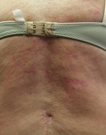
Recently, a 57-year-old woman was informed by her husband that she has a discolored area of skin on her back. This discovery followed a prolonged period of back pain, for which she was prescribed an NSAID. When this yielded no relief, she started sleeping with an electric heating pad, held in place by an elastic bandage, to ease the pain.
She reports mild itching in the affected area. She claims to be in good health otherwise and is not taking any medication.
EXAMINATION
The patient is in no acute distress but has obvious difficulty walking without pain. Covering her back, from the bra strap down, is modest erythema, a reticular pattern of modest hyperpigmentation, and focal areas of mild scaling.
No tenderness or increased warmth is detected on palpation. No notable changes are seen elsewhere on her skin.
What is the diagnosis?
This condition, termed erythema ab igne (EAI), was first described in 18th century women who worked while seated in front of a fire all day—hence the name, which translates to “redness from fire.” Over time, this exposure produced permanent changes on the anterior portions of their legs—similar to those seen in this patient.
Today, the condition can develop in a variety of contexts, all of which involve prolonged, repeated exposure to infrared radiation (eg, electric blankets, hot water bottles, electric space heaters, laptop computers). EAI can also affect the faces and arms of bakers, chefs, welders, and silversmiths working in close proximity to radiation.
This type of heat increases vasodilation in the superficial venous plexus (located in the papillary dermis), which causes hemosiderin to leak into the superficial dermis over time. This permanently stains the skin with a characteristic reticular pattern that mimics the affected vascular pattern. Generally, the darker the patient’s skin, the darker the pattern appears.
EAI more commonly affects women than men, and its diagnosis should prompt further investigation for underlying conditions. Musculoskeletal problems, chronic infection, anemia, or hypothyroidism could be involved.
Treatment, besides avoidance of radiation, includes topical application of retinoids or use of lasers.
In this case, the changes were discovered early enough to be at least partially reversible. In more advanced cases, focal areas of scaling can coalesce into papules or nodules that can undergo malignant transformation to squamous cell carcinoma.
TAKE-HOME LEARNING POINTS
- Erythema ab igne (EAI) is a fairly common condition caused by close proximity to infrared radiation (eg, heating pads, laptop computers, electric space heaters).
- This radiation causes chronic vasodilatation of the superficial venous plexus, which leads to leakage of hemosiderin pigment, permanently staining the skin in a reticular pattern.
- EAI can affect the faces and arms of bakers, welders, chefs, and silversmiths.
- Treatment can be attempted with topical application of retinoids or with use of lasers.
Together Since Birth
A 51-year-old Hispanic man presents for evaluation of a lesion he was born with. It grew as he did and was never more than a cosmetic problem until recently, when it became enlarged and sensitive. His family members convinced him to have it evaluated by primary care, who referred him to dermatology.
The patient denies any health problems other than type 2 diabetes, which is adequately controlled. His dark skin almost always tans, rather than burns, with sun exposure.
EXAMINATION
The lesion is hard to miss, measuring 1.8 cm x 5.5 mm and located prominently in the upper left nasolabial fold. It is quite firm, brownish red, hair-bearing, and round, with fine mammilations on the surface. It sits on a base only slightly smaller than the lesion’s surface. The rest of the patient’s skin is unremarkable.

Given that the lesion has changed and is quite sizeable, it is excised with a curving elliptiform incision along relaxed skin tension lines, paralleling the nasolabial fold, and sent for pathologic examination.
What is the diagnosis?
The pathology report confirmed the benign nature of this congenital melanocytic nevus (CMN). CMNs are hamartomatous lesions that form when collections of melanocytes (normal pigment cells) congregate in one location. These cells migrate during the embryonal stage of development—but some never make it to their final destination.
While CMNs are almost always benign, the history of change in this case made excision compelling. Given the lesion’s likely extension well under the surface of the skin, a shave removal would not suffice—and would leave a noticeable scar.
Despite their low potential for malignancy, CMNs—like any lesion removed from the body—must be sent for pathologic examination. The general rule is: the larger the congenital lesion, the greater the malignant potential.
Remember, too, that not all skin cancers are melanomas. Virtually any cell in the skin (sweat glands, neural tissue, sebaceous glands, smooth muscle, and even hair follicles) can undergo malignant transformation, so microscopic examination is mandatory for all removed lesions, except tiny tags or warts.
Interestingly, CMNs are more common on patients with lighter skin. However, besides the case patient, there were also several family members reporting a history of similar lesions. They were all thrilled with the patient’s cosmetic outcome—and more so with the benign report.
TAKE-HOME LEARNING POINTS
- Congenital melanocytic nevi (CMNs) are collections of melanocytes in one area.
- CMNs have little malignant potential, but morphology (eg, color, shape, symptoms) or size may dictate the need for removal or biopsy.
- Deep shave or excision are the best options for removal; patient age, lesion location, potential for scarring, and surgical ability of the provider influence choice.
- Since melanocytes can end up in other extracutaneous locations, primary melanomas can develop almost anywhere—including the lungs, the gut, or even the eyes.
A 51-year-old Hispanic man presents for evaluation of a lesion he was born with. It grew as he did and was never more than a cosmetic problem until recently, when it became enlarged and sensitive. His family members convinced him to have it evaluated by primary care, who referred him to dermatology.
The patient denies any health problems other than type 2 diabetes, which is adequately controlled. His dark skin almost always tans, rather than burns, with sun exposure.
EXAMINATION
The lesion is hard to miss, measuring 1.8 cm x 5.5 mm and located prominently in the upper left nasolabial fold. It is quite firm, brownish red, hair-bearing, and round, with fine mammilations on the surface. It sits on a base only slightly smaller than the lesion’s surface. The rest of the patient’s skin is unremarkable.
Given that the lesion has changed and is quite sizeable, it is excised with a curving elliptiform incision along relaxed skin tension lines, paralleling the nasolabial fold, and sent for pathologic examination.
What is the diagnosis?
The pathology report confirmed the benign nature of this congenital melanocytic nevus (CMN). CMNs are hamartomatous lesions that form when collections of melanocytes (normal pigment cells) congregate in one location. These cells migrate during the embryonal stage of development—but some never make it to their final destination.
While CMNs are almost always benign, the history of change in this case made excision compelling. Given the lesion’s likely extension well under the surface of the skin, a shave removal would not suffice—and would leave a noticeable scar.
Despite their low potential for malignancy, CMNs—like any lesion removed from the body—must be sent for pathologic examination. The general rule is: the larger the congenital lesion, the greater the malignant potential.
Remember, too, that not all skin cancers are melanomas. Virtually any cell in the skin (sweat glands, neural tissue, sebaceous glands, smooth muscle, and even hair follicles) can undergo malignant transformation, so microscopic examination is mandatory for all removed lesions, except tiny tags or warts.
Interestingly, CMNs are more common on patients with lighter skin. However, besides the case patient, there were also several family members reporting a history of similar lesions. They were all thrilled with the patient’s cosmetic outcome—and more so with the benign report.
TAKE-HOME LEARNING POINTS
- Congenital melanocytic nevi (CMNs) are collections of melanocytes in one area.
- CMNs have little malignant potential, but morphology (eg, color, shape, symptoms) or size may dictate the need for removal or biopsy.
- Deep shave or excision are the best options for removal; patient age, lesion location, potential for scarring, and surgical ability of the provider influence choice.
- Since melanocytes can end up in other extracutaneous locations, primary melanomas can develop almost anywhere—including the lungs, the gut, or even the eyes.
The Lesion with Legs
No one in the family is certain when this 6-year-old boy first developed the red spot on his nose, but it is increasingly noticeable. And with school picture day approaching, they would like the redness to resolve.
Their primary care provider reassured them, at length, that it was benign and would eventually resolve without treatment. The lesion causes no symptoms and is purely a cosmetic concern.
The child is otherwise healthy and has been since birth.
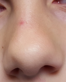
EXAMINATION
A pinpoint red dot can be seen on the upper nasal bridge, just to the right of the midline. Tiny linear red “legs” extend from the central dot, like spokes on a wheel. In aggregate, the lesion measures about 3 mm in diameter. There is no palpable component.
However, the entire lesion is blanchable: Pinpoint pressure on the central dot causes it to blanch, and as the pressure is released, the legs of the lesion refill immediately from the center outward. When a glass slide is pressed against the lesion (a process called diascopy) and then released, the same process occurs.

What is the diagnosis?
Spider angiomas (SAs), originally called nevus araneus, are actually neither angiomas nor nevi. Instead, they are telangiectasias formed from a superficial arteriole whose surrounding sphincter muscle has failed. The “legs” are tiny veins that carry blood away from the central lesion; this is why they blanch so readily with central pressure and refill from the center outward.
SAs affect 10% to 15% of children and occur only in areas along the path of the superior vena cava. Besides the face, they can be found on the trunk, arms, and dorsal hands.
When solitary, these lesions are benign and can be either left to clear on their own or removed by laser or electrodessication. The presence of three or more lesions warrants further investigation, since multiple SAs can signify underlying disease (eg, cirrhosis of the liver, thyrotoxicosis, or systemic sclerosis).
This patient was not inclined to let us treat the lesion; within a few years, a teasing comment from a classmate or two may change his mind! But with a little luck, it will disappear on its own eventually.
TAKE-HOME LEARNING POINTS
- Spider angiomas (SAs) are actually dilated telangiectatic arterioles manifesting as blanchable, bright red, pinpoint papules with venous “legs” that extend from the center.
- SAs affect 10% to 15% of all children and are confined to areas drained by the superior vena cava.
- Pressure on the lesion with a glass slide (a process called diascopy) causes total blanching; this, along with the clinical findings, confirms the diagnosis.
- Most SAs resolve on their own eventually, but they can be destroyed by laser or electrodessication.
- The presence of more than three SAs should prompt a search for potential causes, such as liver disease, thyrotoxicosis, or systemic sclerosis.
No one in the family is certain when this 6-year-old boy first developed the red spot on his nose, but it is increasingly noticeable. And with school picture day approaching, they would like the redness to resolve.
Their primary care provider reassured them, at length, that it was benign and would eventually resolve without treatment. The lesion causes no symptoms and is purely a cosmetic concern.
The child is otherwise healthy and has been since birth.
EXAMINATION
A pinpoint red dot can be seen on the upper nasal bridge, just to the right of the midline. Tiny linear red “legs” extend from the central dot, like spokes on a wheel. In aggregate, the lesion measures about 3 mm in diameter. There is no palpable component.
However, the entire lesion is blanchable: Pinpoint pressure on the central dot causes it to blanch, and as the pressure is released, the legs of the lesion refill immediately from the center outward. When a glass slide is pressed against the lesion (a process called diascopy) and then released, the same process occurs.
What is the diagnosis?
Spider angiomas (SAs), originally called nevus araneus, are actually neither angiomas nor nevi. Instead, they are telangiectasias formed from a superficial arteriole whose surrounding sphincter muscle has failed. The “legs” are tiny veins that carry blood away from the central lesion; this is why they blanch so readily with central pressure and refill from the center outward.
SAs affect 10% to 15% of children and occur only in areas along the path of the superior vena cava. Besides the face, they can be found on the trunk, arms, and dorsal hands.
When solitary, these lesions are benign and can be either left to clear on their own or removed by laser or electrodessication. The presence of three or more lesions warrants further investigation, since multiple SAs can signify underlying disease (eg, cirrhosis of the liver, thyrotoxicosis, or systemic sclerosis).
This patient was not inclined to let us treat the lesion; within a few years, a teasing comment from a classmate or two may change his mind! But with a little luck, it will disappear on its own eventually.
TAKE-HOME LEARNING POINTS
- Spider angiomas (SAs) are actually dilated telangiectatic arterioles manifesting as blanchable, bright red, pinpoint papules with venous “legs” that extend from the center.
- SAs affect 10% to 15% of all children and are confined to areas drained by the superior vena cava.
- Pressure on the lesion with a glass slide (a process called diascopy) causes total blanching; this, along with the clinical findings, confirms the diagnosis.
- Most SAs resolve on their own eventually, but they can be destroyed by laser or electrodessication.
- The presence of more than three SAs should prompt a search for potential causes, such as liver disease, thyrotoxicosis, or systemic sclerosis.