User login
My, How You've Grown
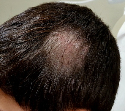
Six years ago, a lesion appeared on this now 39-year-old woman’s forehead. It grew steadily to its current size, impinging on her brow line. Although it has been asymptomatic, the patient is concerned about malignancy, since she has a significant personal and family history of skin cancer. She has had several lesions removed from her face and back over the years.
EXAMINATION
There is a 2.2-cm, roughly round, white, cicatricial, concave lesion on the patient’s lower right forehead, extending into the brow line. Around the periphery are several 2- to 5-mm eroded papules. There are no palpable nodes on the head or neck.
Several scars are seen elsewhere on the patient’s face and back, consistent with her history. Her type II/VI skin is quite fair and sun-damaged.
A 6-mm deep punch biopsy of the lesion is obtained.

What is the diagnosis?
Biopsy reveals a morpheaform basal cell carcinoma (BCC; also known as cicatricial BCC) with perineural involvement that extends to the margin of the sample. While BCCs are almost never fatal, if ignored, their relentless growth can be problematic. This case illustrates that, along with the wide variety of morphologic presentations.
Of the different types of BCC, the most common are nodular. These present as pearly (ie, translucent) papules or nodules, with or without focal erosion or frank ulceration; they often have prominent telangiectasias coursing over their surfaces. BCCs can also appear as rashes (superficial BCC) that may not attract attention.
This patient’s lesion is one of the least common variations: It combines features of a morpheaform (scarlike) BCC with focal noduloulcerative papular lesions studding its periphery. The concavity of the scarlike portion, along with its prolonged presence, predicted deep involvement of adjacent tissue—confirmed by the biopsy results.
At a minimum, this patient will need Mohs micrographic surgical removal, with closure by skin graft or secondary intention. Given the deep perineural involvement, surgery alone may not clear the cancer; radiation therapy may be necessary.
TAKE-HOME LEARNING POINTS
- Morpheaform basal cell carcinoma (BCC), also known as cicatricial BCC, can present as a white, scarlike patch, often with an atrophic surface.
- This type of BCC is more aggressive than most, often requiring Mohs surgery.
- There are at least three other types of BCC, most of which involve nonhealing ulcerative papules or nodules.
- This patient’s history of sun-caused skin cancers makes recurrence likely.
Six years ago, a lesion appeared on this now 39-year-old woman’s forehead. It grew steadily to its current size, impinging on her brow line. Although it has been asymptomatic, the patient is concerned about malignancy, since she has a significant personal and family history of skin cancer. She has had several lesions removed from her face and back over the years.
EXAMINATION
There is a 2.2-cm, roughly round, white, cicatricial, concave lesion on the patient’s lower right forehead, extending into the brow line. Around the periphery are several 2- to 5-mm eroded papules. There are no palpable nodes on the head or neck.
Several scars are seen elsewhere on the patient’s face and back, consistent with her history. Her type II/VI skin is quite fair and sun-damaged.
A 6-mm deep punch biopsy of the lesion is obtained.
What is the diagnosis?
Biopsy reveals a morpheaform basal cell carcinoma (BCC; also known as cicatricial BCC) with perineural involvement that extends to the margin of the sample. While BCCs are almost never fatal, if ignored, their relentless growth can be problematic. This case illustrates that, along with the wide variety of morphologic presentations.
Of the different types of BCC, the most common are nodular. These present as pearly (ie, translucent) papules or nodules, with or without focal erosion or frank ulceration; they often have prominent telangiectasias coursing over their surfaces. BCCs can also appear as rashes (superficial BCC) that may not attract attention.
This patient’s lesion is one of the least common variations: It combines features of a morpheaform (scarlike) BCC with focal noduloulcerative papular lesions studding its periphery. The concavity of the scarlike portion, along with its prolonged presence, predicted deep involvement of adjacent tissue—confirmed by the biopsy results.
At a minimum, this patient will need Mohs micrographic surgical removal, with closure by skin graft or secondary intention. Given the deep perineural involvement, surgery alone may not clear the cancer; radiation therapy may be necessary.
TAKE-HOME LEARNING POINTS
- Morpheaform basal cell carcinoma (BCC), also known as cicatricial BCC, can present as a white, scarlike patch, often with an atrophic surface.
- This type of BCC is more aggressive than most, often requiring Mohs surgery.
- There are at least three other types of BCC, most of which involve nonhealing ulcerative papules or nodules.
- This patient’s history of sun-caused skin cancers makes recurrence likely.
Sunny Side's Up
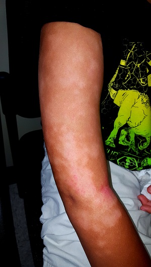
A year ago, this 60-year-old man noticed an asymptomatic lesion on the dorsum of his right hand. When it grew in size over the course of a few months, he showed it to his primary care provider, who believed it to be a wart and froze it with liquid nitrogen. This reduced its size, but only temporarily. It has since been treated with topical and oral antibiotics to no avail.
The patient has had several basal cell carcinomas removed from his face, arms, and trunk in the past.
EXAMINATION
On the mid dorsum of the patient’s right hand is a 1.5-cm ovoid nodule with a smooth surface and very firm feel. It appears in the context of fully sun-exposed, sun-damaged skin. Several scars are noted in the area, consistent with his history of sun-caused skin cancers.

The lesion is removed by deep shave biopsy, and the base curetted. The entire lesion is sent to pathology.
What’s the diagnosis?
The pathology report shows a low-grade, well-differentiated squamous cell carcinoma (SCC)—in this case, a keratoacanthoma (KA). This common form of SCC is usually found on the sun-exposed skin of older patients. The lesions can range in size from 3 mm to 3 cm or larger and are usually round to oval and dome-like, with symmetrical architecture and, often, a central keratotic core. The differential includes cysts, warts, and seborrheic keratosis.
Histologically, KAs are composed of uniformly staining (blue) cells of similar size and shape (connoting relative benignancy), to which we apply the term well-differentiated. Poorly-differentiated cellular composition manifests with cells of different sizes, shapes, and colors; these characteristics suggest more aggressive malignancy.
Even though KAs are skin cancers, they are quite low-grade, which means they rarely metastasize; if left alone, they can resolve completely over time. However, their odd appearance and rapid growth are usually concerning enough to prompt their removal.
When suspected KAs are removed, it’s essential that the entire lesion be submitted for pathologic examination. This allows for the architecture of the entire lesion—its cellular composition and margins—to be evaluated. When only part of the lesion is removed for biopsy, the diagnosis will be “squamous cell carcinoma, well differentiated, without evidence of invasion.” In the minds of many dermatology providers, this diagnosis demands excision—but a KA lesion completely removed by shave biopsy is considered cured.
Histologic examination of these lesions is not always as straightforward as in this case. KAs can be poorly differentiated or demonstrate focal areas of invasion, which justifies excision with margins.
TAKE-HOME LEARNING POINTS
- Keratoacanthoma (KA) is an extremely common low-grade squamous cell carcinoma most often seen on directly sun-exposed skin (eg, hands, arms, face, ears) of older, sun-damaged patients.
- KA typically manifests as a round to oval, dome-like, firm nodule, often with a central keratotic core and a history of rapid growth.
- It’s important to remove these lesions in one piece (eg, by deep shave biopsy) because identification is based on architecture and cellular composition.
- The pathology report will show a well-differentiated squamous cell carcinoma with architecture consistent with KA.
- Although some believe that excision is necessary, a deep shave biopsy performed with clear margins is adequate treatment.
A year ago, this 60-year-old man noticed an asymptomatic lesion on the dorsum of his right hand. When it grew in size over the course of a few months, he showed it to his primary care provider, who believed it to be a wart and froze it with liquid nitrogen. This reduced its size, but only temporarily. It has since been treated with topical and oral antibiotics to no avail.
The patient has had several basal cell carcinomas removed from his face, arms, and trunk in the past.
EXAMINATION
On the mid dorsum of the patient’s right hand is a 1.5-cm ovoid nodule with a smooth surface and very firm feel. It appears in the context of fully sun-exposed, sun-damaged skin. Several scars are noted in the area, consistent with his history of sun-caused skin cancers.
The lesion is removed by deep shave biopsy, and the base curetted. The entire lesion is sent to pathology.
What’s the diagnosis?
The pathology report shows a low-grade, well-differentiated squamous cell carcinoma (SCC)—in this case, a keratoacanthoma (KA). This common form of SCC is usually found on the sun-exposed skin of older patients. The lesions can range in size from 3 mm to 3 cm or larger and are usually round to oval and dome-like, with symmetrical architecture and, often, a central keratotic core. The differential includes cysts, warts, and seborrheic keratosis.
Histologically, KAs are composed of uniformly staining (blue) cells of similar size and shape (connoting relative benignancy), to which we apply the term well-differentiated. Poorly-differentiated cellular composition manifests with cells of different sizes, shapes, and colors; these characteristics suggest more aggressive malignancy.
Even though KAs are skin cancers, they are quite low-grade, which means they rarely metastasize; if left alone, they can resolve completely over time. However, their odd appearance and rapid growth are usually concerning enough to prompt their removal.
When suspected KAs are removed, it’s essential that the entire lesion be submitted for pathologic examination. This allows for the architecture of the entire lesion—its cellular composition and margins—to be evaluated. When only part of the lesion is removed for biopsy, the diagnosis will be “squamous cell carcinoma, well differentiated, without evidence of invasion.” In the minds of many dermatology providers, this diagnosis demands excision—but a KA lesion completely removed by shave biopsy is considered cured.
Histologic examination of these lesions is not always as straightforward as in this case. KAs can be poorly differentiated or demonstrate focal areas of invasion, which justifies excision with margins.
TAKE-HOME LEARNING POINTS
- Keratoacanthoma (KA) is an extremely common low-grade squamous cell carcinoma most often seen on directly sun-exposed skin (eg, hands, arms, face, ears) of older, sun-damaged patients.
- KA typically manifests as a round to oval, dome-like, firm nodule, often with a central keratotic core and a history of rapid growth.
- It’s important to remove these lesions in one piece (eg, by deep shave biopsy) because identification is based on architecture and cellular composition.
- The pathology report will show a well-differentiated squamous cell carcinoma with architecture consistent with KA.
- Although some believe that excision is necessary, a deep shave biopsy performed with clear margins is adequate treatment.
Talk About Premature Balding...
Several months ago, this 8-year-old boy began losing hair from his scalp. Other than mild itching, there are no associated symptoms. The patient has no pets at home, but he spends his after-school hours with his cousin, who does.
The child is allergy prone but otherwise healthy. No one else in the family (ie, his two younger siblings) has been similarly affected.
EXAMINATION
About three-quarters of the hair is missing from a 6-cm oval patch on the parietal scalp. A few short hairs remain. The skin in this area is slightly edematous, with focal areas of broken, scaly skin.
Palpation of the head and neck reveals adenopathy in the nuchal area of the affected side. A KOH prep is performed with a #10 blade; the sample includes hairs as well as skin.
What is the diagnosis?
Examination of the sample revealed endothrix, in which fungal spores and hyphae are found inside the broken-off hairs, especially near the roots. A fungal culture confirmed the presence of Trychophyton tonsurans.
T tonsurans is the most common culprit in tinea captitis cases in the United States. This dermatophytic infection of the scalp is a common diagnosis in children, who typically contract it from other children. (Some causative species—such as Microsporum audouinii—spread via contact with animals, but these organisms are generally rare in the US.) Tinea capitis is seen more frequently in boys than in girls, and African-American patients are especially at risk.
Tinea capitis infects the deep hair shaft but spares the skin. Diagnosis requires a combination of clinical signs and identification of the organism in the hair shaft; the latter will also help to guide treatment. In contrast, tinea corporis is diagnosed by clinical features and KOH examination of external scales where the organism resides. Traditionally, infected hairs have needed to be removed for KOH exam—but practical experience has shown that a vigorous scrape that captures infected hairs can accomplish the same thing.
The results of fungal culture may take a month or more to finalize; in the interim, patients such as this one may be treated with griseofulvin (10 mg/kg/d) and application of topical ciclopirox cream bid to reduce infectivity. Total clearance will take at least two months.
Tinea capitis has several forms including inflammatory (which manifests with a large, swollen, inflamed mass) and black dot (named for the tips of broken hair shafts that remain in the affected areas). The differential includes psoriasis, alopecia areata, and seborrhea.
TAKE-HOME LEARNING POINTS
- Tinea capitis is a dermatophytic infection of the scalp usually caused by the dermatophytes Trychophyton tonsurans or T rubrum.
- These infections involve the hair shaft below the skin line, rather than the surface of the skin.
- The organisms that commonly cause tinea capitis in the US typically spread through contact with another person.
- Diagnosis can be made from clinical findings only, including reactive adenopathy. KOH and culture can be necessary in questionable cases, and because of the length of treatment.
Being a Teenager Is Rough Enough
Since she was about 2 years old, this now 14-year-old girl has had asymptomatic bumps on her arms and face. They are most noticeable in the wintertime and a bit less conspicuous during humid months. Besides the appearance of the lesions, the patient is annoyed by the rough feel of them, which is unaffected by her use of OTC moisturizers and lotions.
Her mother recalls having the same problem as a child but says it improved with time. The patient’s immediate family members are all atopic, with seasonal allergies and eczema.
EXAMINATION
The patient is Hispanic with type IV skin. Dense patches of brown, tiny, rough, papulofollicular lesions cover the surface of both posterior triceps. They are also visible on the patient’s anterior thighs and the posterior two-thirds of her face.

What is the diagnosis?
DISCUSSION
Keratosis pilaris (KP) is an inherited condition that affects 30% to 50% of all children without respect to race or gender. It can be a problem unto itself, or it can be part of the atopic diathesis in patients with seasonal allergies, dry skin, and eczema.
KP is caused by an excessive production of keratin that plugs the follicles, often trapping tiny fine hairs inside and resulting in a firm follicular papule. The distribution exhibited in this case is quite typical, as is the brown color on the patient’s arms (common in those with darker skin). KP can also affect the skin on other convex areas (eg, buttocks, deltoids, and thighs). It spares glabrous skin completely. A common variant is rubra facei, characterized by redness and bumps on the posterior two-thirds of the face.
Though it cannot be cured, the condition can be controlled with keratolytics containing salicylic acid, urea, or glycolic acid, or with pure emollients, which trap moisture in the skin. For the occasional itch, topical steroid creams or ointments can be helpful.
Patient education is a key aspect of treatment. Patients can be reassured of the condition’s benignancy, and they may be relieved to know that most KP patients see major improvement as they reach their third decade of life (and beyond).
TAKE-HOME LEARNING POINTS
- Keratosis pilaris (KP) is an extremely common condition inherited by autosomal dominant mode that affects more than 30% of children.
- Excessive keratin production is the cause of KP; it plugs follicular orifices, trapping fine hairs inside and causing crops of firm scaly papules to develop on triceps, deltoids, anterior thighs, and other areas (eg, the face).
- KP is considered part of the minor diagnostic criteria for atopy, but it can also be a standalone condition.
- Treatment of KP is far from perfect, but improvement is seen with the use of either keratolytics or emollients. Anything that dries it out more (eg, long, hot showers) or irritates individual lesions (eg, picking) is to be avoided.
- Most KP patients report improvement after the third decade of life.
Since she was about 2 years old, this now 14-year-old girl has had asymptomatic bumps on her arms and face. They are most noticeable in the wintertime and a bit less conspicuous during humid months. Besides the appearance of the lesions, the patient is annoyed by the rough feel of them, which is unaffected by her use of OTC moisturizers and lotions.
Her mother recalls having the same problem as a child but says it improved with time. The patient’s immediate family members are all atopic, with seasonal allergies and eczema.
EXAMINATION
The patient is Hispanic with type IV skin. Dense patches of brown, tiny, rough, papulofollicular lesions cover the surface of both posterior triceps. They are also visible on the patient’s anterior thighs and the posterior two-thirds of her face.
What is the diagnosis?
DISCUSSION
Keratosis pilaris (KP) is an inherited condition that affects 30% to 50% of all children without respect to race or gender. It can be a problem unto itself, or it can be part of the atopic diathesis in patients with seasonal allergies, dry skin, and eczema.
KP is caused by an excessive production of keratin that plugs the follicles, often trapping tiny fine hairs inside and resulting in a firm follicular papule. The distribution exhibited in this case is quite typical, as is the brown color on the patient’s arms (common in those with darker skin). KP can also affect the skin on other convex areas (eg, buttocks, deltoids, and thighs). It spares glabrous skin completely. A common variant is rubra facei, characterized by redness and bumps on the posterior two-thirds of the face.
Though it cannot be cured, the condition can be controlled with keratolytics containing salicylic acid, urea, or glycolic acid, or with pure emollients, which trap moisture in the skin. For the occasional itch, topical steroid creams or ointments can be helpful.
Patient education is a key aspect of treatment. Patients can be reassured of the condition’s benignancy, and they may be relieved to know that most KP patients see major improvement as they reach their third decade of life (and beyond).
TAKE-HOME LEARNING POINTS
- Keratosis pilaris (KP) is an extremely common condition inherited by autosomal dominant mode that affects more than 30% of children.
- Excessive keratin production is the cause of KP; it plugs follicular orifices, trapping fine hairs inside and causing crops of firm scaly papules to develop on triceps, deltoids, anterior thighs, and other areas (eg, the face).
- KP is considered part of the minor diagnostic criteria for atopy, but it can also be a standalone condition.
- Treatment of KP is far from perfect, but improvement is seen with the use of either keratolytics or emollients. Anything that dries it out more (eg, long, hot showers) or irritates individual lesions (eg, picking) is to be avoided.
- Most KP patients report improvement after the third decade of life.

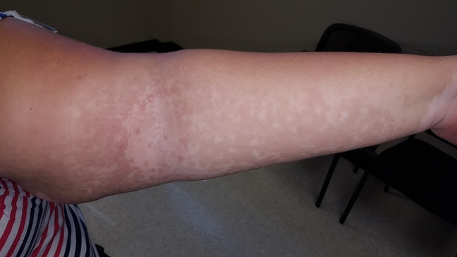
Wanna See My Not-Tan Lines?
A 16-year-old African-American girl is brought in by her mother for evaluation of skin changes affecting both arms: small, round, slightly scaly, 2- to 3-cm patches on the triceps, antecubitals, and deltoids. The changes manifested in early spring and worsened with the arrival of summer.
The condition has been previously diagnosed as vitiligo by her primary care provider and as fungal infection by an urgent care provider. Nystatin cream and clotrimazole cream have had no effect.
The patient’s history includes eczema, extensive atopy manifesting with seasonal allergies, and childhood asthma. Her siblings also had these problems.
EXAMINATION
Extensive, mottled hypopigmentation is noted on the skin of both arms, in stark contrast to the patient’s type V skin. Very little scale is seen. There are focal areas of slight erythema around the antecubital folds.
What is the diagnosis?
DISCUSSION
This phenomenon is so common in dermatology clinics that it’s a rare day when we don’t see it. This form of hypopigmentation is pityriasis alba (PA), in which areas of eczema don’t tan at all while the surrounding skin darkens with sun exposure. The lateral aspects of the arms are often affected (sparing the sun-protected medial aspects), as are the sides of the face and the posterior neck. The contrast is striking, especially on those with darker skin.
PA occurs mostly in children and young adults, becoming less frequent with age. It differs from vitiligo in that PA involves seasonal, partial pigment loss; vitiligo by contrast manifests with complete pigment loss (leaving utterly white skin) that is almost always permanent.
Treatment consists of sun protection, moisturization to prevent eczema, and use of class IV steroid creams or ointments when lesions appear. Even without treatment, PA usually clears during the winter months—when the surrounding skin loses its tan—only to recur the following spring.
TAKE-HOME LEARNING POINTS
- Pityriasis alba (PA) occurs when patches of eczema fail to tan, producing marked contrast between them and the normal surrounding skin; it is often mistaken for fungal infection.
- PA favors the antecubital, deltoid, and lateral tricep areas, as well as the lateral face.
- PA is more common in atopic individuals who are prone to eczema and appears more dramatic in those with darker skin.
- Vitiligo is a major item in the differential, but color loss with PA is only partial (rather than permanent) and almost always resolves in the winter.
- Once color is lost with PA, treatment is largely ineffective. It is then best to use preventive measures (eg, sunscreen and moisturizer), plus/minus topical steroid creams, for the eczema.

Shedding Light on the Problem
A 49-year-old man presents to dermatology for evaluation of an itchy rash that manifested several months ago. Attempts to eradicate or ameliorate it—which have included topical and systemic steroids and oral antibiotics (minocycline)—have had no effect. A biopsy ordered by his primary care provider (PCP) showed nonspecific changes, termed “dermatitis” in the report.
The patient denies any history of atopy, such as seasonal allergies or eczema. His only medical problem is moderate hypertension, for which his PCP prescribed hydrochlorothiazide. He started taking the drug a few weeks before the rash appeared.
He claims to be in good health otherwise, with no weakness or weight loss. He has never smoked.
EXAMINATION
A bright red, blanchable, maculopapular rash is notably confined to the patient’s sun-exposed skin; it sharply spares the areas covered by clothing and the watch on his left wrist. His palms, soles, scalp, and face are also spared. No nail changes are noted.
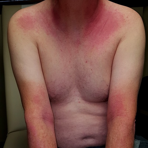
Otherwise, the patient appears well and is able to rise from a seated position without difficulty. No nodes are palpable in the head or neck, and there is no organomegaly detected on abdominal examination.
What is the diagnosis?
Photosensitivity to hydrochlorothiazide was the obvious culprit, so the patient was advised to stop using that product (after consulting his PCP). He’ll remain off the medication for at least two months, then present for follow-up.
There are some potentially troubling items in the differential, particularly dermatomyositis (DM). Patients with DM may exhibit a sunburn-like rash, but they will additionally demonstrate muscle weakness and chronic fatigue. A significant proportion of their complaints are driven by an occult carcinoma (eg, stomach, lung, colon, breast). While it’s doubtful that this patient has DM, his follow-up may include a fresh biopsy, blood work, anteroposterior and lateral chest films, and possibly a colonoscopy.
Another item in the differential is lupus. However, the original biopsy yielded no suggestive findings (eg, interface dermatitis), nor did the patient have any complaints referable to the disease.
This case nicely demonstrates the concept that it is equally important to note which areas are affected by and spared by a skin condition. With that in mind, we can at least establish that sunlight is a major factor in the genesis of this rash. Unfortunately, that still leaves room for conjecture as to the diagnosis.
TAKE-HOME LEARNING POINTS
- Rashes confined to sun-exposed skin can be a symptom of systemic disease, such as lupus or dermatomyositis.
- Various drugs—including hydrochlorothiazide, NSAIDs, sulfas, and certain tetracyclines—can also cause photosensitivity reactions.
- Hydrochlorothiazide is one of the more common drugs to cause such a rash, which may take weeks to clear after cessation of use.
- If terminating hydrochlorothiazide doesn’t help, skin biopsy and labs (especially creatine kinase and immunoglobulins) are the next step in determining the problem.
A 49-year-old man presents to dermatology for evaluation of an itchy rash that manifested several months ago. Attempts to eradicate or ameliorate it—which have included topical and systemic steroids and oral antibiotics (minocycline)—have had no effect. A biopsy ordered by his primary care provider (PCP) showed nonspecific changes, termed “dermatitis” in the report.
The patient denies any history of atopy, such as seasonal allergies or eczema. His only medical problem is moderate hypertension, for which his PCP prescribed hydrochlorothiazide. He started taking the drug a few weeks before the rash appeared.
He claims to be in good health otherwise, with no weakness or weight loss. He has never smoked.
EXAMINATION
A bright red, blanchable, maculopapular rash is notably confined to the patient’s sun-exposed skin; it sharply spares the areas covered by clothing and the watch on his left wrist. His palms, soles, scalp, and face are also spared. No nail changes are noted.
Otherwise, the patient appears well and is able to rise from a seated position without difficulty. No nodes are palpable in the head or neck, and there is no organomegaly detected on abdominal examination.
What is the diagnosis?
Photosensitivity to hydrochlorothiazide was the obvious culprit, so the patient was advised to stop using that product (after consulting his PCP). He’ll remain off the medication for at least two months, then present for follow-up.
There are some potentially troubling items in the differential, particularly dermatomyositis (DM). Patients with DM may exhibit a sunburn-like rash, but they will additionally demonstrate muscle weakness and chronic fatigue. A significant proportion of their complaints are driven by an occult carcinoma (eg, stomach, lung, colon, breast). While it’s doubtful that this patient has DM, his follow-up may include a fresh biopsy, blood work, anteroposterior and lateral chest films, and possibly a colonoscopy.
Another item in the differential is lupus. However, the original biopsy yielded no suggestive findings (eg, interface dermatitis), nor did the patient have any complaints referable to the disease.
This case nicely demonstrates the concept that it is equally important to note which areas are affected by and spared by a skin condition. With that in mind, we can at least establish that sunlight is a major factor in the genesis of this rash. Unfortunately, that still leaves room for conjecture as to the diagnosis.
TAKE-HOME LEARNING POINTS
- Rashes confined to sun-exposed skin can be a symptom of systemic disease, such as lupus or dermatomyositis.
- Various drugs—including hydrochlorothiazide, NSAIDs, sulfas, and certain tetracyclines—can also cause photosensitivity reactions.
- Hydrochlorothiazide is one of the more common drugs to cause such a rash, which may take weeks to clear after cessation of use.
- If terminating hydrochlorothiazide doesn’t help, skin biopsy and labs (especially creatine kinase and immunoglobulins) are the next step in determining the problem.

Blood in Urine, Rash on Trunk
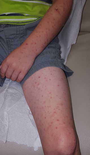
Several days ago, a 14-year-old boy suddenly became ill with abdominal pain, fever, and arthralgia. Within 12 hours, a rash developed that covered most of his trunk, arms, and legs but spared his face, palms, and soles. It quickly flared bright red; some lesions were tender to touch. The patient’s legs and scrotum became edematous, and he lost his appetite. The patient developed diarrhea, and bright red blood was seen in his stools.
He was taken to the local emergency department, where examination revealed a fever of 101.5°F, an elevated white blood cell count, and a small amount of blood in his urine. Stool cultures were ordered, and the patient was placed on an unknown antibiotic.
The next day, he consulted his pediatrician, who referred him to dermatology.
EXAMINATION
Today, the patient is afebrile and in no acute distress. He still has a florid rash on his trunk, arms, and legs consisting of very evenly distributed, purpuric lesions that average 3 mm in diameter. A few are palpable, and none are blanchable. A punch biopsy is performed, and an entire lesion is obtained and submitted for pathologic examination.
What is the diagnosis?
The report showed leukocytoclastic vasculitis, in which activated lymphocytes attack the inner lining of blood vessels, causing them to leak blood into the surrounding interstitial spaces. Besides the extravasated red blood cells, nuclear dust (remnants of the attacking lymphocytes) is also seen.
These biopsy findings, in context with the patient’s history, help to confirm the diagnosis of Henoch-Schönlein purpura (HSP), an IgA-mediated disease that causes widespread vasculitis of small vessels throughout the body. Besides affecting the skin, this process can injure the gastrointestinal tract, joints, kidneys, and even lungs. As this case illustrates, it almost always presents with a palpable, purpuric, widespread rash, abdominal pain, fever, joint pain, and bloody stools.
HSP is seen primarily in children; in the US, 75% of cases occur in those ages 2 to 5. The most consistent presenting symptoms in this population include rash, abdominal pain, and joint pain. When fever is present, it is typically mild.
A variety factors can trigger HSP, including medications (eg, penicillin, NSAIDs, sulfa) and infection (with organisms such as mycoplasma, mononucleosis, strep, Legionella)—but many cases are simply idiopathic. History of upper respiratory infection, pharyngitis, or intestinal infection is found in 75% of young HSP patients. Antecedent vaccinations have also been reported as a potential trigger.
The diagnosis of HSP is primarily clinical, based on a combination of signs and symptoms and the exclusion of other items in the differential. Besides bloodwork to rule out end-organ (eg, renal) damage, a skin biopsy of the purpuric rash is necessary to establish the type of vasculitis.
Fortunately, most HSP patients recover uneventfully; the exception is the occasional patient with renal complications. The case patient successfully recovered following treatment with oral antibiotics (for presumed strep) and a three-week course of prednisone.
TAKE-HOME LEARNING POINTS
- A purpuric rash should prompt a punch biopsy to search for vasculitis.
- A widespread, palpable, purpuric rash accompanied by systemic symptoms of abdominal pain, arthralgia, fever, and malaise is suggestive of serious disease. In younger patients, Henoch-Schönlein purpura (HSP) should be a major suspect.
- Drugs, bugs, and vaccinations are all possible triggers for HSP.
- Once the diagnosis of HSP is made, monitoring for end-organ damage is essential.
Lost Weight, Gained Rash?
A 25-year-old African-American woman presents for evaluation of an asymptomatic rash she has had for several months. It manifested shortly after she began an exercise program to help her lose weight. Her primary care provider made a presumptive diagnosis of tinea versicolor (TV), but the rash persists despite treatment attempts with topical selenium sulfide shampoo and a 10-day course of fluconazole (200 mg/d).
The patient denies having endocrine problems, such as diabetes. However, she states that given her weight and family history, she was warned about the possibility.
EXAMINATION
The patient is obese and has type V skin. Her rash is dark brown and feels slightly rough. It appears solid brown on the central back and chest, peripherally becoming sparser and more reticular (net-like). It extends to involve the flexural surfaces of both arms.
What is the diagnosis?
This is a fairly typical case of confluent and reticulated papillomatosis (CRP), also known as Gougerot-Carteaud syndrome. CRP is rare, mainly affecting young adults with darker skin after puberty. It can manifest in both genders. Originally believed to be a variant of acanthosis nigricans, CRP is now considered a distinct diagnostic entity.
At first glance, the appearance of CRP mimics that of TV. But the rough feel, reticular look, and dark color of CRP (which results from an increase in melanosomes) are totally missing in TV. Histologic studies of CRP show abnormal keratinocyte differentiation and maturation, a picture markedly at odds with that of TV.
TV, a result of the commensal yeast organism Malassezia furfur (M furfur) metabolizing normal sebum and leaving behind azelaic acid, causes color changes in the skin. But M furfur is not involved in CRP, and therefore the condition does not respond to oral or topical antiyeast medications.
The most effective treatment for CRP is minocycline (100 mg bid for 10 d). Long-term treatment includes weight loss and reduction of ambient heat.
TAKE-HOME LEARNING POINTS
- Confluent and reticulated papillomatosis (CRP) affects the trunk and extremities of obese patients with darker skin.
- In contrast with tinea versicolor (TV), CRP has a rough texture and reticulated look, especially on the periphery of the involved areas.
- Biopsy can help distinguish CRP from its lookalikes; it shows abnormal keratinocyte differentiation and maturation, as well as increased melanosomes.
- Oral minocycline is the best treatment, along with weight loss and reduction of ambient heat.
The Itch That Won't Quit
A 68-year-old woman with a very itchy rash is referred to dermatology for evaluation. She reports the itching to be constant—24 hours a day, seven days a week—but particularly severe at bedtime.
The rash has been totally unresponsive to numerous treatment attempts over the past year, including topical steroids (triamcinolone 0.1% cream bid), oral antibiotics (trimethoprim/sulfa), and oral steroids (prednisone).
The patient lives alone, apart from the occasional overnight visit from her grandchild.
EXAMINATION
The widespread rash spares only the patient’s legs below the knees. It is comprised of sparsely distributed excoriated foci, some surrounded by oval-to-round scales. The patient scratches the sites throughout the examination.
During a shave biopsy of one of the lesions on the patient’s arm, she mentions that occasionally lesions also manifest on her hands and fingers. Closer examination reveals a few unremarkable, scaly, 1- to 3-mm papules on her volar wrists. These are scraped with a #10 blade and placed on a slide, which is covered, filled with potassium hydroxide 10%, and examined under 10x magnification.

What is the diagnosis?
After a lengthy search, a single scabies adult (scabies sarcoptei var humanus) was found embedded in the scales. Scabies is one of the two most common ectoparasitic infestations in this country (the other being head lice).
Paradoxically, it is one of the most over- and under-diagnosed medical conditions worldwide. It is transmitted from person to person and can only be acquired from close, prolonged contact with another human who has the condition. This case illustrates some of the difficulties involved in making the diagnosis.
While it is vital to consider scabies in the differential for constant, severe itching, there are situations in which it can be ruled out. People who live alone and/or avoid physical contact with other people cannot get scabies. It can only be acquired from an infected person—not from a dog, cat, or inanimate object. In this case, the patient lived alone, but she hosted sleepovers with her grandchild—the likely source of this infestation.
Scabies can manifest as an eczematoid rash that will not respond to topical or systemic steroids. Conversely, when eczema patients are misdiagnosed with scabies, permethrin cream worsens the condition. Therefore, once scabies is considered in the differential, a KOH prep is indicated for a definitive diagnosis.
In terms of treatment, it does little good to simply treat the patient in question. The entire family (and/or close contacts) needs to be treated simultaneously—but before that, the source of the scabies needs to be identified. Failure to address all of these factors often leads to “treatment failure.”
The case patient was successfully treated with a combination of permethrin cream and oral ivermectin, according to the standard regimen (two treatments, seven to 10 days apart).
TAKE-HOME LEARNING POINTS
- Scabies can only be acquired from close, prolonged contact with another human who has the condition.
- Intractable itching and failure to respond to treatment (ie, topical and systemic steroids) are its dependable diagnostic features.
- Scraping suspected scabetic lesions (tiny vesicles, dried papules, or—if you’re lucky—a burrow) and examining them under 10x microscopy is preferable for confirmation of the diagnosis.
- The whole family must be treated in synchrony with the patient.
- It is essential to identify the source of the scabies (eg, spouse, boyfriend/girlfriend, child) to successfully eradicate the problem.
A 68-year-old woman with a very itchy rash is referred to dermatology for evaluation. She reports the itching to be constant—24 hours a day, seven days a week—but particularly severe at bedtime.
The rash has been totally unresponsive to numerous treatment attempts over the past year, including topical steroids (triamcinolone 0.1% cream bid), oral antibiotics (trimethoprim/sulfa), and oral steroids (prednisone).
The patient lives alone, apart from the occasional overnight visit from her grandchild.
EXAMINATION
The widespread rash spares only the patient’s legs below the knees. It is comprised of sparsely distributed excoriated foci, some surrounded by oval-to-round scales. The patient scratches the sites throughout the examination.
During a shave biopsy of one of the lesions on the patient’s arm, she mentions that occasionally lesions also manifest on her hands and fingers. Closer examination reveals a few unremarkable, scaly, 1- to 3-mm papules on her volar wrists. These are scraped with a #10 blade and placed on a slide, which is covered, filled with potassium hydroxide 10%, and examined under 10x magnification.
What is the diagnosis?
After a lengthy search, a single scabies adult (scabies sarcoptei var humanus) was found embedded in the scales. Scabies is one of the two most common ectoparasitic infestations in this country (the other being head lice).
Paradoxically, it is one of the most over- and under-diagnosed medical conditions worldwide. It is transmitted from person to person and can only be acquired from close, prolonged contact with another human who has the condition. This case illustrates some of the difficulties involved in making the diagnosis.
While it is vital to consider scabies in the differential for constant, severe itching, there are situations in which it can be ruled out. People who live alone and/or avoid physical contact with other people cannot get scabies. It can only be acquired from an infected person—not from a dog, cat, or inanimate object. In this case, the patient lived alone, but she hosted sleepovers with her grandchild—the likely source of this infestation.
Scabies can manifest as an eczematoid rash that will not respond to topical or systemic steroids. Conversely, when eczema patients are misdiagnosed with scabies, permethrin cream worsens the condition. Therefore, once scabies is considered in the differential, a KOH prep is indicated for a definitive diagnosis.
In terms of treatment, it does little good to simply treat the patient in question. The entire family (and/or close contacts) needs to be treated simultaneously—but before that, the source of the scabies needs to be identified. Failure to address all of these factors often leads to “treatment failure.”
The case patient was successfully treated with a combination of permethrin cream and oral ivermectin, according to the standard regimen (two treatments, seven to 10 days apart).
TAKE-HOME LEARNING POINTS
- Scabies can only be acquired from close, prolonged contact with another human who has the condition.
- Intractable itching and failure to respond to treatment (ie, topical and systemic steroids) are its dependable diagnostic features.
- Scraping suspected scabetic lesions (tiny vesicles, dried papules, or—if you’re lucky—a burrow) and examining them under 10x microscopy is preferable for confirmation of the diagnosis.
- The whole family must be treated in synchrony with the patient.
- It is essential to identify the source of the scabies (eg, spouse, boyfriend/girlfriend, child) to successfully eradicate the problem.
How Many Shades of Gray?
The hyperpigmentation on both of this 56-year-old man’s legs is asymptomatic. But it has steadily worsened, causing him a great deal of distress.
A few months before the dyschromia manifested, he underwent orthopedic surgery and developed a postop infection. He was prescribed minocycline. As of today, he has been taking a 100-mg bid dose for three months.
He is otherwise healthy and not taking any other medications.
EXAMINATION
From the knees down, both legs display marked circumferential, bluish gray hyperpigmentation. It is not seen on any other areas (eg, arms, face, sclerae, or trunk).

What is the diagnosis?
This type of bluish gray hyperpigmentation is an uncommon idiosyncratic effect of minocycline ingestion. Although long-term, high-dose use of the drug is usually responsible, hyperpigmentation has been reported with short-term use at relatively low doses. The most common presentation is in acne patients taking minocycline who notice color changes in their gums, sclerae, and nail beds.
Four distinct variations of hyperpigmentation have been described: a slate gray color on the face, a circumscribed distribution on arms and legs, a diffuse muddy brown discoloration on sun-exposed skin, and development on the thorax in old scars. In addition to affecting the skin, it can stain internal organs, heart valves, joints, and bones. Biopsy will reveal pigment granules in dendritic cells and extracellularly in the dermis.
In some cases, the discoloration can be permanent, but it typically clears upon cessation of the offending drug. In this particular case, the benefits of minocycline outweighed any concern about the dyschromia. Alternately, a 755-nm Q-switched alexandrite laser has been used successfully.
There are numerous causes of dyschromia, including other drugs (eg, antimalarials, amiodarones, gold and silver salts) and medical conditions (eg, Addison disease, onchronosis).
TAKE-HOME LEARNING POINTS
- Though uncommon, there is an association between minocycline use and various forms of hyperpigmentation.
- The discoloration ranges from slate gray to muddy brown and can be seen on the sclerae, face, gums, trunk, and legs, as well as in scars.
- Though more common with long-term, high-dose use of the drug, dyschromia has been reported after as little as three months of therapy.
- Other drugs that can cause hyperpigmentation include antimalarials, amiodarone, and silver and gold salts.
- Most cases resolve upon cessation of the drug; for those that don’t, a 755-nm Q-switched alexandrite laser is effective.
The hyperpigmentation on both of this 56-year-old man’s legs is asymptomatic. But it has steadily worsened, causing him a great deal of distress.
A few months before the dyschromia manifested, he underwent orthopedic surgery and developed a postop infection. He was prescribed minocycline. As of today, he has been taking a 100-mg bid dose for three months.
He is otherwise healthy and not taking any other medications.
EXAMINATION
From the knees down, both legs display marked circumferential, bluish gray hyperpigmentation. It is not seen on any other areas (eg, arms, face, sclerae, or trunk).
What is the diagnosis?
This type of bluish gray hyperpigmentation is an uncommon idiosyncratic effect of minocycline ingestion. Although long-term, high-dose use of the drug is usually responsible, hyperpigmentation has been reported with short-term use at relatively low doses. The most common presentation is in acne patients taking minocycline who notice color changes in their gums, sclerae, and nail beds.
Four distinct variations of hyperpigmentation have been described: a slate gray color on the face, a circumscribed distribution on arms and legs, a diffuse muddy brown discoloration on sun-exposed skin, and development on the thorax in old scars. In addition to affecting the skin, it can stain internal organs, heart valves, joints, and bones. Biopsy will reveal pigment granules in dendritic cells and extracellularly in the dermis.
In some cases, the discoloration can be permanent, but it typically clears upon cessation of the offending drug. In this particular case, the benefits of minocycline outweighed any concern about the dyschromia. Alternately, a 755-nm Q-switched alexandrite laser has been used successfully.
There are numerous causes of dyschromia, including other drugs (eg, antimalarials, amiodarones, gold and silver salts) and medical conditions (eg, Addison disease, onchronosis).
TAKE-HOME LEARNING POINTS
- Though uncommon, there is an association between minocycline use and various forms of hyperpigmentation.
- The discoloration ranges from slate gray to muddy brown and can be seen on the sclerae, face, gums, trunk, and legs, as well as in scars.
- Though more common with long-term, high-dose use of the drug, dyschromia has been reported after as little as three months of therapy.
- Other drugs that can cause hyperpigmentation include antimalarials, amiodarone, and silver and gold salts.
- Most cases resolve upon cessation of the drug; for those that don’t, a 755-nm Q-switched alexandrite laser is effective.