User login
Genetic Analysis of a Family with Migraine, Vertigo, and Motion Sickness
Migraine-associated vertigo and motion sickness may involve distinct susceptibility genes, according to a new study. Researchers identified a large American family of 29 individuals of which 17 members suffered from at least 1 of the following disorders: migraine, vertigo, or motion sickness. Many suffered from several simultaneously. Family members were phenotyped for each condition and analyzed separately. Among the findings:
- A novel locus for migraine, 9q13-q22 was identified.
- Suggestive LOD scores localized to different chromosomes for each phenotype; vertigo (chromosome 18, LOD score 1.82) and motion sickness (chromosome 4, LOD score 2.09).
Peddareddygari LR, et al. Genetic analysis of a large family with migraine, vertigo, and motion sickness. [Published online ahead of print July 1, 2019]. Can J Neuro Sci. doi: 10.1017/cjn.2019.64.
Ubrogepant shows acute migraine efficacy in triptan nonresponders
PHILADELPHIA – The oral, small molecule, calcitonin gene-related peptide receptor antagonist ubrogepant, currently under regulatory review for approval as a treatment for acute migraine headache, was as effective for migraine relief in patients with a history of triptan ineffectiveness as it was in patients for whom triptans had been effective, based on a post hoc analysis of data collected from 1,798 patients enrolled in two phase 3 trials.
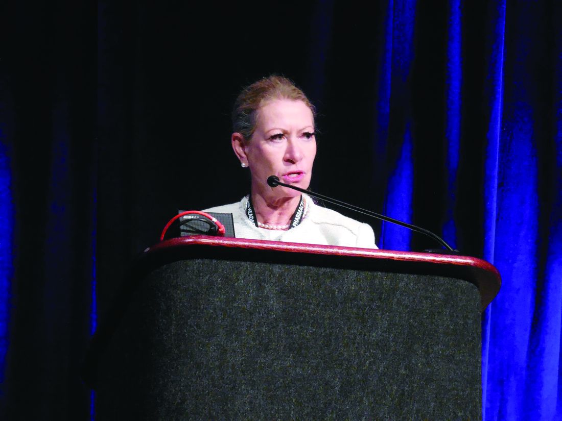
In the roughly one-quarter of all patients who had a clinical history consistent with triptan ineffectiveness, one 50-mg dose of ubrogepant led to pain freedom 2 hours after treatment in 16% of patients, compared with a response rate of 8% in placebo-treated patients. Ubrogepant’s effectiveness rate that was about the same as seen in patients with a history of triptan effectiveness, who were about 37% of the study population, Susan Hutchinson, MD, said at the annual meeting of the American Headache Society.
Among those with a history of triptan effectiveness, 20% were pain free after 2 hours, compared with 11% of placebo-treated controls, said Dr. Hutchinson, a family physician and headache specialist who practices in Irvine, Calif. Both of these between-group differences were statistically significant. The remaining patients included in the analysis had no triptan history, and among these patients a single, 50-mg dose of ubrogepant produced pain freedom at 2 hours in 24% of patients, versus an 18% response in placebo-treated controls, a difference that fell short of statistical significance.
The analysis Dr. Hutchinson reported came from data collected in two pivotal trials of ubrogepant for treatment of an acute migraine headache, the ACHIEVE I and ACHIEVE II trials, which together randomized more than 2,600 migraine patients eligible for the study’s modified intention-to-treat analysis, and 1,798 patients from that analysis who received a 50-mg dose of ubrogepant or placebo. In March 2019, the company developing ubrogepant, Allergan, announced that the Food and Drug Administration had accepted the company’s application for marketing approval of ubrogepant as a treatment for acute migraine largely based on data from ACHIEVE I and ACHIEVE II.
The researchers defined a history of triptan ineffectiveness as a patient who never used a triptan because of a warning, precaution, or contraindication, a patient who recently used triptans but did not achieve pain freedom within 2 hours more than half the time taking the drugs, or a patient who no longer used triptans because of adverse effects or lack of efficacy. Patients who had used triptans in the past and had complete pain relief within 2 hours more than half the time were deemed triptan-effective patients.
The analysis also looked at two other endpoints in addition to complete pain freedom: complete relief of the most-bothersome symptom of the migraine headache (photophobia for most patients), which resolved in 39% and 36% of the triptan-effective and triptan-ineffective patients, respectively, compared with placebo response rates of 27% and 23%; both between-group differences were statistically significant. A third measure of efficacy was some degree of pain relief after 2 hours, which occurred in 62% of patients in whom triptans were effective and 55% in those in whom triptans were ineffective, which were again statistically significant higher rates than among patients who received placebo.
Safety findings from the two ubrogepant pivotal trials showed good drug tolerability, with no treatment-related serious adverse events, and a profile of modest numbers of treatment-related and total adverse events similar to what occurred among patients who received placebo.
ACHIEVE I and II were funded by Allergan, the company developing ubrogepant. Dr. Hutchinson has been an adviser to and a speaker on behalf of Allergan and several other companies.
SOURCE: Blumenfeld AM. Headache. 2019 June;59[S1]:1-208, Abstract IOR02.
PHILADELPHIA – The oral, small molecule, calcitonin gene-related peptide receptor antagonist ubrogepant, currently under regulatory review for approval as a treatment for acute migraine headache, was as effective for migraine relief in patients with a history of triptan ineffectiveness as it was in patients for whom triptans had been effective, based on a post hoc analysis of data collected from 1,798 patients enrolled in two phase 3 trials.
In the roughly one-quarter of all patients who had a clinical history consistent with triptan ineffectiveness, one 50-mg dose of ubrogepant led to pain freedom 2 hours after treatment in 16% of patients, compared with a response rate of 8% in placebo-treated patients. Ubrogepant’s effectiveness rate that was about the same as seen in patients with a history of triptan effectiveness, who were about 37% of the study population, Susan Hutchinson, MD, said at the annual meeting of the American Headache Society.
Among those with a history of triptan effectiveness, 20% were pain free after 2 hours, compared with 11% of placebo-treated controls, said Dr. Hutchinson, a family physician and headache specialist who practices in Irvine, Calif. Both of these between-group differences were statistically significant. The remaining patients included in the analysis had no triptan history, and among these patients a single, 50-mg dose of ubrogepant produced pain freedom at 2 hours in 24% of patients, versus an 18% response in placebo-treated controls, a difference that fell short of statistical significance.
The analysis Dr. Hutchinson reported came from data collected in two pivotal trials of ubrogepant for treatment of an acute migraine headache, the ACHIEVE I and ACHIEVE II trials, which together randomized more than 2,600 migraine patients eligible for the study’s modified intention-to-treat analysis, and 1,798 patients from that analysis who received a 50-mg dose of ubrogepant or placebo. In March 2019, the company developing ubrogepant, Allergan, announced that the Food and Drug Administration had accepted the company’s application for marketing approval of ubrogepant as a treatment for acute migraine largely based on data from ACHIEVE I and ACHIEVE II.
The researchers defined a history of triptan ineffectiveness as a patient who never used a triptan because of a warning, precaution, or contraindication, a patient who recently used triptans but did not achieve pain freedom within 2 hours more than half the time taking the drugs, or a patient who no longer used triptans because of adverse effects or lack of efficacy. Patients who had used triptans in the past and had complete pain relief within 2 hours more than half the time were deemed triptan-effective patients.
The analysis also looked at two other endpoints in addition to complete pain freedom: complete relief of the most-bothersome symptom of the migraine headache (photophobia for most patients), which resolved in 39% and 36% of the triptan-effective and triptan-ineffective patients, respectively, compared with placebo response rates of 27% and 23%; both between-group differences were statistically significant. A third measure of efficacy was some degree of pain relief after 2 hours, which occurred in 62% of patients in whom triptans were effective and 55% in those in whom triptans were ineffective, which were again statistically significant higher rates than among patients who received placebo.
Safety findings from the two ubrogepant pivotal trials showed good drug tolerability, with no treatment-related serious adverse events, and a profile of modest numbers of treatment-related and total adverse events similar to what occurred among patients who received placebo.
ACHIEVE I and II were funded by Allergan, the company developing ubrogepant. Dr. Hutchinson has been an adviser to and a speaker on behalf of Allergan and several other companies.
SOURCE: Blumenfeld AM. Headache. 2019 June;59[S1]:1-208, Abstract IOR02.
REPORTING FROM AHS 2019
Can Remote Electrical Neuromodulation Help Acute Migraine?
Remote Electrical Neuromodulation (REN) may provide relief of migraine pain and most bothersome symptoms compared to placebo, a new study found. The efficacy and safety of a REN device for acute treatment of migraine was assessed. The randomized, double-blind, multicenter study was conducted at 7 sites in the United States and 5 sites in Israel. The study included 252 adults meeting the International Classification of Headache Disorders criteria for migraine with 2 to 8 migraine headaches per month, and the patients were randomized 1:1 to active or sham stimulation. Among the findings:
- Active stimulation was more effective than sham stimulation in achieving pain relief, pain-free, and MBS relief at 2 hours post-treatment.
- The pain relief of the active treatment was sustained 48 hours post-treatment.
- Incidence of device-related adverse events was low and similar between treatment groups.
Yarnitsky D, et al. Remote electrical neuromodulation (REN) relieves acute migraine: A randomized, double-blind, placebo-controlled, multicenter trial. [Published online ahead of print May 9, 2019]. Headache. doi: 10.1111/head.13551.
Multiple Cranial Nerve Blocks in Patients With Chronic Headache
Multiple cranial nerve blocks may provide an efficacious, well tolerated and reproducible transitional treatment for chronic headache disorders, a new study found. The uncontrolled, open-label study included 119 patients with chronic cluster headache, chronic migraine, short-lasting unilateral neuralgiform attack disorders, new daily persistent headaches, hemicrania continua and chronic paroxysmal hemicrania. All had failed to respond to greater occipital nerve blocks. Researchers found:
- Response rate for the entire cohort was 55.4%: Chronic cluster headache (69.2%), chronic migraine (49%), short-lasting unilateral neuralgiform attack disorders (56.3%), new daily persistent headache (10%), hemicrania continua (83.3%), and chronic paroxysmal hemicrania (25%).
- Time to benefit was between 0.5 and 33.58 hours.
- Benefit was maintained up to 4 weeks in over half of responders in all groups except chronic migraine and paroxysmal hemicrania.
Miller S, et al. Multiple cranial nerve blocks for the transitional treatment of chronic headaches. [Published online ahead of print May 13, 2019]. Cephalalgia. doi: 10.1177/0333102419848121.
Brainstem Raphe and Depression in Patients With Migraine
Transcranial sonography (TCS) signal alteration of brainstem raphe (BR) can be a biomarker for depression in migraine but is not associated with migraine headache itself, a new study found. Forty-two patients who had migraine without aura and 40 healthy controls were recruited for the study. Echogenicity of lentiform nuclei (LN), caudate nuclei (CN), substantia nigra (SN) and BR, width of the frontal horns of the lateral ventricles, and the third ventricle were assessed with TCS. Researchers found:
- There were no significant differences between migraineurs and controls in the width of front horn of the lateral ventricle, width of third ventricle, as well as in the echogenicity of SN, CN, LN and BR.
- More patients with migraine were detected with increased echogenicity of CN and LN compared with controls.
- Patients with hypoechogenic BR had significantly higher Hamilton Rating Scale for Depression (HAM-D) and Hospital Anxiety and Depression Scale depression subscale (HADS-D) scores than those with normal BR signal.
Tao WW, et al. Hypoechogenicity of brainstem raphe correlates with depression in migraine patients. [Published online ahead of print May 15, 2019]. J Headache Pain. doi: 10.1186/s10194-019-1011-2.
Periodontal Inflammation in Patients with Migraine
Periodontal inflammation is associated with increased circulating levels of calcitonin gene-related peptide (CGRP) in patients with chronic migraine, a new study found. The cohort included 102 chronic migraineurs and 77 age- and sex-matched individuals free of headache/migraine. Full-mouth periodontal parameters were recorded and the periodontal inflamed surface area (PISA) was calculated to quantify the periodontal inflammatory status for each participant. Researchers found:
- In the chronic migraine group, patients with periodontitis had greater levels of serum CGRP and IL-6, while nonsignificant differences were observed with IL-10 concentrations vs those without periodontitis.
- PISA was independently associated with CGRP in patients with chronic migraine.
Leira Y, et al. Periodontal inflammation is related to increased serum calcitonin gene-related peptide (CGRP) levels in patients with chronic migraine. [Published online ahead of print May 9, 2019]. J Periodontol. doi: 10.1002/JPER.19-0051.
Dry Eye Symptoms in Individuals with Migraine
Individuals with migraine demonstrated a different dry eye (DE) symptom, yet a similar DE sign profile when compared with those without migraine, a new study found. The prospective cross-sectional study of individuals with DE symptoms evaluated symptoms and signs of DE, including symptoms suggestive of nerve dysfunction. Among the details:
- Of 250 individuals, 31 met International Classification of Headache Disorders criteria for migraine based on a validated screen.
- Those with migraine were significantly younger and more likely to be female vs controls.
- Individuals with migraine had more severe DE symptoms and ocular pain vs controls.
- DE symptoms in those with migraine may be driven by nerve dysfunction as opposed to ocular surface abnormalities.
Farhangi M, et al. Individuals with migraine have a different dry eye symptom profile than individuals without migraine. [Published online ahead of print April 30, 2019]. Br J Opthalmol. doi: 10.1136/bjophthalmol-2018-313471.
Atypical Interactions of Cortical Networks in Chronic Migraine
Atypical Interactions of Cortical Networks in Chronic Migraine
The severity of headache is associated with opposite connectivity patterns in frontal executive and dorsal attentional networks in patients with chronic migraine, a new study found. Twenty patients with chronic migraine (CM) without preventive therapy, or acute medication overuse underwent 3T MRI scans and were compared to a group of 20 healthy controls (HC). Researchers used MRI to collect resting-state data in 3 selected networks, identified using group independent component analysis (ICA): the default mode network (DMN), the executive control network (ECN), and the dorsal attention system (DAS). They found:
- Compared to HC, patients with CM had significantly reduced functional connectivity between the DMN and the ECN.
- The DAS showed significantly stronger functional connectivity (FC) with the DMN and weaker FC with the ECN.
- The higher the severity of the headache, the increased strength of DAD connectivity, and the lower the strength of the ECN connectivity.
Coppola G, et al. Aberrant interactions of cortical networks in chronic migraine: A resting-state fMRI study. [Published online ahead of print May 28, 2019]. Neurology. doi: 10.1212/WNL.0000000000007577.
Static and Dynamic Functional Connectivity in Migraine
Resting-state functional imaging revealed static functional connectivity and dynamic functional connectivity differences between migraine and persistent post-traumatic headache for regions involved in pain processing, a new study found. The case-control study integrated the static functional connectivity and dynamic functional connectivity patterns of 59 a priori selected regions of interest involved in pain processing. Pairwise connectivity differences between migraine (n=33) and persistent post-traumatic headache (n=44) were determined and compared to healthy controls (n=36) with ANOVA and subsequent t-tests. Researchers found:
- Significant differences in static functional connectivity between migraine and persistent post-traumatic headache were found for 17 region pairs.
- Significant differences in dynamic functional connectivity between migraine and persistent post-traumatic headache were found for 10 region pairs.
- These differences in functional connectivity may be indicative of pathophysiology associated with migraine vs persistent post-traumatic headache.
Dumkrieger G, et al. Static and dynamic functional connectivity differences between migraine and persistent post-traumatic headache: A resting-state magnetic resonance imaging study. [Published online ahead of print May 1, 2019]. Cephalalgia. doi: 10.1177/0333102419847728.
Characteristics of Adolescents with Prolonged Headache
High levels of disability and low quality of life (QOL) were reported among children and adolescents with migraine utilizing infusion treatment, a new study found. Patients aged 6-19 years treated in an outpatient headache infusion center were included. A subset of these patients completed a behavioral health evaluation (treatment group) and were compared with a control group of similar age and gender to patients not seeking infusion treatment. Among the findings:
- 284 patients were included in the study (n=227 treatment group; n=57 controls).
- There was a promising difference in the Pediatric Pain Coping Inventory (PPCI) Distraction subscale, with a mean rank score of 61.90 for the treatment group vs 50.21 for the control group.
- There was a statistically significant difference on the Social Support subscale, with a mean rank score of 65.92 for the treatment group vs 46.26 for the control group.
- Patient-reported data also revealed a significantly higher level of disability among those seeking infusion treatment compared to the non-infusion group.
Woods K, et al. Psychosocial and demographic characteristics of children and adolescents with headache presenting for treatment in a headache infusion center. Headache. 2019;59(6):858-868. doi: 10.1111/head.13537.