User login
Take a Bite of the Big Apple at the VEITHsymposium
New York City is a global destination renowned for its unparalleled variety of things to do and see to match all tastes and interests. In your time between and after the VEITHsymposium sessions, you can enjoy a Broadway show, take in some of the city’s famous museums, go on a shopping spree, or dine at some of the best restaurants in the world. Whether with family or on your own, there is something for everyone.
For a quick look at current entertainment and dining options, pick up a copy of TimeOut New York or visit www.timeout.com/newyork, where you’ll find current listings for what is playing at theaters off and on Broadway as well as live music, special events, shopping, fine dining, and more.
For a more comprehensive listing of Broadway and off-Broadway shows, visit www.Broadway.com.
For the sports fan, you can find a complete online calendar listing of sporting events that are going during the VEITHsymposium (www.nycgo.com/things-to-do/events-in-nyc/sports-calendar). Pick up the New York Knicks basketball team playing the Detroit Pistons on Nov. 16 at Madison Square Garden. College basketball will also be featured at the Garden with the Champions Classic on Nov. 15.
If you’re a classical music enthusiast, you should make time to attend a performance at Lincoln Center (www.lincolncenter.org), which plays host to the New York Philharmonic and the Metropolitan Opera. During the VEITHsymposium, the operas “Aida,” “La Boheme,” and “Manon Lescaut” are all being performed.
And for the forever young crowd, Billy Joel is in concert at Madison Square Garden on Nov. 19.
No matter where your music tastes lie, for the most up-to-date accounting of musical performances in the city, pick up a copy of the free weekly newspaper, the Village Voice, or visit it online (www.villagevoice.com).
For your exercise pleasure and a literal change of pace, there are a number of interesting walking tours available, including ones that amble through the historic lower East Side, the Metropolitan Opera, and Broadway theaters (www.walksofnewyork.com).
The New York food scene is both vast and varied, so to choose your dining from fine to fun, you can visit the venerable New York Times website (www.nytimes.com/reviews/dining) to plan your eating experience by rating, price, neighborhood, and choice of cuisine.
You will find several notable museums and landmarks within a few blocks of the VEITHsymposium. The Museum of Modern Art is the closest to the New York Hilton Midtown, at 11 West 53rd St., between Fifth and Sixth Avenues. MoMA houses more than 150,000 paintings, sculptures, drawings, prints, photographs, architectural models and drawings, and design objects. The Metropolitan Museum of Art is a bit farther away (1000 Fifth Ave. at 82nd St.). The holdings include 2 million works spanning 5,000 years of art history from around the world from Egyptian mummies to Rembrandts and Picassos.
The American Museum of Natural History is on the Upper West Side of Central Park (at 79th St.) and has something for museum goers of all ages: dinosaurs, fossils, stuffed specimens, minerals, gems, and human cultural artifacts.
The memorial at the site of the World Trade Center towers stands witness to a national tragedy and to the spirit of the people of New York. The names of nearly 3,000 men, women, and children killed in the attacks of Sept. 11, 2001, and Feb. 26, 1993, are inscribed on the parapets surrounding the two memorial pools (www.911memorial.org/visit).
In addition the famous Central Park, don’t overlook the less-noted Bryant Park (between 40th and 42nd Streets and Fifth and Sixth Avenues), which offers an opportunity to do some holiday shopping at the annual Holiday Shops fair. This outdoor, European-style market features everything from handcrafted items to gourmet treats.
If you’re a first-time visitor, the Empire State Building, the Statue of Liberty, and Ellis Island remain the must-see symbols of New York. Be prepared for long lines to get to the top of the Empire State Building (www.esbnyc.com). Dress warmly for the ferry ride from Battery Park (www.statuecruises.com) to the Statue of Liberty.
Above all, while you’re at the VEITHsymposium, be sure that you and your guests enjoy some of the many pleasures that New York has to offer. It’s your kind of town.
Welcome to the 2016 VEITHsymposium
Welcome to the 43rd annual Vascular & Endovascular, Issues, Techniques and Horizons Symposium (VEITHsymposium). This year’s program promises to be one of the best, most comprehensive, and most thought-provoking of any of our meetings. This year we celebrate our 43rd anniversary and have introduced several improvements.
Nearly 600 international clinician/educators have gathered to provide attendees with the latest topics and advances that are important to the global vascular community. These data span the breadth of vascular diseases, diagnostic procedures, medical treatments, interventional procedures and open surgical advances for treating vascular disease. As is the hallmark of the VEITHsymposium, the 5-day program will run from dawn to dusk daily and will be fully captured in our online library.
Top vascular experts from around the world will provide updates on the latest clinical trials and offer insight into the real-life application of the most recent data to close the gap between the current state of knowledge and actual clinical practice.
Controversial issues will be approached from multiple perspectives to ensure a balanced, unbiased exposure of topics and to provide audience members with the information they need to make informed choices in their own practices.
This year our meeting continues its increased emphasis on venous disease. Three full days of the meeting are developments in venous disease of all sorts and active endovascular treatments in this rapidly expanding area of opportunity.
Some of the program’s other hot topics will be the continuing controversies surrounding parallel grafts (chimneys, and snorkel and sandwich grafts); multilayer open stents versus fenestrated and branched endografts; new developments in carotid stenting; new developments in the treatment of aortic dissections; a day devoted to the management of arteriovenous malformations (AVMs); new developments in the endovascular treatment of lower-extremity ischemia, particularly below the knee; the latest developments in EVAR and TEVAR including experiences with a plethora of new endovascular grafts and devices that have appeared on the scene in the last year; and improvements in the medical treatment of vascular disease and vascular patients undergoing surgery and other interventions. Important issues to vascular specialists and outpatient vascular treatment will also be highlighted.
This year’s program will include a special session all-day Tuesday, focused in the morning on management options for pulmonary embolism led by Dr. Michael R. Jaff. The afternoon part of the day will focus on new developments in the management of acute and chronic large vein occlusion, and will be led by Dr. Kenneth Ouriel.
This year there will also be sessions devoted to crucial issues for vascular specialists including changing relationships with government and the FDA and how to survive under new reimbursement rules and regulations including Obamacare. Our physician/educators will also offer a glimpse into some new techniques and technologies that have been available overseas, but are just gaining approval in the United States, such as drug-eluting balloons and stents.
Attendees will notice some other exciting changes or additions to this year’s program. We have included a new Job Fair Program on Friday in the Americas Hall 1 on the 3rd floor. In addition, there will be more breaks in the schedule to encourage exploration of state-of-the-art technology, products, and services available in the Exhibit areas and Pavilions. The Exhibit Halls are crowded with displays and booths of particular interest to vascular surgeons and vascular specialists. The Pavilions and Exhibits also offer attendees the chance to meet faculty and to network with other attendees and industry partners. This is one place to learn more about exciting new technologies and developments in our field.
Other new additions to our meeting this year will be an exciting Abbott Pavilion in the Americas Hall as well as an expanded Innovations and Investment Summit which facilitates interaction between innovators, industry and investors. This non-CME Session will be held from 8 AM to 3 PM on Thursday, November 17th in the Gramercy Suites on the 2nd floor, and will be led by Kenneth Ouriel, Jean Bismuth and Chris Cheng.
Also new this year will be an expanded VEITHsymposium mobile app, provided courtesy of Cook Medical. Download the app for your iPhone, iPad or Android phone or tablet! Search the App Store (iPhone/iPad) or Google Play Store (Android) for “VEITHsymposium 2016” and install the app. You will be able to access the complete program, create your personal program, add your notes, view the location of sessions and exhibitors on the floor plan, and much more. After you have installed the app and opened it for the first time, you can continue to use it offline. To receive the latest updates and announcements, you will need to be connected to the internet.
In addition, there will be expanded Associate Faculty programs which will give younger and less well-known vascular specialists the opportunity to present their work at the podium with leading experts as session moderators.
Again this year, an Online Library will be available for a minimal fee of $75 for clinical meeting attendees and will include access to talks, slides, videos, and panels from the meeting. This Library will enable all attendees to see and hear key presentations they may miss because of the concurrent sessions or other reasons. This library will be available 10-14 days after the meeting. Attendees should note in their program talks they wish to hear but could not, and then revisit the missed talks on the Online Library which is indexed exactly to the program. The talks are also indexed in the Library by presenter, topic, or session. This Library is a great resource for study, research or review for any purpose.
On behalf of all the meeting Co-Chairmen and our entire staff, we greatly appreciate you coming to our meeting. We hope it is our best meeting ever and that you find it educational, most useful and exciting so that you return next year.
Homans Lecture: Celebrating the past and looking to the future
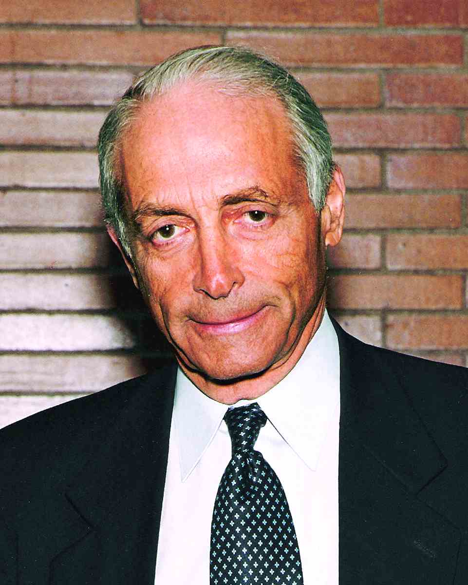
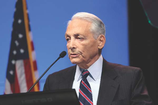
NATIONAL HARBOR, MD – “Specialties are like species,” said Frank J. Veith, MD, “they must evolve or go extinct.”
Dr. Veith of the New York University Langone Medical Center made this comparison in his 2016 Homans Lecture on the topic of “The future of vascular surgery,” at this year’s annual meeting hosted by the Society for Vascular Surgery.
Dr. Veith reviewed the history of vascular surgery, touched on its present status, and speculated on its potentially bright future. The vascular specialty has evolved dramatically over the past decades, especially in the area of embracing the endovascular revolution, said Dr. Veith, with that revolution putting vascular surgery at the forefront of research to develop new techniques.
His witnessing such innovations as those developed by Dr. Juan Parodi, and being a part of the early history of endovascular surgery, convinced Dr. Veith of its long-term importance to the development and survival of the specialty.
In his 1996 SVS Presidential Address, he predicted that 40%-70% of the open operations being done then would be replaced by endovascular procedures. “Accordingly, to survive, I recommended that vascular surgeons become endocompetent, learn how to do these procedures, and embrace them.” Dr. Veith added that, although his recommendation was not greeted with open arms by everyone, endovascular techniques moved forward.
In fact, “vascular surgeons often lead in developing many evolving endovascular procedures that are currently the standard of care,” he said.
Dr. Veith pointed out that a wide variety of conditions are now amenable to endovascular treatment, although some, including carotid disease, remain controversial. He listed examples of those conditions that he felt were still best treated with open surgery: thoracic outlet and entrapment syndromes, some ascending aorta and arch lesions, a few rare aneurysms not suited for endovascular treatment, some Takayasu’s lesions, some congenital and genetic aortic and renal artery lesions, some infected arteries and arterial grafts, a rare recurrent or complex lower-extremity lesion, some carotid lesions, and some failed endovascular treatments.
“Our specialty has embraced the endovascular revolution and become endocompetent,” he said. “It is why vascular surgery is doing as well as it is today.” He added. “Vascular surgery is presently an exciting, vibrant specialty in the United States.”
Dr. Veith noted, “Well-trained vascular surgeons are the only ones who can provide the most appropriate, full spectrum of care for patients with vascular disease, outside the head and the heart – whether that treatment be medical, endovascular, or open. There are abundant numbers of patients who require our skills. In addition, we use fascinating technology and have good industry relationships. And finally, many patients regard their vascular surgeon as a key doctor who they see regularly. As a result of these advantages, many bright medical students and general surgery residents are choosing to train as vascular surgeons. Vascular surgery should be flourishing.”
However, despite the fact vascular surgery is an exciting and vibrant specialty, and the best for treating vascular disease outside of the heart and the brain, the vascular specialty has significant problems competing with other specialties, he said.
He blamed in part the size and structure of the specialty, in particular with regard to its competition.
“Vascular surgery competes, as it always has, with general and cardiac surgeons. However, general surgeons have become less competitive, but cardiac surgeons have become more in need of work, and thus more active beyond the heart and thoracic aorta – as their open operations are replaced by coronary stents and transcatheter valves. More importantly, as vascular treatments become increasingly endovascular, vascular surgery will be competing with interventional radiology and, importantly, interventional cardiology.”
He outlined a number of major challenges these other disciplines create, in part, because of the DRG/RVU/dollar orientation of institutions, and the fact that most institutions still consider vascular surgery a subspecialty of general or cardiac surgery, or a subordinate part of a Heart & Vascular Center, with administrative control of these centers rarely in the hands of vascular surgeons. Moreover, when institutional resources – like angiography suites or hybrid operating rooms are distributed, the interests of vascular surgery are often represented by a general or cardiac surgeon – or worse a cardiologist,” he added.
He stated that these conditions limit vascular surgery’s ability to get its fair share of institutional resources.
“The competitive playing field is not level, and vascular surgeons are disadvantaged in the Darwinian struggle to survive,” he stated.
“To survive, vascular surgery needs to unify, recognize this inequity, and fix it. This can only be done if all vascular surgeons engage vigorously in this issue. We need equal administrative status with cardiac and general surgery in our institutions,” Dr. Veith advised.
In discussing the technological future, Dr. Veith said that by 2026, 75%-95% of all vascular cases requiring more than medical therapy will be treated endovascularly, with perhaps 5% in a hybrid fashion (open plus endovascular), and between 5% and 15% being treated fully by open surgery. This shift away from open surgery is and will continue to cause challenges in training and patient access to open treatment.
He asked the question: How should vascular surgery deal with the decreasing numbers of complex open procedures and who should do them?
“One solution is to have centers to which these patients are sent and in which vascular surgeons seeking this skill can get adequate open training,” he answered.
But the technological future he painted was bright. Not only was the future likely to be filled with new advances in medical therapy, but he also highlighted computer-assisted 3-D–device navigational tools to aid endovascular treatment; advances in robotic guidance to decrease radiation exposure and facilitate device placement; computer-enhanced simulation to improve training and, when patient specific, to allow procedure planning and rehearsal; and even 3-D printed modeling of lesions and blood vessels.
He predicted that the endovascular problems of intimal hyperplasia will be overcome by antiproliferative drugs in all vascular beds – once the best way of getting the best drug to the proper location is found – and that computer-enabled remote monitoring of flows within grafts and stents, perhaps using miniaturized piezoelectric sensors, will allow corrective treatment before occlusion occurs.
Dr. Veith stated that, in his view, to take its proper place, vascular surgery should rise above its subspecialty status in the shadow of general surgery and in its competition with cardiology.
This “will help vascular surgery to flourish and be recognized as the main specialty devoted to patients with noncardiac vascular diseases. Vascular surgery can then fulfill its potential for a brighter future. More importantly, patients and society will be the ultimate beneficiaries,” he concluded.
Dr. Veith reported that he had no conflicts to disclose with regard to his remarks.
On Twitter @VascularTweets
AT THE 2016 VASCULAR ANNUAL MEETING
Aortic Session to Focus on Evolving Treatments
With treatments evolving rapidly for aortic diseases, best approaches and optimal use of technology will be the focus of “New Developments in Diseases of the Aorta and their Treatment,” on Saturday afternoon.
“As we keep pace with developments in stent graft technology, it is critical to also hone our understanding of aortic aneurysm pathology and make sure the technology fits the disease and not vice versa,” said co-moderator Dr. Thomas S. Maldonado, chief of vascular surgery at Manhattan VA Hospital, New York University Medical Center, New York. “The session features a number of interesting talks on aortic and iliac artery morphology as it relates to EVAR.”
The session will be co-moderated by Dr. Nicholas J.W. Cheshire, chief of vascular surgery for Aortic Centre, Royal Brompton Hospital in London.
Dr. Maldonado said attendees will learn about the role of preoperative neck thrombus and aortic sac morphology in planning endovascular repair as well as innovative solutions for complex iliac anatomy during EVAR. The session will also address the critical topic of EVAR for treatment of aortic aneurysm rupture and aortic trauma.
“Despite enthusiasm for endovascular management of ruptured AAA and aortic trauma, many centers lack cohesive protocols and strategies,” Dr. Maldonado said. “Experts will share their paradigms and tips and tricks for management of emergent EVAR for rupture and trauma.”
The session will be broken into two primary themes. The first half will address aortic aneurysm morphology and preoperative assessment of anatomy with the goal of achieving optimal endovascular repair, Dr. Maldonado said. The second half of the session will focus on emergent or urgent repair of aortic morphology, specifically aortic aneurysm rupture and blunt abdominal aortic trauma.
“This is an area that is hotly contested because traditionally, we’ve all been taught that ruptured aneurysm should be treated with open repair as expeditiously as possible,” he said. “The idea that perhaps endovascular repair – especially when there may be some need for preoperative imaging that might delay repair – is why there is some controversy as to whether endovasular repair has superiority over open repair. I think there is a lot of valuable insight in some of the talks in this session relating to” these issues.
At the start of the session, Dr. Maldonado will discuss quantitation of aortic neck and sac thrombosis and whether neck thrombosis is a risk factor for unsuccessful EVAR. Next, Dr. Benjamin M. Jackson of the Hospital of the University of Pennsylvania, Philadelphia, will address whether saccular AAAs should be treated differently from other AAAs and size criteria physicians should consider. Dr. Barend M.E. Mees of Maastricht University Medical Center, the Netherlands, will then conduct a presentation on back table reversed limb endografts (zenith and endurant) and technique/indications pertaining to treating challenging aorto-lliac pathology. Next up, Dr. Irwin V. Mohan of Westmead Hospital in Sydney, Australia, will discuss the lack of seasonality for ruptured AAAs. Presenters Dr. Zoran Rancic, Dr. Dieter O. Mayer, and Dr. Mario L. Lachat, all of the Clinic for Cardiovascular Surgery, University Hospital Zürich, Switzerland, will then host a dedicated workshop focused on improving treatment of ruptured AAAs. Finally, Dr. Zachary M. Arthurs of San Antonio Military Medical Center, San Antonio, Texas, will address blunt abdominal aortic injury, including incidence, etiology, diagnosis and treatment.
Session 108: New Developments in Diseases of the Aorta and their Treatment
Saturday, 3:05 p.m. – 3:35 p.m.
Grand Ballroom East, 3rd Floor
Coordinated Care is Key to Good Post-Intervention Outcomes
While drugs and other medical technologies might be effective in treating cardiovascular issues, a potentially overlooked component is the regular interaction a patient has with his primary care physician, according to Dr. Philip Goodney, who will be discussing research findings during his Saturday presentation, “Why Your Patient’s Primary Care Physician and Internist Are as Responsible as You for the Outcomes of Your Lower Extremity Revascularization.”
“We’ve had some convincing data, whether it’s in the care of diabetes or in the care of high cholesterol or the overall management of cardiovascular disease, that our interventions, whether it’s an operation, whether it’s a stent or combination thereof, tend to last longer and our patients tend to have fewer complications if they are followed closely by a primary care physician,” said Dr. Goodney, associate professor at Dartmouth-Hitchcock Medical Center in Lebanon, N.H..
He said that simple things, such as having a primary care physician ensuring that hemoglobin A1C is regularly checked and monitored, statins as needed are provided when indicated, and regular visits are conducted to monitor chronic illness are occurring can go a long way to improve outcomes.
“While there’s not been good data to support this before, many have thought that the results of our operations tend to be better if patients have primary care physicians that follow them closely,” Dr. Goodney said.
With the support of the Society for Vascular Surgery and the National Instutites of Health, research was undertaken to see if there was evidence to support this notion that better primary care monitoring and follow-up leads to better patient outcomes.
“We did a series of analyses, both within the Vascular Quality Initiative, which is a large quality improvement dataset that is supported by the Society for Vascular Surgery, as well large analyses of patients in Medicare, and found that patients who are commonly seen by their primary care physicians were less likely to have complications after major thoracic aneurysm surgery, were less likely to have complications after procedures to help their lower extremities, and were also more likely to have better results from vascular care,” Dr. Goodney said.
“We thought this was pretty compelling evidence that vascular surgeons should really have, as partners, primary care physicians who actively help manage these complex patients,” he continued. “An effective partnership should translate into better results.”
The research generally showed that rates of complication were often 20%-30% lower in patients who had good care coordination, he said, something that could have the ancillary benefit of lowering health care costs overall.
“I think it speaks to what is sort of low-hanging fruit, if you will,” Dr. Goodney said. “We spend a lot of time in cardiovascular care looking at advanced devices and the latest stent or the latest drug-coated balloon. That might be a pretty expensive way to get better results. But I think what our data show is the answer might be right under our noses and it might be a little simpler than we think.”
He acknowledged that while the solution might be straightforward, “getting care coordination is certainly a challenge. However, I think what our data suggest is that there’s really a bit of a pot of gold at the end of the rainbow if we can make sure these patients get the coordinated care they need.”
Session 102: Why Your Patient’s Primary Care Physician and Internist Are as Responsible as You for the Outcome of Your Lower Extremity Revascularization
Saturday 6:45 a.m. - 6:50 a.m.
Grand Ballroom East, 3rd Floor
Innovations to Prevent Spinal Cord Ischemia
Ischemic spinal cord injury remains one of the most dreaded complications of open and endovascular complex aortic aneurysm repair, with affected patients facing significantly worse survival rates than those who escape this grave outcome. But recent work is advancing efforts to prevent SCI, as experts will discuss Friday morning in “More About Complex Aneurysm Treatment and Spinal Ischemia.”
We know much more about preventing SCI than we did a decade ago, according to moderator Dr. Gustavo Oderich, who is a professor of surgery at Mayo Clinic Medical College in Rochester, Minn. For example, work by Dr. Randall Griepp and colleagues at Mount Sinai Hospital, New York, has shown that spinal cord perfusion can adapt through an extensive network of collateral vessels, and that SCI rates drop when surgeons use staged aortic coverage to promote collateral enlargement and recruitment, Dr. Oderich said. “The pathophysiology of SCI includes hemodynamic compromise or embolization, which has been increasingly recognized as an important factor during complex aortic repair,” he added. “It is clear that we have a better understanding of the etiology of SCI and possible preventive measures, but that the complication can’t be avoided in every patient.”
The session will be co-moderated by Dr. Christian Etz, a professor at Leipzig University and an attending surgeon at Leipzig Heart Center in Germany. Attendees will learn how staged repair can reduce the risk of permanent SCI, paraplegia, and mortality after endovascular thoraco-abdominal repair. Staging can involve a number of strategies, including proximal thoracic coverage with stent-graft, perfusion branches, or incomplete endovascular repair by leaving one of the side branches or the contralateral iliac limb unstented, Dr. Oderich noted. Dr. Etz also will discuss his group’s alternative, which is to preemptively perform ischemic preconditioning through coil embolization of intercostal arteries.
“We termed this approach ‘minimally invasive segmentaI artery coil embolization’ (MISACE), and published our first-in-human-experience with it recently,” said Dr. Etz. “I believe this solution has the potential to eliminate SCI or permanent paraplegia once and for all in the next five to 10 years.”
Other experts will describe how to optimize patient selection – a key step in preventing SCI, said Dr. Oderich. “Endovascular procedures should likely be avoided in patients with ‘shaggy aorta’ – that is, extensive atherosclerotic debris – and poor collateral networks to the spine,” he added. Surgeons also should confirm that the patient has adequate iliofemoral access to avoid intraoperative complications and to prevent prolonged pelvic and lower limb ischemia, he said.
There also have been advances in routine protocols to prevent SCI during endovascular repair of thoracoabdominal aortic aneurysms. While a few investigators still debate some of these protocols, most groups now support routine cerebrospinal fluid drainage, permissive hypertension and mean arterial pressure augmentation, revascularization of the left subclavian and internal iliac arteries when indicated, aggressive transfusion in the early postoperative period, and prevention of systemic hypotension, said Dr. Oderich.
Presenters also will discuss preserving internal iliac and vertebral artery flow, which has been shown to help reduce rates of SCI and to increase the chances of recovery when patients do sustain injury. In addition, Dr. Michael Jacobs will discuss intraoperative monitoring of motor and somatosensory evoked potentials, which can help clinicians immediately recognize drops in perfusion of the spine and lower extremities and respond through such maneuvers as increasing MAP, decreasing CSF pressure, early limb reperfusion, and incomplete endovascular repair.
Attendees also will hear Dr. Stephan Haulon, of the Université de Lille and CHRU Lille, France, discuss how to achieve early pelvic and lower limb reperfusion by immediately restoring flow after placing a distal bifurcated component stent graft and iliac limbs. “This has been shown by Haulon and colleagues to improve outcomes of endovascular TAAA repair,” Dr. Oderich said. “The Mayo Clinic group has also recently reported on their protocol to use standardized maneuvers during the repair to prevent SCI.” In the next decade, investigators will continue honing and developing techniques to optimize spinal cord perfusion by recruiting and adapting the collateral vasculature, he added.
Dr. Etz added “This unique session brings together all the world-renowned experts in the field for the very first time. Attendees will really get a completely new understanding of this very complex matter, and will ultimately learn how to prevent SCI in their own patients.”
Session 78: More About Complex Aneurysm Treatment and Spinal Cord Ischemia
8:00 a.m. – 9:07 a.m.
Grand Ballroom East, 3rd Floor
Hybrid ORs and Imaging Techniques: Advances in Minimally Invasive Procedures
Hybrid operating rooms are constructed and equipped for both open and minimally invasive surgery, enabling vascular surgeons to perform highly complex procedures while potentially improving accuracy and efficiency. On Friday morning, experts will discuss these increasingly popular blended surgical theaters, the state-of-the-art imaging technologies they incorporate, and ways to reduce associated radiation exposure in “New Developments in Hybrid Operating Suites and Improvements in Imaging.”
“Endovascular techniques have transformed our practice for the better, but have raised new challenges. We’re treating more and more challenging anatomies by endovascular means, with the potential for exposure to higher doses of ionizing radiation for staff and patients, higher contrast volume dose for patients, and the chances of complications,” said moderator Dr. Frans L. Moll, a professor of vascular surgery and head of the department of surgery at University Medical Center Utrecht in the Netherlands.
Fortunately, technological and technical advances are helping lessen these safety concerns, Dr. Moll said. For example, clinicians are incorporating pre-acquired diagnostic imaging into image fusion techniques to support catheter navigation, and are starting to use x-ray equipment that is designed for the OR environment and that employs lower radiation dose protocols than standard machines. Studies show that these novel x-ray imaging technologies can significantly decrease radiation exposure of both patients and staff who work in hybrid ORs, without reductions in procedure or fluoroscopy time.
The session will be co-moderated by Dr. Daniel Clair, professor of surgery at Cleveland Clinic Lerner College of Medicine, Case Western University. Attendees will learn about the expanding role of advanced imaging in hybrid procedures, said Dr. Moll. Notably, three-dimensional imaging and image fusion techniques are enabling endovascular surgeons to “see what one could see in open surgery,” he added. “These advancements will give physicians more insight and confidence in the treatment of complex anatomy in a minimally invasive setting.”
At the beginning of the session, Dr. Neal Cayne, of NYU Langone Medical Center, New York City, and Dr. Alan Lumsden, of Houston Methodist, Houston, Texas, will discuss how fusion can reduce exposure to radiation and contrast, and how advances in fusion techniques can prevent registration errors that result from anatomical distortions caused by wires and sheaths. Dr. Thomas Carrell, of King’s College, London, will then describe a new automated three-dimensional fusion overlay system that works with both fixed and mobile digital fluoroscopes and re-registers in response to movement of the patient or OR table.
Despite rising demand for hybrid ORs, “questions remain about the need for these technologies in everyday practice,” said Dr. Clair. Experts also are debating the best use of interventional robotic technology and CT imaging during interventions, and the optimal use of these technologies for image registration. Nonetheless, advances in hybrid room imaging are cutting radiation exposure and operative time and improving outcomes, said Dr. Clair. “Over time, the use of these technologies will be invaluable to clinicians,” he added.
Next, Dr. Lieven Maene, of OLV Hospital Aalst, Belgium, will offer “tips and tricks” for optimizing the hybrid OR to reduce radiation dose and protect patient’s kidneys, followed by an update by Dr. Timothy Resch, of Skåne University Hospital in Malmö, Sweden, on how rotation angio-CT can improve endovascular procedures compared with standard digital subtraction angiography and pressure measurements.
To close the session, Dr. Stephan Haulon, of CHRU de Lille, France, will discuss new assets for hybrid suites, and Dr. Rodney White, of the UCLA School of Medicine and Harbor-UCLA Medican Center, Los Angeles, will recommend ways to train staff in hybrid ORs, particularly when using equipment during complex aortic procedures.
“Ongoing improvements in technology and methods should help us overcome the challenges” of increased radiation and contrast exposure, Dr. Moll said. He looks forward in the next decade to continued advances in ‘”clean technologies, as I call them – low-radiation or even radiation-free technologies to help us do our job in a safer way for patients and staff. And further progress in three-dimensional imaging will help us treat patients with challenging anatomies even better.”
Session 87: New Developments in Hybrid Operating Suites and Improvements in Imaging
Friday, 9:37 a.m. – 11:11 a.m.
Grand Ballroom West, 3rd Floor
New Coil Technology Gets the Job Done
Technologies to be discussed during Thursday’s session “New Devices and Concepts for Embolization, Thrombectomy and Thrombolysis,” are the newest, most advanced, and most promising in a field and will give the audience practical information for implementing these innovations in clinical practice.
Devices used during these procedures “were archaic for a while,” said session moderator Dr. Nicholas Morrissey, a vascular surgeon at The New York Presbyterian Hospital and associate professor of surgery at Columbia University College of Physicians and Surgeons, both in New York. “We relied on old techniques. Recently, some newer devices have launched us into an era where things can be done a lot quicker and safer.
“It’s a great session because it is going to expose people to technologies that are much more advanced than they realized,” he said, “but simple enough that people will leave that session and be able to treat their patients the next day differently and more effectively.”
The program, which will be co-moderated by Dr. Christopher Kwolek, director of vascular and endovascular surgery at Massachusetts General Hospital and associate professor of surgery at Harvard Medical School in Boston, begins with two presentations about Penumbra’s new peripheral occlusion device (POD) coils. Dr. Frank Arko of the Sanger Heart and Vascular Institute of Charlotte, N.C., will discuss the technical aspects and clinical experience with these coils, and Dr. Claudio Schonholz of the Medical University of South Carolina Heart & Vascular Center in Charleston will discuss experience with POD coil embolization in the aneurysm coiling efficiency (ACE) multicenter study.
“This coil has an extra little wire at the end that allows you to anchor it into a blood vessel,” Dr. Morrissey said. Unlike previous types of coils that had to be injected via catheter, devices such as the POD coil “offer more of a controlled release,” he said. “You can pack them in a little tighter and only release them when you’re sure they’re in the right place. That eliminates the risk of them breaking off and going down to the wrong artery. It will probably help shorten operating room times and improve outcomes when we’re trying to embolize something therapeutically, which is something we’re doing more and more these days.”
The session also features three talks about the Indigo Catheter Thrombectomy System, which will look at a novel way to remove clots from medium-sized and small arteries and discuss results from the PRISM trial; advantages of the device in treating ALI; and how the system reduces the need for thrombolytics and decreases the risk and cost of treatment for ALI.
“This technique, originally used in clearing clots to the brain, suctions out the clot,” Dr. Morrissey said. “It’s a very effective tool for medium-sized blood vessels. You can use the device to clear out pathways without having to give large amounts of aggressive clot-busting medications, so you minimize the risk of bleeding. You also can get it done through a very small sheath without having to make an incision in the patient, and can get it done quickly.”
Also included in the lineup are presentations about technical tips and tricks to help manage bleeding associated with catheter-directed thrombolysis and for when thrombosis complicates retrograde access for complex lower extremity interventions. The afternoon will conclude with a talk by Dr. Martin Bjorck of University Hospital in Uppsala, Sweden, suggesting that there is no advantage to giving heparin with intra-arterial thrombolysis.
“We’re excited about these technologies: They’re taking what we’ve done now for a long time and bringing it to a more sophisticated level,” Dr. Morrissey said. “Our session highlights two important messages. First, if you have a technology that gets the job done but is cumbersome and a little bit dangerous, taking it to the next level can improve its safety profile and its efficacy, allowing the technology to be brought to patients who may not have been able to have it before. As a result, you can potentially decrease OR times, increasing patient safety and decreasing costs to the health care system. Fine tuning the technology to modernize it is going to make a tremendous difference.
“In addition, by doing good quality studies and investigating the status quo, you may actually find something that’s less invasive and less risky, and can achieve the same effect without exposing patients to the same risk.”
Session 62: New Devices and Concepts for Embolization, Thrombectomy, and Thrombolysis
Thursday, 4:48 p.m. – 5:44 p.m.
Grand Ballroom West, 3rd Floor
The Rise of the Machines: Vascular Robotics and Guidance Systems
Efforts to develop robotically assisted laparoscopic techniques have had limited success, and some notable failures. At the same time, advances in robotic catheterization systems have spurred interest in their use during endovascular surgery. On Thursday afternoon, Dr. Hans Coveliers of VU Medical Center in Amsterdam, the Netherlands, will join other experts in reviewing the state of vascular robotics and future developments in the field.
Dr. Coveliers calls laparoscopic robotic aortic surgery “a failed experiment,” adding that robotically assisted laparoscopic repair of aortic aneurysms is no more effective than manual laparoscopic techniques. In one study, use of a robotic system cut down on suturing time during aortic anastomosis, but the complexity of the device significantly lengthened total operating times, and robotically treated patients fared no better than those who underwent a manual, fully laparoscopic approach.
Other vascular surgeons also have reported steep learning curves and longer operating times because of the intricacy of these devices, and these drawbacks have eroded their potential cost savings, according to health technology assessments.
Safety is also a concern. “In particular, robot-assisted laparoscopic surgery for aortic aneurysmal disease has shown some catastrophic cases,” Dr. Coveliers said. There have been reports of laparoscopic robotic aortic repairs that had to be converted because of bleeding from the anastomosis. And in a recent review of 12 years of safety reports from the Food and Drug Administration, LexisNexis, and PACER, investigators linked the controversial da Vinci robotic surgical system to 245 adverse events and 71 deaths.
“All cases that are suitable for robotic vascular surgery can be treated with simple endovascular techniques in a faster, safer, and thus better way,” Dr. Coveliers emphasized. As a result, vascular surgeons are shifting their focus from robot-aided arterial anastomosis to robotically assisted endoluminal vessel cannulation, he said. “This is more in line with the current developments in endovascular surgery, where advanced catheterization skills are required for vessel cannulation in cases of complex endovascular aneurysm repair,” he added.
Because robotic systems offer better steerability over manual tracking systems or conventional cannulation techniques, they can potentially simplify treatment of complex EVAR cases, according to Dr. Coveliers. Studies have reported that robotic endovascular surgery cut total procedure time by a factor of six, shaved about 12 minutes off fluoroscopic exposure time, and reduced the number of movements required in robotic surgery by up to 10 times. A review of these studies concluded that robotic endovascular systems offer better catheter stability, a shorter learning curve, and better performance while cannulating tortuous vessels, compared with conventional endovascular techniques.
But it remains unclear whether current robotic systems can consistently streamline EVARs and benefit patients. Studies of phantom and animal models have posted strong results, but more clinical trials in humans are needed, according to reviewers.
Earlier this year, for example, reviewers scrutinized 18 studies of four robotic navigation systems and concluded that only one system potentially outperformed manual cannulation during complex vascular cannulation tasks, and only when performed in a rigid experimental environment. Average tracking accuracy was 1 mm under those controlled conditions, but target registration error exceeded 3 mm when measured in a non-rigid environment. The reviewers concluded that it was unclear if current systems save time and materials while improving clinical outcomes in complex EVAR.
But such proof is just around the corner, according to Dr. Coveliers. “In the future,” he said, “manual steering of catheters and guide wires for cannulation of any target vessel will be obsolete, and will be replaced by robotic navigation systems that reduce fluoroscopy time, radiation dose and contrast volumes to a minimum.”
Session 53: Vascular Robotics and Guidance Systems
3:52 p.m. – 3:57 p.m.
Grand Ballroom East, 3rd Floor
Venous Disease: Use the Right Scoring Tool to Choose the Right Treatment
Endovenous interventions have revolutionized the treatment of venous disease, enabling many more physicians to effectively treat many more patients. But there’s a flip side to this valuable coin – one that will be explored in Thursday’s session “Venous Clinical Examination and Hemodynamics.”
“Since the advent of the endovenous revolution, the treatment of venous disease has become popular,” said Dr. Jose Almeida, who will moderate the session with Dr. Lowell S. Kabnick. “But many physicians have only one tool in the toolbox – saphenous vein ablation. They have little understanding of the underlying pathophysiology, and therefore almost reflexively treat all patients whom they see with saphenous vein ablation. Physicians from disparate specialties are now treating venous disease; therefore, we are seeing great discontinuity in the care being rendered.”
The goal of the session, said Dr. Almeida, medical director of the Miami Vein Center, is to get more physicians observing the guidelines put forth from the American Venous Forum and Society for Vascular Surgery.
He will discuss how to frame a treatment plan using two clinical assessment scores, both created by the American Venous Forum: CEAP and the Venous Clinical Severity Score (VCSS). The CEAP is a purely clinical measure, taking into account clinical manifestation, etiology, anatomy, and pathophysiologic dysfunction. The VCSS, on the other hand, describes the degree to which both the disease and its treatment affect the patient’s daily life. Given at baseline and after treatment, the VCSS focuses on the patient’s perspective of how treatment improved those symptoms.
Dr. Kabnick will discuss a new outcomes assessment tool in development – the Varicose Vein Symptom Quick (VVSymQ) score. VVSymQ is a patient-reported electronic diary of five items that assess symptoms of varicose veins (heaviness, achiness, swelling, throbbing and itching). In practice settings, patients are asked to complete the entry daily for 10 days before and after surgery, Dr. Kabnick said.
All of these tools are meaningful ways to understand how venous insufficiency affects a patient, said Dr. Kabnick of New York Langone Medical Center. “One score allows us to understand disease severity, and the others let us see if we really helped the patient with our treatment.”
Dr. Neil Khilnani of Weill Cornell Medical Center, N.Y., will examine the use of duplex ultrasound mapping to identify venous reflux pathways. Dr. Andrew Nicolaides of Imperial College, London, will touch on another imaging modality –- air plethysmography. This is most often employed in patients who have more advanced stages of venous insufficiency. It can help clarify the ultrasound exam when venous reflux or outflow obstruction are suspected but not clearly visualized.
“Air plethysmography is a research tool right now,” Dr. Kabnick said. “But we would like to see it adopted into mainstream testing.”
Dr. Raghu Kolluri, director of vascular medicine at Riverside Methodist Hospital, Columbus,Ohio, will discuss the symptoms of venous disease.
Dr. Seshadri Raju of the Rane Center, Jackson, Miss., will tackle the biggest controversy in venous insufficiency right now – the debate over where pathophysiology begins.
“This controversy really lies in the hemodynamics,” Dr. Kabnick said. The more traditional theory of descending pathology considers dilation and backward flow in the saphenous vein as the driving force behind varicosities. An emerging school of thought, however, suggests an ascending evolution of disease, which begins in the smallest peripheral veins and progresses upward into the saphenous vein. This creates a backward flow that would be initially reversible.
“It will be interesting for the attendees to be introduced to these philosophies,”
Dr. Kabnick said. “In the United States, we generally believe venous insufficiency is a combination of ascending and descending pathophysiology, and we mostly try to treat what we think of as the underlying problem, which is a truncal vein.”
Outside the United States, two other techniques are often employed: ASVAL (ambulatory selective varices ablation under local anesthesia) and CHIVA (conservative hemodynamic treatment for chronic venous insufficiency). Both of these preserve the saphenous vein by targeting collateral veins for intervention (sclerotherapy, ligation, or removal). Dr. Sylvain Chastanet of the Riviera Vein Institute, Nice, France, will discuss the ASVAL technique, and Dr. Paolo Zamboni of the University of Ferrara, Italy, will describe CHIVA.
Session 63: Venous Clinical Examination and Hemodynamics
7:30 a.m. – 8:30 a.m.


