User login
Bacterial conjunctivitis: A review for internists
Bacterial conjunctivitis is common in children and adults presenting with a red eye. Although most cases are self-limited, appropriate antimicrobial treatment accelerates resolution and reduces complications. It is critical to differentiate bacterial conjunctivitis from other types of conjunctivitis and more serious vision-threatening conditions so that patients can be appropriately treated and, if necessary, referred to an ophthalmologist.
This paper is an overview of how to diagnose and manage bacterial conjunctivitis for the office-based internist.
CAUSES VARY BY AGE
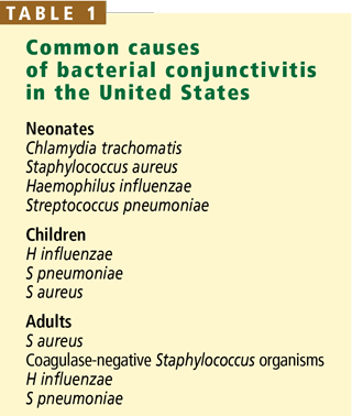
In neonates, conjunctivitis is predominantly bacterial, and the most common organism is Chlamydia trachomatis. Chlamydial conjuctivitis typically presents with purulent unilateral or bilateral discharge about a week after birth in children born to mothers who have cervical chlamydial infection. Many infants with chlamydial conjunctivitis develop chlamydial pneumonitis: approximately 50% of infants with chlamydial pneumonitis have concurrent conjunctivitis or a recent history of conjunctivitis.1
Neisseria gonorrhoeae is a rare cause of neonatal conjunctivitis. The onset is somewhat earlier than in chlamydial conjunctivitis, ie, in the first week of life, and this organism classically causes severe “hyperacute” conjunctivitis with profuse discharge and may result in corneal involvement and perforation. Routine antibiotic prophylaxis at birth has markedly reduced its incidence and complications.
Other bacteria that can cause neonatal conjunctivitis include Staphylococcus aureus, Haemophilus influenzae, and Streptococcus pneumoniae.2
In children, bacterial conjunctivitis is most often caused by H influenzae or S pneumoniae, which accounted for 29% and 20% of cases, respectively, in a prospective study in Israel.3 Whether patients had been vaccinated against H influenzae in this study is unclear.
H influenzae conjunctivitis spreads easily in schools and households. It is associated with concurrent upper respiratory tract infections and otitis media (conjunctivitis-otitis syndrome): 45% to 73% of patients with purulent conjunctivitis also have ipsilateral otitis media.4
S pneumoniae, the second most common cause of bacterial conjunctivitis in children, is a common cause in epidemic outbreaks among young adults. Newly described unencapsulated pneumococcal strains caused outbreaks that affected 92 recruits at a military training facility and 100 students at Dartmouth University.5S pneumoniae is also associated with conjunctivitis-otitis syndrome, accounting for approximately 23% of culture-proven cases.4
Moraxella species, S aureus, and coagulase-negative staphylococci are less common causes of bacterial conjunctivitis in children.6–8
In adults, the most common causes of bacterial conjunctivitis are S aureus and H influenzae. Conjunctivitis caused by S aureus is often recurrent and associated with chronic ble-pharoconjunctivitis (inflammation of the eyelid and conjunctiva). The conjunctivae are colonized by S aureus in 3.8% to 6.3% of healthy adults.9–11 In addition, about 20% of people normally harbor S aureus continually in the nasal passages, and another 60% harbor it intermittently; in both cases, the bacteria may be a reservoir for recurrent ocular infection.12
Other organisms that commonly cause conjunctivitis in adults are S pneumoniae, coagulase-negative staphylococci, and Moraxella and Acinetobacter species.13
HOSPITAL-ACQUIRED CONJUNCTIVITIS
Little has been published about hospital-acquired conjunctivitis. In a neonatal intensive care unit, the most common organisms isolated in patients with conjunctivitis were coagulase-negative staphylococci, S aureus, and Klebsiella species.14 We found that about 30% of children who developed bacterial conjunctivitis after 2 days of hospitalization at Cleveland Clinic harbored gram-negative organisms. In addition, in patients who were found to have conjunctivitis caused by Staphylococcus species, the rate of methicillin resistance was higher in those hospitalized for more than 2 days than those with Staphylococcus species who were hospitalized for less than 2 days. This suggests that the bacterial pathogens encountered in hospitalized children with conjunctivitis differ from those found in the outpatient setting.15
EYE DISORDERS PREDISPOSE TO INFECTION
The conjunctiva is a transparent membrane that covers the sclera and lines the inside of the eyelid. It is a protective barrier against invading pathogens and lubricates the ocular surface by secreting components of the tear film (although the lacrimal glands contribute more to the tear film).
Several unique anatomic and functional features of the ocular surface help prevent bacterial infection in the healthy eye. The tear film contains secreted immunoglobulins, lysozyme, complement, and multiple antibacterial enzymes, and it is continuously being flushed and renewed, creating a physically and immunologically adverse environment for bacterial growth.
Disorders involving the eyelids or tear film such as chronic dry eye and lagophthalmos (in which the eye cannot close completely) may predispose the eye to frequent infections. Also, an adjacent focus of infection, such as inflammation of the lacrimal gland (dacryocystitis), can cause recurrent or chronic conjunctivitis.16
CLINICAL FEATURES OF BACTERIAL CONJUNCTIVITIS
Bacterial conjunctivitis is commonly classified according to its clinical presentation: hyperacute, acute, or chronic.
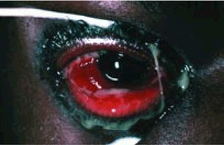
Hyperacute bacterial conjunctivitis presents with the rapid onset of conjunctival injection, eyelid edema, severe, continuous, and copious purulent discharge, chemosis, and discomfort or pain.
N gonorrhoeae is a frequent cause of hyperacute conjunctivitis in sexually active patients; the patient usually also has N gonorrhoeae genital infection, which is often asymptomatic. N gonorrhoeae conjunctivitis also occurs in neonates, as noted above. The cornea is frequently involved, and untreated cases can progress within days to corneal perforation. Unlike most other types of conjunctivitis, gonococcal conjunctivitis should be treated as a systemic disease, with both systemic and topical antibacterial therapy.2
Acute bacterial conjunctivitis typically presents abruptly with red eye and purulent drainage without significant eye pain, discomfort, or photophobia. Visual acuity does not typically decrease unless large amounts of discharge intermittently obscure vision.
Chronic bacterial conjunctivitis, ie, red eye with purulent discharge persisting for longer than a few weeks, is generally caused by Chlamydia trachomatis or is associated with a nidus for infection such as in dacryocystitis.
BACTERIAL CONJUNCTIVITIS VS OTHER CAUSES OF A RED EYE
Clinical signs and symptoms of infection with certain organisms have been extensively described, but a meta-analysis17 found no evidence that these textbook features help to distinguish between bacterial and viral causes of conjunctivitis. Instead, whether a bacterial cause was likely was best determined from just three features: having both eyes glued shut in the morning had an odds ratio of 15:1 in predicting a positive bacterial culture, and either itching or previous conjunctivitis made a bacterial cause less likely.18
In general, however, viral conjunctivitis typically presents as an itchy red eye with mild watery discharge. Many patients have signs and symptoms of a viral upper respiratory tract infection (eg, cough, runny nose, congestion) and have been in contact with a sick person. Ipsilateral preauricular lymphadenopathy is common in viral conjunctivitis and strongly suggests this diagnosis.19
Viral conjunctivitis is often epidemic and is easily contagious. Several epidemics have been traced to eyecare facilities. Adenovirus conjunctivitis is extremely contagious and can be transmitted both between people and via inanimate objects; it has been reported to be spread by workers in health care facilities.20
Allergic conjunctivitis is also common. Patients typically report itching and redness of both eyes in response to an allergen exposure. Other allergic symptoms may be present, such as allergic rhinosinusitis, asthma, or atopic dermatitis in response to seasonal or perennial environmental allergens.
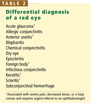
Other causes of a red eye. Many patients with a red eye have conjunctivitis, but other conditions can also present in a similar manner. Whether a patient has a serious vision-threatening condition (eg, acute-angle closure glaucoma, microbial keratitis, or anterior uveitis) can usually be determined with a focused ophthalmologic history and physical examination. Any alarming clinical features such as severe pain, decreased vision, or a hazy cornea in a patient with a red eye should alert the clinician to a more serious condition and prompt a referral to an ophthalmologist for an urgent evaluation (Table 2). A complete review for internists on how to manage a red eye was recently published in this journal.21
TREATMENT
Systemic treatment needed for gonococcal or chlamydial infections
The US Centers for Disease Control and Prevention recommend treating gonococcal conjunctivitis with ceftriaxone (Rocephin) 1 g in a single intramuscular dose plus topical saline lavage of the eye.22,23 Sexual partners of the patient should be referred for evaluation and treatment, as should mothers of affected neonates and the mother’s sexual partners.
Chlamydial conjunctivitis is also treated with systemic antibiotics. In neonates, the treatment is the same as for pneumonia caused by C trachomatis: erythromycin taken orally for 14 days. In adults, it can be treated with a single oral dose of azithromycin (Zithromax) 1 g.
Some authors recommend that H influenzae conjunctivitis also be treated with systemic antibiotics, as it is frequently associated with concurrent otitis media.24
Topical antibiotics hasten cure
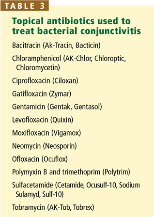
Other types of bacterial conjunctivitis usually resolve spontaneously: early placebo-controlled studies found that more than 70% of cases of bacterial conjunctivitis resolve within 8 days.25 However, treatment with antibacterial agents leads to a faster clinical and microbiological cure26 and reduces the chance of rare complications27 and of transmitting the infection.
Is culture necessary?
A predictable set of organisms accounts for most cases of bacterial conjunctivitis in outpatients, so many physicians start therapy empirically without culturing the conjunctiva. But in the hospital the organisms and their antibiotic resistance patterns are more varied, so culturing the conjunctiva before starting broad-spectrum therapy may be warranted.15 For an outpatient with possible hyperacute conjunctivitis, it is reasonable to perform a Gram stain in the office if the facilities exist, but it is not essential because urgent referral to an ophthalmologist is warranted regardless of the results to rule out corneal involvement.
Unfortunately, antibiotic resistance is increasing even among outpatients. Susceptibility of the most common ocular pathogens to ophthalmic antimicrobial agents has dropped dramatically: S pneumoniae and S aureus have developed high rates of resistance.30 Recent data also suggest that treatment with topical ophthalmic antibiotics can induce resistance among colonizing bacteria in nonocular locations.31 Widespread systemic treatment with azithromycin or tetracycline for control of endemic trachoma in two villages in Nepal resulted in increased rates of antibiotic resistance among nasopharyngeal isolates of S pneumoniae. S aureus is developing resistance to methicillin and to fluoroquinolones, such as levofloxacin (Levaquin).32,33 But fluoroquinolones are still effective against most bacteria that cause conjunctivitis or keratitis, and because they penetrate the cornea well, they should be used if clinical features suggest corneal involvement. Remember also that most patients recover without treatment even if the organism has appreciable antibiotic resistance.28
Corticosteroids should be avoided
Although corticosteroid drops (either alone or combined with antibiotic drops) may quickly relieve symptoms, some conditions that present as a red eye with watery discharge, such as herpetic keratitis, worsen with corticosteroid use. We recommend that internists avoid prescribing corticosteroid drops.
Remove contact lenses, replace eye drops
Contact lenses should be taken out until an infection is completely resolved. Disposable lenses should be thrown away. Nondisposable lenses should be cleaned thoroughly as recommended by the manufacturer, and a new lens case should be used.
Patients who use prescription eye drops for glaucoma should continue to use them, but the bottles should be replaced in case they have been contaminated by inadvertent contact with the eye.
Over-the-counter lubricating eye drops may be continued if desired, but a fresh bottle or vial should be used.
WHEN TO REFER
Red flags indicating that a patient may have a serious vision-threatening condition that requires urgent referral to an ophthalmologist include severe eye pain or headache, photophobia, decreased vision, or contact lens use. Patients with hyperacute cases should also be referred at once to rule out corneal involvement, although the internist should start treatment for gonorrhea. In addition, patients with apparent bacterial conjunctivitis that does not improve after 24 hours of antibiotic treatment should also be referred to an ophthalmologist.
- Tipple MA, Beem MO, Saxon EM. Clinical characteristics of the afebrile pneumonia associated with Chlamydia trachomatis infection in infants less than 6 months of age. Pediatrics 1979; 63:192–197.
- De Toledo AR, Chandler JW. Conjunctivitis of the newborn. Infect Dis Clin North Am 1992; 6:807–813.
- Buznach N, Dagan R, Greenberg D. Clinical and bacterial characteristics of acute bacterial conjunctivitis in children in the antibiotic resistance era. Pediatr Infect Dis J 2005; 24:823–828.
- Bodor FF. Conjunctivitis-otitis syndrome. Pediatrics 1982; 69:695–698.
- Crum NF, Barrozo CP, Chapman FA, Ryan MA, Russell KL. An outbreak of conjunctivitis due to a novel unencapsulated Streptococcus pneumoniae among military trainees. Clin Infect Dis 2004; 39:1148–1154.
- Gigliotti F, Williams WT, Hayden FG, et al. Etiology of acute conjunctivitis in children. J Pediatr 1981; 98:531–536.
- Weiss A, Brinser JH, Nazar-Stewart V. Acute conjunctivitis in childhood. J Pediatr 1993; 122:10–14.
- Block SL, Hedrick J, Tyler R, et al. Increasing bacterial resistance in pediatric acute conjunctivitis (1997–1998). Antimicrob Agents Chemother 2000; 44:1650–1654.
- Singer TR, Isenberg SJ, Apt L. Conjunctival anaerobic and aerobic bacterial flora in paediatric versus adult subjects. Br J Ophthalmol 1998; 72:448–451.
- Kato T, Hayasaka S. Methicillin-resistant Staphylococcus aureus and methicillin-resistant coagulase-negative staphylococci from conjunctivas of preoperative patients. Jpn J Ophthalmol 1998; 42:461–465.
- Nakata K, Inoue Y, Harada J, et al. A high incidence of Staphylococcus aureus colonization in the external eyes of patients with atopic dermatitis. Ophthalmology 2000; 107:2167–2171.
- Kluytmans J, van Belkum A, Verbrugh H. Nasal carriage of Staphylococcus aureus: epidemiology, underlying mechanisms, and associated risks. Clin Microbiol Rev 1997; 10:505–520.
- Kowalski RP, Karenchak LM, Romanowski EG. Infectious disease: changing antibiotic susceptibility. Ophthalmol Clin North Am 2003; 16:1–9.
- Haas J, Larson E, Ross B, See B, Saiman L. Epidemiology and diagnosis of hospital acquired conjunctivitis among neonatal intensive care unit patients. Pediatr Infect Dis J 2005; 24:586–589.
- Tarabishy AB, Hall GS, Procop GW, Jeng BH. Bacterial culture isolates from hospitalized pediatric patients with conjunctivitis. Am J Ophthalmol 2006; 142:678–680.
- Limberg MB. A review of bacterial keratitis and bacterial conjunctivitis. Am J Ophthalmol 1991; 112:2S–9S.
- Rietveld RP, van Weert HC, ter Riet G, Bindels PJ. Diagnostic impact of signs and symptoms in acute infectious conjunctivitis: systematic literature search. BMJ 2003; 327:789.
- Rietveld RP, ter Riet G, Bindels PJ, Sloos JH, van Weert HC. Predicting bacterial cause in infectious conjunctivitis: cohort study on informativeness of combinations of signs and symptoms. BMJ 2004; 329:206–210.
- Syed NA, Hyndiuk RA. Infectious conjunctivitis. Infect Dis Clin North Am 1992; 6:789–805.
- Azar MJ, Dhaliwal DK, Bower KS, Kowalski RP, Gordon YJ. Possible consequences of shaking hands with your patients with epidemic keratoconjunctivitis. Am J Ophthalmol 1996; 121:711–712.
- Galor A, Jeng BH. The red eye: a primer for the internist. Cleve Clin J Med 2008; 75:137–144.
- Centers for Disease Control and Prevention,Workowski KA, Berman SM. Sexually transmitted diseases treatment guidelines, 2006. MMWR Recomm Rep 2006; 55( RR11):1–94. http://www.cdc.gov/mmwr/preview/mmwrhtml/rr5511a1.htm. Erratum in: MMWR Recomm Rep 2006; 55:997. Accessed 4/30/08.
- Haimovici R, Roussel TJ. Treatment of gonococcal conjunctivitis with single-dose intramuscular ceftriaxone. Am J Ophthalmol 1989; 107:511–514.
- Bodor FF. Systemic antibiotics for treatment of the conjunctivitis-otitis media syndrome. Pediatr Infect Dis J 1989; 8:287–290.
- Gigliotti F, Hendley JO, Morgan J, Michaels R, Dickens M, Lohr J. Efficacy of topical antibiotic therapy in acute conjunctivitis in children. J Pediatr 1984; 104:623–626.
- Sheikh A, Hurwitz B. Topical antibiotics for acute bacterial conjunctivitis: Cochrane systematic review and meta-analysis update. Br J Gen Pract 2005; 55:962–964.
- Aung T, Chan TK. Nosocomial Klebsiella pneumoniae conjunctivitis resulting in infectious keratitis and bilateral corneal perforation. Cornea 1998; 17:558–561.
- Baum J, Barza M. The evolution of antibiotic therapy for bacterial conjunctivitis and keratitis: 1970–2000. Cornea 2000; 19:659–672.
- Schlech BA, Alfonso E. Overview of the potency of moxifloxacin ophthalmic solution 0.5% (VIGAMOX). Surv Ophthalmol 2005; 50:S7–S15.
- Chalita MR, Höfling-Lima AL, Paranhos A, Schor P, Belfort R. Shifting trends in in vitro antibiotic susceptibilities for common ocular isolates during a period of 15 years. Am J Ophthalmol 2004; 137:43–51.
- Gaynor BD, Chidambaram JD, Cevallos V, et al. Topical ocular antibiotics induce bacterial resistance at extraocular sites. Br J Ophthalmol 2005; 89:1097–1099.
- Marangon FB, Miller D, Muallem MS, Romano AC, Alfonso EC. Ciprofloxacin and levofloxacin resistance among methicillin-sensitive Staphylococcus aureus isolates from keratitis and conjunctivitis. Am J Ophthalmol 2004; 137:453–458.
- Goldstein MH, Kowalski RP, Gordon YJ. Emerging fluoroquinolone resistance in bacterial keratitis: a 5-year review. Ophthalmology 1999; 106:1313–1318.
Bacterial conjunctivitis is common in children and adults presenting with a red eye. Although most cases are self-limited, appropriate antimicrobial treatment accelerates resolution and reduces complications. It is critical to differentiate bacterial conjunctivitis from other types of conjunctivitis and more serious vision-threatening conditions so that patients can be appropriately treated and, if necessary, referred to an ophthalmologist.
This paper is an overview of how to diagnose and manage bacterial conjunctivitis for the office-based internist.
CAUSES VARY BY AGE
In neonates, conjunctivitis is predominantly bacterial, and the most common organism is Chlamydia trachomatis. Chlamydial conjuctivitis typically presents with purulent unilateral or bilateral discharge about a week after birth in children born to mothers who have cervical chlamydial infection. Many infants with chlamydial conjunctivitis develop chlamydial pneumonitis: approximately 50% of infants with chlamydial pneumonitis have concurrent conjunctivitis or a recent history of conjunctivitis.1
Neisseria gonorrhoeae is a rare cause of neonatal conjunctivitis. The onset is somewhat earlier than in chlamydial conjunctivitis, ie, in the first week of life, and this organism classically causes severe “hyperacute” conjunctivitis with profuse discharge and may result in corneal involvement and perforation. Routine antibiotic prophylaxis at birth has markedly reduced its incidence and complications.
Other bacteria that can cause neonatal conjunctivitis include Staphylococcus aureus, Haemophilus influenzae, and Streptococcus pneumoniae.2
In children, bacterial conjunctivitis is most often caused by H influenzae or S pneumoniae, which accounted for 29% and 20% of cases, respectively, in a prospective study in Israel.3 Whether patients had been vaccinated against H influenzae in this study is unclear.
H influenzae conjunctivitis spreads easily in schools and households. It is associated with concurrent upper respiratory tract infections and otitis media (conjunctivitis-otitis syndrome): 45% to 73% of patients with purulent conjunctivitis also have ipsilateral otitis media.4
S pneumoniae, the second most common cause of bacterial conjunctivitis in children, is a common cause in epidemic outbreaks among young adults. Newly described unencapsulated pneumococcal strains caused outbreaks that affected 92 recruits at a military training facility and 100 students at Dartmouth University.5S pneumoniae is also associated with conjunctivitis-otitis syndrome, accounting for approximately 23% of culture-proven cases.4
Moraxella species, S aureus, and coagulase-negative staphylococci are less common causes of bacterial conjunctivitis in children.6–8
In adults, the most common causes of bacterial conjunctivitis are S aureus and H influenzae. Conjunctivitis caused by S aureus is often recurrent and associated with chronic ble-pharoconjunctivitis (inflammation of the eyelid and conjunctiva). The conjunctivae are colonized by S aureus in 3.8% to 6.3% of healthy adults.9–11 In addition, about 20% of people normally harbor S aureus continually in the nasal passages, and another 60% harbor it intermittently; in both cases, the bacteria may be a reservoir for recurrent ocular infection.12
Other organisms that commonly cause conjunctivitis in adults are S pneumoniae, coagulase-negative staphylococci, and Moraxella and Acinetobacter species.13
HOSPITAL-ACQUIRED CONJUNCTIVITIS
Little has been published about hospital-acquired conjunctivitis. In a neonatal intensive care unit, the most common organisms isolated in patients with conjunctivitis were coagulase-negative staphylococci, S aureus, and Klebsiella species.14 We found that about 30% of children who developed bacterial conjunctivitis after 2 days of hospitalization at Cleveland Clinic harbored gram-negative organisms. In addition, in patients who were found to have conjunctivitis caused by Staphylococcus species, the rate of methicillin resistance was higher in those hospitalized for more than 2 days than those with Staphylococcus species who were hospitalized for less than 2 days. This suggests that the bacterial pathogens encountered in hospitalized children with conjunctivitis differ from those found in the outpatient setting.15
EYE DISORDERS PREDISPOSE TO INFECTION
The conjunctiva is a transparent membrane that covers the sclera and lines the inside of the eyelid. It is a protective barrier against invading pathogens and lubricates the ocular surface by secreting components of the tear film (although the lacrimal glands contribute more to the tear film).
Several unique anatomic and functional features of the ocular surface help prevent bacterial infection in the healthy eye. The tear film contains secreted immunoglobulins, lysozyme, complement, and multiple antibacterial enzymes, and it is continuously being flushed and renewed, creating a physically and immunologically adverse environment for bacterial growth.
Disorders involving the eyelids or tear film such as chronic dry eye and lagophthalmos (in which the eye cannot close completely) may predispose the eye to frequent infections. Also, an adjacent focus of infection, such as inflammation of the lacrimal gland (dacryocystitis), can cause recurrent or chronic conjunctivitis.16
CLINICAL FEATURES OF BACTERIAL CONJUNCTIVITIS
Bacterial conjunctivitis is commonly classified according to its clinical presentation: hyperacute, acute, or chronic.
Hyperacute bacterial conjunctivitis presents with the rapid onset of conjunctival injection, eyelid edema, severe, continuous, and copious purulent discharge, chemosis, and discomfort or pain.
N gonorrhoeae is a frequent cause of hyperacute conjunctivitis in sexually active patients; the patient usually also has N gonorrhoeae genital infection, which is often asymptomatic. N gonorrhoeae conjunctivitis also occurs in neonates, as noted above. The cornea is frequently involved, and untreated cases can progress within days to corneal perforation. Unlike most other types of conjunctivitis, gonococcal conjunctivitis should be treated as a systemic disease, with both systemic and topical antibacterial therapy.2
Acute bacterial conjunctivitis typically presents abruptly with red eye and purulent drainage without significant eye pain, discomfort, or photophobia. Visual acuity does not typically decrease unless large amounts of discharge intermittently obscure vision.
Chronic bacterial conjunctivitis, ie, red eye with purulent discharge persisting for longer than a few weeks, is generally caused by Chlamydia trachomatis or is associated with a nidus for infection such as in dacryocystitis.
BACTERIAL CONJUNCTIVITIS VS OTHER CAUSES OF A RED EYE
Clinical signs and symptoms of infection with certain organisms have been extensively described, but a meta-analysis17 found no evidence that these textbook features help to distinguish between bacterial and viral causes of conjunctivitis. Instead, whether a bacterial cause was likely was best determined from just three features: having both eyes glued shut in the morning had an odds ratio of 15:1 in predicting a positive bacterial culture, and either itching or previous conjunctivitis made a bacterial cause less likely.18
In general, however, viral conjunctivitis typically presents as an itchy red eye with mild watery discharge. Many patients have signs and symptoms of a viral upper respiratory tract infection (eg, cough, runny nose, congestion) and have been in contact with a sick person. Ipsilateral preauricular lymphadenopathy is common in viral conjunctivitis and strongly suggests this diagnosis.19
Viral conjunctivitis is often epidemic and is easily contagious. Several epidemics have been traced to eyecare facilities. Adenovirus conjunctivitis is extremely contagious and can be transmitted both between people and via inanimate objects; it has been reported to be spread by workers in health care facilities.20
Allergic conjunctivitis is also common. Patients typically report itching and redness of both eyes in response to an allergen exposure. Other allergic symptoms may be present, such as allergic rhinosinusitis, asthma, or atopic dermatitis in response to seasonal or perennial environmental allergens.
Other causes of a red eye. Many patients with a red eye have conjunctivitis, but other conditions can also present in a similar manner. Whether a patient has a serious vision-threatening condition (eg, acute-angle closure glaucoma, microbial keratitis, or anterior uveitis) can usually be determined with a focused ophthalmologic history and physical examination. Any alarming clinical features such as severe pain, decreased vision, or a hazy cornea in a patient with a red eye should alert the clinician to a more serious condition and prompt a referral to an ophthalmologist for an urgent evaluation (Table 2). A complete review for internists on how to manage a red eye was recently published in this journal.21
TREATMENT
Systemic treatment needed for gonococcal or chlamydial infections
The US Centers for Disease Control and Prevention recommend treating gonococcal conjunctivitis with ceftriaxone (Rocephin) 1 g in a single intramuscular dose plus topical saline lavage of the eye.22,23 Sexual partners of the patient should be referred for evaluation and treatment, as should mothers of affected neonates and the mother’s sexual partners.
Chlamydial conjunctivitis is also treated with systemic antibiotics. In neonates, the treatment is the same as for pneumonia caused by C trachomatis: erythromycin taken orally for 14 days. In adults, it can be treated with a single oral dose of azithromycin (Zithromax) 1 g.
Some authors recommend that H influenzae conjunctivitis also be treated with systemic antibiotics, as it is frequently associated with concurrent otitis media.24
Topical antibiotics hasten cure
Other types of bacterial conjunctivitis usually resolve spontaneously: early placebo-controlled studies found that more than 70% of cases of bacterial conjunctivitis resolve within 8 days.25 However, treatment with antibacterial agents leads to a faster clinical and microbiological cure26 and reduces the chance of rare complications27 and of transmitting the infection.
Is culture necessary?
A predictable set of organisms accounts for most cases of bacterial conjunctivitis in outpatients, so many physicians start therapy empirically without culturing the conjunctiva. But in the hospital the organisms and their antibiotic resistance patterns are more varied, so culturing the conjunctiva before starting broad-spectrum therapy may be warranted.15 For an outpatient with possible hyperacute conjunctivitis, it is reasonable to perform a Gram stain in the office if the facilities exist, but it is not essential because urgent referral to an ophthalmologist is warranted regardless of the results to rule out corneal involvement.
Unfortunately, antibiotic resistance is increasing even among outpatients. Susceptibility of the most common ocular pathogens to ophthalmic antimicrobial agents has dropped dramatically: S pneumoniae and S aureus have developed high rates of resistance.30 Recent data also suggest that treatment with topical ophthalmic antibiotics can induce resistance among colonizing bacteria in nonocular locations.31 Widespread systemic treatment with azithromycin or tetracycline for control of endemic trachoma in two villages in Nepal resulted in increased rates of antibiotic resistance among nasopharyngeal isolates of S pneumoniae. S aureus is developing resistance to methicillin and to fluoroquinolones, such as levofloxacin (Levaquin).32,33 But fluoroquinolones are still effective against most bacteria that cause conjunctivitis or keratitis, and because they penetrate the cornea well, they should be used if clinical features suggest corneal involvement. Remember also that most patients recover without treatment even if the organism has appreciable antibiotic resistance.28
Corticosteroids should be avoided
Although corticosteroid drops (either alone or combined with antibiotic drops) may quickly relieve symptoms, some conditions that present as a red eye with watery discharge, such as herpetic keratitis, worsen with corticosteroid use. We recommend that internists avoid prescribing corticosteroid drops.
Remove contact lenses, replace eye drops
Contact lenses should be taken out until an infection is completely resolved. Disposable lenses should be thrown away. Nondisposable lenses should be cleaned thoroughly as recommended by the manufacturer, and a new lens case should be used.
Patients who use prescription eye drops for glaucoma should continue to use them, but the bottles should be replaced in case they have been contaminated by inadvertent contact with the eye.
Over-the-counter lubricating eye drops may be continued if desired, but a fresh bottle or vial should be used.
WHEN TO REFER
Red flags indicating that a patient may have a serious vision-threatening condition that requires urgent referral to an ophthalmologist include severe eye pain or headache, photophobia, decreased vision, or contact lens use. Patients with hyperacute cases should also be referred at once to rule out corneal involvement, although the internist should start treatment for gonorrhea. In addition, patients with apparent bacterial conjunctivitis that does not improve after 24 hours of antibiotic treatment should also be referred to an ophthalmologist.
- Tipple MA, Beem MO, Saxon EM. Clinical characteristics of the afebrile pneumonia associated with Chlamydia trachomatis infection in infants less than 6 months of age. Pediatrics 1979; 63:192–197.
- De Toledo AR, Chandler JW. Conjunctivitis of the newborn. Infect Dis Clin North Am 1992; 6:807–813.
- Buznach N, Dagan R, Greenberg D. Clinical and bacterial characteristics of acute bacterial conjunctivitis in children in the antibiotic resistance era. Pediatr Infect Dis J 2005; 24:823–828.
- Bodor FF. Conjunctivitis-otitis syndrome. Pediatrics 1982; 69:695–698.
- Crum NF, Barrozo CP, Chapman FA, Ryan MA, Russell KL. An outbreak of conjunctivitis due to a novel unencapsulated Streptococcus pneumoniae among military trainees. Clin Infect Dis 2004; 39:1148–1154.
- Gigliotti F, Williams WT, Hayden FG, et al. Etiology of acute conjunctivitis in children. J Pediatr 1981; 98:531–536.
- Weiss A, Brinser JH, Nazar-Stewart V. Acute conjunctivitis in childhood. J Pediatr 1993; 122:10–14.
- Block SL, Hedrick J, Tyler R, et al. Increasing bacterial resistance in pediatric acute conjunctivitis (1997–1998). Antimicrob Agents Chemother 2000; 44:1650–1654.
- Singer TR, Isenberg SJ, Apt L. Conjunctival anaerobic and aerobic bacterial flora in paediatric versus adult subjects. Br J Ophthalmol 1998; 72:448–451.
- Kato T, Hayasaka S. Methicillin-resistant Staphylococcus aureus and methicillin-resistant coagulase-negative staphylococci from conjunctivas of preoperative patients. Jpn J Ophthalmol 1998; 42:461–465.
- Nakata K, Inoue Y, Harada J, et al. A high incidence of Staphylococcus aureus colonization in the external eyes of patients with atopic dermatitis. Ophthalmology 2000; 107:2167–2171.
- Kluytmans J, van Belkum A, Verbrugh H. Nasal carriage of Staphylococcus aureus: epidemiology, underlying mechanisms, and associated risks. Clin Microbiol Rev 1997; 10:505–520.
- Kowalski RP, Karenchak LM, Romanowski EG. Infectious disease: changing antibiotic susceptibility. Ophthalmol Clin North Am 2003; 16:1–9.
- Haas J, Larson E, Ross B, See B, Saiman L. Epidemiology and diagnosis of hospital acquired conjunctivitis among neonatal intensive care unit patients. Pediatr Infect Dis J 2005; 24:586–589.
- Tarabishy AB, Hall GS, Procop GW, Jeng BH. Bacterial culture isolates from hospitalized pediatric patients with conjunctivitis. Am J Ophthalmol 2006; 142:678–680.
- Limberg MB. A review of bacterial keratitis and bacterial conjunctivitis. Am J Ophthalmol 1991; 112:2S–9S.
- Rietveld RP, van Weert HC, ter Riet G, Bindels PJ. Diagnostic impact of signs and symptoms in acute infectious conjunctivitis: systematic literature search. BMJ 2003; 327:789.
- Rietveld RP, ter Riet G, Bindels PJ, Sloos JH, van Weert HC. Predicting bacterial cause in infectious conjunctivitis: cohort study on informativeness of combinations of signs and symptoms. BMJ 2004; 329:206–210.
- Syed NA, Hyndiuk RA. Infectious conjunctivitis. Infect Dis Clin North Am 1992; 6:789–805.
- Azar MJ, Dhaliwal DK, Bower KS, Kowalski RP, Gordon YJ. Possible consequences of shaking hands with your patients with epidemic keratoconjunctivitis. Am J Ophthalmol 1996; 121:711–712.
- Galor A, Jeng BH. The red eye: a primer for the internist. Cleve Clin J Med 2008; 75:137–144.
- Centers for Disease Control and Prevention,Workowski KA, Berman SM. Sexually transmitted diseases treatment guidelines, 2006. MMWR Recomm Rep 2006; 55( RR11):1–94. http://www.cdc.gov/mmwr/preview/mmwrhtml/rr5511a1.htm. Erratum in: MMWR Recomm Rep 2006; 55:997. Accessed 4/30/08.
- Haimovici R, Roussel TJ. Treatment of gonococcal conjunctivitis with single-dose intramuscular ceftriaxone. Am J Ophthalmol 1989; 107:511–514.
- Bodor FF. Systemic antibiotics for treatment of the conjunctivitis-otitis media syndrome. Pediatr Infect Dis J 1989; 8:287–290.
- Gigliotti F, Hendley JO, Morgan J, Michaels R, Dickens M, Lohr J. Efficacy of topical antibiotic therapy in acute conjunctivitis in children. J Pediatr 1984; 104:623–626.
- Sheikh A, Hurwitz B. Topical antibiotics for acute bacterial conjunctivitis: Cochrane systematic review and meta-analysis update. Br J Gen Pract 2005; 55:962–964.
- Aung T, Chan TK. Nosocomial Klebsiella pneumoniae conjunctivitis resulting in infectious keratitis and bilateral corneal perforation. Cornea 1998; 17:558–561.
- Baum J, Barza M. The evolution of antibiotic therapy for bacterial conjunctivitis and keratitis: 1970–2000. Cornea 2000; 19:659–672.
- Schlech BA, Alfonso E. Overview of the potency of moxifloxacin ophthalmic solution 0.5% (VIGAMOX). Surv Ophthalmol 2005; 50:S7–S15.
- Chalita MR, Höfling-Lima AL, Paranhos A, Schor P, Belfort R. Shifting trends in in vitro antibiotic susceptibilities for common ocular isolates during a period of 15 years. Am J Ophthalmol 2004; 137:43–51.
- Gaynor BD, Chidambaram JD, Cevallos V, et al. Topical ocular antibiotics induce bacterial resistance at extraocular sites. Br J Ophthalmol 2005; 89:1097–1099.
- Marangon FB, Miller D, Muallem MS, Romano AC, Alfonso EC. Ciprofloxacin and levofloxacin resistance among methicillin-sensitive Staphylococcus aureus isolates from keratitis and conjunctivitis. Am J Ophthalmol 2004; 137:453–458.
- Goldstein MH, Kowalski RP, Gordon YJ. Emerging fluoroquinolone resistance in bacterial keratitis: a 5-year review. Ophthalmology 1999; 106:1313–1318.
KEY POINTS
- Viral conjunctivitis typically presents as an itchy red eye with mild watery discharge. Many patients have signs and symptoms of a viral upper respiratory tract infection (eg, cough, runny nose, congestion) and have been in contact with a sick person.
- Having both eyes glued shut in the morning had an odds ratio of 15:1 in predicting a positive bacterial culture, whereas either itching or previous conjunctivitis made a bacterial cause less likely.
- In adults, Neisseria gonorrhoeae causes hyperacute conjunctivitis and is associated with concurrent, often asymptomatic genital infection. Gonococcal conjunctivitis should be treated with a single dose of ceftriaxone (Rocephin) 1 g intramuscularly plus saline eye-washing.
- Corticosteroid drops should not be prescribed for a red eye before consultation with an ophthalmologist because these drops may worsen some conditions.
Red eye for the internist: When to treat, when to refer
Many patients present to internists because of redness in the eye. The possible causes range from benign (which generally can be handled by an internist) to vision-threatening (which need prompt or emergency referral to an ophthalmologist).
HISTORY HELPS IDENTIFY THE CAUSE
The internist should ascertain:
- Whether one or both eyes are affected
- The duration of symptoms
- Previous eye and medical problems
- The type of discharge (watery or purulent), if present
- Whether the patient has any visual changes, pain, or photosensitivity.
Refer patients to an ophthalmologist for further evaluation if they use contact lenses or if they have had trauma to the eye, vision changes, severe pain, or systemic symptoms such as nausea, vomiting, or severe headache.
BASIC EYE EXAMINATION
Examine:
- Visual acuity
- Pupil size and reaction to light
- The pattern and location of the redness
- The cornea and anterior segment for gross abnormalities such as corneal opacities, hypopyon (a layer of inflammatory cells in the anterior chamber), and hyphema (hemorrhage in the anterior chamber) (Use a penlight.)
- The preauricular lymph nodes. Preauricular lymphadenopathy, detected by palpation, suggests but is not specific for viral conjunctivitis.
- Funduscopy has little value in evaluating a red eye.
Refer immediately anyone who has marked purulent discharge or abnormalities in the cornea or anterior segment.
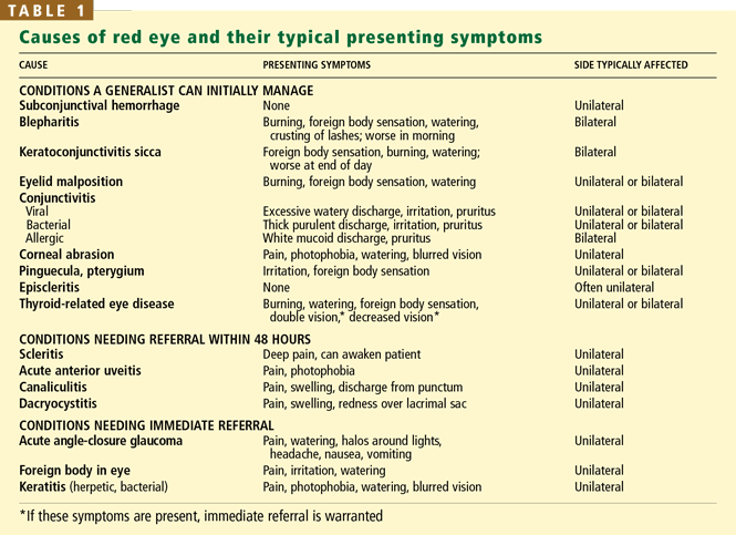
CONDITIONS A GENERALIST CAN INITIALLY MANAGE
Subconjunctival hemorrhage
Subconjunctival hemorrhages are harmless and do not cause pain or vision changes. No treatment is required, and the blood resorbs within a few weeks. However:
- Measure the blood pressure—uncontrolled hypertension can present with subconjunctival hemorrhage.1
- If the patient is on an antithrombotic agent, test the prothrombin and activated partial thromboplastin times.
- If the patient has recurrent unexplained episodes of subconjunctival hemorrhage, look for a bleeding disorder such as von Willebrand disease, hemophilia, or autoimmune thrombocytopenic purpura.
Blepharitis
Blepharitis, a common condition, is inflammation of the eyelid margins. Anterior blepharitis affects the eyelashes and anterior eyelid margin and is most often caused by a low-grade staphylococcal infection or seborrheic dermatitis. Posterior blepharitis involves the orifices of the slender sebaceous glands of the eyelids (the meibomian glands) and is often associated with acne rosacea.
Symptoms include ocular burning, a sensation that a foreign body is in the eye, and watering. Symptoms are typically worse in the morning and gradually improve throughout the day. Although the onset is sudden in some patients, blepharitis is usually chronic—often lifelong—and starts insidiously.
A sign of anterior blepharitis is crusting around the eyelashes. Patients with concomitant seborrheic dermatitis also have oily skin and flaking from the eyebrows and scalp. Signs of posterior blepharitis are oil inspissation around the meibomian gland openings, telangiectasias of the eyelid margin, and accompanying acne rosacea (skin pustules, telangiectasias, and erythema).
Treat both forms with eyelid hygiene: applying warm compresses to the eyelid margins, followed by gentle massage to remove the debris from the eyelashes and meibomian glands. This is done two to four times daily until acute symptoms resolve, then once daily. Because blepharitis is chronic, eyelid hygiene must be continued indefinitely to prevent acute exacerbations.
Posterior blepharitis that does not respond to hygiene can be also treated with oral tetracycline, which is believed to improve meibomian gland function and alter bacterial colonization.
Some patients also have tear deficiency, which can be addressed with tear replacement therapy (see below).2,3
Keratoconjunctivitis sicca (dry eye)
Dryness can cause mild eye redness. Patients typically report a foreign body sensation, burning, and paradoxically, watering. Symptoms often worsen as the day progresses and are most prominent at night.
Dryness can be due to:
- Local disturbances in the tear film such as aqueous deficiency
- An abnormal eyelid position
- Systemic autoimmune conditions such as Sjögren syndrome
- Hormonal changes (eg, in menopause)
- Excessively dry environments (eg, winter)
- Medications, including anticholinergics, antihistamines, antidepressants (eg, tricyclics), and antihypertensives (eg, beta-blockers).
Staining the cornea with fluorescein highlights small epithelial defects; rose bengal highlights devitalized cells.
Treat initially with artificial tears (eg, Refresh Tears, GenTeal, Systane, Bion Tears) and ointments (eg, Refresh Liquigel, Lacri-Lube). Dry eye has an inflammatory component; cyclosporine ophthalmic 0.05% (Restasis) may increase tear production and improve symptoms.4
Refer patients with symptoms that do not respond to therapy. An ophthalmologist can place silicone plugs in the canaliculi, a procedure with a 75% success rate for improving dry-eye symptoms.5 Plugs must be carefully fitted: loose ones can spontaneously dislodge, and tight ones can irritate the eye.
Eyelid malposition
Entropion (in-turning of the eyelid) causes eyelashes to rub on the cornea. Ectropion (outward turning of the eyelid) results in tear-film abnormalities and corneal exposure. Both conditions are most commonly caused by aging but may be secondary to scarring or to mechanical, paralytic, or congenital conditions. Definitive treatment involves surgery to restore the normal eyelid position. Several techniques have high success rates.1
Lagophthalmos (inability to fully close the eyes) is caused by orbicularis muscle dysfunction, which may be secondary to Bell palsy, stroke, or neurosurgical procedures that disrupt the facial nerve. The exposed cornea is prone to dryness and irritation. Treatments include artificial tears, lubricating ointments, and surgery—gold weight placement or suturing the eyelid margins (tarsorrhaphy).
Floppy eyelid syndrome refers to a lax upper eyelid that may evert during contact with the pillow during sleep, resulting in irritation and inflammation of the upper palpebral conjunctiva. Signs and symptoms are unilateral eye irritation, burning, and a ropy mucous discharge, which is usually worse in the morning. The upper eyelid is lax and easily everted when pulled toward the eyebrow. Most patients are obese, have obstructive sleep apnea, and sleep on the affected side.
Tell the patient to tape the affected eyelid shut or wear a protective eye shield in bed to prevent rubbing the eye on the pillow. Definitive treatment is surgery to tighten the lax upper eyelid.6
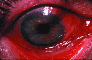
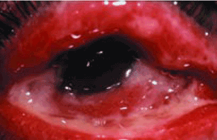
Conjunctivitis
Conjunctivitis involves hyperemia and edema of the bulbar conjunctiva (the part of the conjunctiva covering the eyeball) along with papillary and follicular changes of the palpebral conjunctiva (the inner layer of the eyelids).
Conjunctivitis can be viral, bacterial, or allergic, or due to wearing contact lenses; the cause can usually be distinguished by the history and physical examination.
Viral conjunctivitis, usually caused by an adenovirus, is more common than bacterial conjunctivitis in adults. The patient typically has had a recent upper respiratory tract infection or was exposed to conjunctivitis.
Treat supportively with cool compresses. Symptoms often worsen for a few days, then slowly improve over 1 to 2 weeks.
Viral conjunctivitis is contagious for 2 weeks after the second eye becomes involved, and good hygiene must be maintained to avoid spreading it to coworkers and family members. Those who work with the public, in schools, or in health-care facilities should be given a 2-week leave of absence to avoid spreading the infection to others.
Refer to an ophthalmologist if symptoms do not resolve in 2 weeks, as certain subtypes of adenovirus can cause prolonged symptoms with corneal involvement.7

Treat bacterial conjunctivitis empirically with antibiotic eyedrops (eg, a fluoroquinolone, a polymyxin, or sulfacetamide—several brands available) four times daily for 7 to 10 days, even though most cases are self-limited and do not result in complications. Cultures can be obtained, especially if the patient is in the hospital8 or if the conjunctivitis persists after 1 week of antibiotic therapy.
Refer patients with vision changes or who do not improve after 1 week of treatment.9,10
Treat aggressively with both a topical antibiotic (usually a fluoroquinolone) four times daily and a systemic antibiotic such as ceftriaxone (Rocephin) given as a single 1-g intramuscular injection.11,12 Because one-third of patients with gonorrheal infection also have chlamydial infection, treatment for both diseases is frequently prescribed.
Chlamydial infection, a sexually transmitted disease, can cause chronic follicular conjunctivitis. The genital tract infection may be asymptomatic. Diagnosis is made by swabbing the conjunctiva to culture for Chlamydia trachomatis. Treat systemically with either azithromycin (Zithromax) in a single 1-g oral dose or a 10–14-day course of either doxycycline (Doryx) 100 mg twice daily or erythromycin 250 mg four times daily.13
Allergic conjunctivitis is characterized by bilateral itching that worsens with scratching. Discharge is variable but is usually clear or white and stringy. Many patients have a history of seasonal or perennial allergies.
Remove offending allergens, if possible. Topical mast cell stabilizers and antihistamines relieve symptoms but may exacerbate underlying dry eye symptoms. A combined mast cell stabilizer and antihistamine such as olopatadine (Patanol), ketotifen (Zaditor), or epinastine (Elestat) can be given twice daily.14,15 Artificial tears can treat the associated dryness.
Topical corticosteroids may be used to treat an acute, severe episode but should not be used long-term. In fact, because it is difficult to differentiate between infectious and noninfectious eye conditions, and because treating some infections with corticosteroids by themselves can have grave consequences, we recommend that internists generally refrain from using them.
Oral antihistamines may relieve symptoms but are usually less effective than topical therapy.
Refer if symptoms do not resolve after 2 weeks of topical treatment.
Giant papillary conjunctivitis, most often seen in patients who wear soft contact lenses, presents with bilateral contact lens intolerance, itching, mucous discharge, and giant papillae on the upper palpebral conjunctiva.
Again, promptly refer any patient who wears contact lenses and presents with a red eye, owing to the risk of a vision-threatening corneal infection. The patient should stop wearing contact lenses for about 1 month, after which he or she can be refitted with new soft or gas-permeable lenses and taught better lens hygiene. During an acute episode, topical mast cell stabilizers are helpful for mild irritation, and topical steroids (prednisolone phosphate 1%) are helpful for more severe irritation. Topical steroids should never be used on a long-term basis because of possible adverse effects. Artificial tears can be used for dryness.15
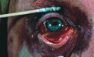
Corneal abrasion
Treat with topical antibiotics to prevent infection until the corneal epithelium has healed.16 However, most abrasions heal rapidly without sequelae because epithelial cells proliferate and migrate rapidly.
Refer if symptoms persist for longer than 48 hours or if pain suddenly worsens after the healing process has started.
Pingueculae and pterygia
A pinguecula is a small, yellow, benign growth on the nasal and temporal conjunctiva near the limbus. A pterygium is a wing-shaped band of fibrovascular tissue originating on the conjunctiva and extending onto the cornea. Both conditions are idiopathic but are believed to arise from chronic sun exposure.
Pingueculae can become inflamed and can cause eye redness and irritation. Treat conservatively with lubrication and judicious use of topical steroids; if irritation persists, pingueculae can be surgically removed.17
Episcleritis
Episcleritis is inflammation of the superficial vessels of the episclera, the connective tissue layer between the conjunctiva and the sclera. It presents with a sectoral area of redness (although it can be diffuse) and is not typically associated with pain, vision changes, or discharge. The condition tends to be recurrent and unilateral, but it can be bilateral or alternating. The underlying pathophysiology is believed to be autoimmune, although a systemic evaluation is often unrevealing.
Episcleritis is treated with topical corticosteroids or oral nonsteroidal anti-inflammatory drugs (NSAIDs); refer if the disease persists or recurs.
Thyroid-related eye disease
Thyroid-associated ophthalmopathy, an autoimmune process, usually occurs in patients with known thyroid disease, although it may develop before other systemic symptoms. Symptoms can include irritation and double vision. Signs are bulging eyes, eyelid retraction, chemosis (swelling of the conjunctiva around the cornea), conjunctival injection, periorbital edema, and limited ocular motility.
Although most cases can be managed with lubrication, vision loss may occur due to corneal exposure or compressive optic neuropathy. Patients with significant visual changes should be referred immediately to an ophthalmologist.18
CONDITIONS NEEDING REFERRAL WITHIN 48 HOURS
Scleritis
Scleritis is inflammation of the deep vessels within the episclera. The red color appears more pronounced and more purplish than in episcleritis and does not blanch after phenylephrine drops are given. The eye is tender to palpation and may be painful enough to awaken the patient from sleep. Vision is not typically affected unless the cornea, anterior chamber, or posterior segment is involved.
Half of patients who have scleritis have an associated systemic disease, eg, rheumatoid arthritis (most common), other autoimmune diseases (Wegener granulomatosis, relapsing polychondritis, inflammatory bowel disease), or infections such as tuberculosis and syphilis.
Therefore, one should search for an underlying systemic condition with a thorough history, physical examination, chest radiography (for sarcoidosis and tuberculosis), and laboratory testing: antineutrophil cytoplasmic antibody test, fluorescent treponemal antibody absorption test, Lyme antibody test, (if in an endemic area), urinalysis, a complete blood count, and a comprehensive metabolic panel.
However, patients should be promptly referred to an ophthalmologist for diagnosis and management. Treatment can depend on the underlying diagnosis, and is often guided by the status of the scleritis. Mild scleritis can be treated with oral NSAIDs; more severe disease should be treated with oral corticosteroids with or without corticosteroid-sparing agents such as methotrexate, mycophenolate (CellCept), cyclophosphamide (Cytoxan, Neosar),19,20 or tumor necrosis factor-alpha antagonists such as infliximab (Remicade) or etanercept (Enbrel).21
Anterior uveitis
Uveitis is inflammation of the uvea (the pigmented layer between the sclera and retina that includes the iris, ciliary body, and choroid). Anterior uveitis is most commonly idiopathic but can be caused by trauma, secondary to herpes virus infection, or associated with the HLA-B27 antigen.
Acute anterior uveitis presents with pain, photophobia, and blurred vision. Perilimbal (circumcorneal) injection overlies the inflamed ciliary body. The pupil is often constricted and poorly reactive to light. Chronic anterior uveitis, defined as lasting more than 6 weeks, typically presents with gradual vision loss and floaters, rather than with the acute pain or severe redness of acute disease. Anterior uveitis is diagnosed by finding cells and flare in the anterior chamber using a slit lamp.
Refer patients to an ophthalmologist immediately to help avoid visual consequences.22,23 Treatment begins with topical corticosteroid drops and can also include oral corticosteroids or long-term immunosuppresion with corticosteroid-sparing agents.
Nasolacrimal infections
Canaliculitis is an inflammation of the canaliculus, the conduit bringing tears from the eye to the nasolacrimal duct. It presents with mild, unilateral eye redness and a slight discharge that can be expressed from the punctum. It is most commonly caused by Actinomyces israelii infection, but Candida and Aspergillus species can also be involved.
Refer to an ophthalmologist for treatment, which involves mechanically removing the granular material from the canaliculi, combined with probing and irrigating the nasolacrimal system with penicillin G solution.
Dacryocystitis is inflammation of the lacrimal sac (the dilated upper end of the nasolacrimal duct) and is caused by obstruction of the duct. Staphylococcus and Streptococcus species are usually involved. Symptoms include unilateral pain, swelling, and redness over the lacrimal sac at the medial canthus of the eye. Purulent discharge can be expressed from the punctum.
Treatment consists of oral antibiotics with gram-positive coverage followed by surgery to open a passage for drainage from the lacrimal sac into the nasal cavity (dacryocystorhinostomy) once the infection has resolved.24
CONDITIONS NEEDING IMMEDIATE REFERRAL
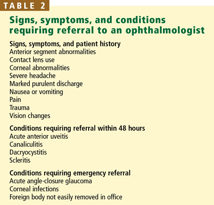
Conditions that require immediate referral to an ophthalmologist can be differentiated from more benign conditions by severe pain or vision loss (Table 2).
Acute angle-closure glaucoma
Patients suffering from an episode of acute angle-closure glaucoma report severe eye pain, seeing halos around lights, headache, nausea, and vomiting. Farsighted people and older people are at greater risk, owing to their eye anatomy. The eyeball is firm to palpation, and the pupil is mid-dilated and poorly reactive to light. The cornea may appear hazy.
Acute angle-closure glaucoma is an emergency and requires immediate lowering of intraocular pressure to avoid permanent vision loss.25
Ocular foreign body
A foreign body lodged in or around the eye causes irritation, redness, and pain. Suspect it in any patient with an appropriate history.
Evert the upper eyelid to search for an occult object and remove any loosely adherent exogenous material on the conjunctiva or sclera. Topical broad-spectrum antibiotic ointments or drops can be started.
Immediately refer any patient with a foreign body that does not dislodge easily for removal and management, or if the patient was working near high-speed objects or with metal on metal (raising the possibility of fragments completely penetrating into the eye).26
Keratitis (corneal inflammation)
Keratitis is inflammation at any level of the cornea.
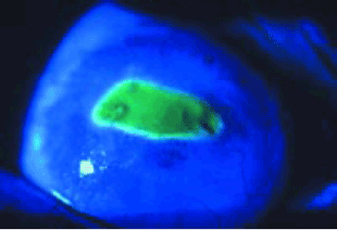
Herpes keratitis presents with unilateral pain, photophobia, and watering. The most common physical finding is a branching ulcer seen with fluorescein staining under Wood’s lamp illumination. Antiviral treatment with an oral medication (acyclovir [Zovirax] 400 mg five times daily) or topical medication (trifluridine 1% [Viroptic] nine times daily) shortens the course of the disease.27,28 Corticosteroid eye-drops should never be given for epithelial herpetic disease without consulting an ophthalmologist.
Bacterial keratitis threatens sight: infection with a virulent bacterium such as Pseudomonas aeruginosa can cause perforation of the cornea within 48 hours. Patients typically report the rapid onset of pain, photophobia, and decreased vision. Common predisposing risk factors include contact lens use and trauma. Examination reveals infiltration, ulceration, and edema of the cornea, and anterior chamber inflammation. Refer immediately to an ophthalmologist for evaluation and management; delaying treatment can have severe visual consequences.29
- Leibowitz HM. The red eye. N Engl J Med 2000; 343:345–351.
- Smith RE, Flowers CW. Chronic blepharitis: a review. CLAO J 1995; 21:200–207.
- McCulley JP, Shine WE. Changing concepts in the diagnosis and management of blepharitis. Cornea 2000; 19:650–658.
- Smith RE. The tear film complex: pathogenesis and emerging therapies for dry eyes. Cornea 2005; 24:1–7.
- Tai MC, Cosar CB, Cohen EJ, Rapuano CJ, Laibson PR. The clinical efficacy of silicone punctal plug therapy. Cornea 2002; 21:135–139.
- McNab AA. Floppy eyelid syndrome and obstructive sleep apnea. Ophthal Plast Reconstr Surg 1997; 13:98–114.
- Alvarenga L, Marinho S, Mark M.Krachmer JH, Mannis MJ, Holland EJ. Viral conjunctivitis, Cornea. 2005: 1. 2. Philadelphia: Elsevier Mosby;629–638.
- Tarabishy AB, Hall GS, Procop GW, Jeng BH. Bacterial culture isolates from hospitalized pediatric patients with conjunctivitis. Am J Ophthalmol 2006; 142:678–680.
- Smith J. Bacterial conjunctivitis. Clin Evid 2004; 12:926–932.
- Sheikh A, Hurwitz B. Topical antibiotics for acute bacterial conjunctivitis: Cochrane systematic review and meta-analysis update. Br J Gen Pract 2005; 55:962–964.
- Ullman S, Roussel TJ, Forster RK. Gonococcal keratoconjunctivitis. Surv Ophthalmol 1987; 32:199–208.
- Deschenes J, Seamone C, Baines M. The ocular manifestations of sexually transmitted diseases. Can J Ophthalmol 1990; 25:177–185.
- Nakagawa H. Treatment of chlamydial conjunctivitis. Ophthalmologica 1997; 211 suppl 1:25–28.
- Owen CG, Shah A, Henshaw K, Smeeth L, Sheikh A. Topical treatments for seasonal allergic conjunctivitis: systematic review and meta-analysis of efficacy and effectiveness. Br J Gen Pract 2004; 54:451–456.
- Stahl JL, Barney NP. Ocular allergic disease. Curr Opin Allergy Clin Immunol 2004; 4:455–459.
- Galor A, Jeng B, Singh A. Current management of corneal abrasion: an evidence based review. Compr Ophthalmol Update 2005; 5:105–111.
- Hirst LW. The treatment of pterygium. Surv Ophthalmol 2003; 48:145–180.
- Mizen TR. Thyroid eye disease. Semin Ophthalmol 2003; 18:243–247.
- Pavesio CE, Meier FM. Systemic disorders associated with episcleritis and scleritis. Curr Opin Ophthalmol 2001; 12:471–478.
- Okhravi N, Odufuwa B, McCluskey P, Lightman S. Scleritis. Surv Ophthalmol 2005; 50:351–363.
- Smith JR, Levinson RD, Holland GN, et al. Differential efficacy of tumor necrosis factor inhibition in the management of inflammatory eye disease and associated rheumatic disease. Arthritis Rheum 2001; 45:252–257.
- Chang JH, Wakefield D. Uveitis: a global perspective. Ocul Immunol Inflamm 2002; 10:263–279.
- Chang JH, McCluskey PJ, Wakefield D. Acute anterior uveitis and HLA-B27. Surv Ophthalmol 2005; 50:364–388.
- Jordan D.Krachmer JH, Mannis MJ, Holland EJ. Dacryoadenitis, dacryocystitis, and canaliculitis, Cornea. 2005: 1. 2. Philadelphia: Elsevier Mosby;541–546.
- Saw SM, Gazzard G, Friedman DS. Interventions for angle-closure glaucoma: an evidence-based update. Ophthalmology 2003; 110:1869–1878.
- Khaw PT, Shah P, Elkington AR. Injury to the eye. BMJ 2004; 328:36–38.
- Suresh PS, Tullo AB. Herpes simplex keratitis. Indian J Ophthalmol 1999; 47:155–165.
- Tullo A. Pathogenesis and management of herpes simplex virus keratitis. Eye 2003; 17:919–922.
- Limberg MB. A review of bacterial keratitis and bacterial conjunctivitis. Am J Ophthalmol 1991; 112:2S–9S.
Many patients present to internists because of redness in the eye. The possible causes range from benign (which generally can be handled by an internist) to vision-threatening (which need prompt or emergency referral to an ophthalmologist).
HISTORY HELPS IDENTIFY THE CAUSE
The internist should ascertain:
- Whether one or both eyes are affected
- The duration of symptoms
- Previous eye and medical problems
- The type of discharge (watery or purulent), if present
- Whether the patient has any visual changes, pain, or photosensitivity.
Refer patients to an ophthalmologist for further evaluation if they use contact lenses or if they have had trauma to the eye, vision changes, severe pain, or systemic symptoms such as nausea, vomiting, or severe headache.
BASIC EYE EXAMINATION
Examine:
- Visual acuity
- Pupil size and reaction to light
- The pattern and location of the redness
- The cornea and anterior segment for gross abnormalities such as corneal opacities, hypopyon (a layer of inflammatory cells in the anterior chamber), and hyphema (hemorrhage in the anterior chamber) (Use a penlight.)
- The preauricular lymph nodes. Preauricular lymphadenopathy, detected by palpation, suggests but is not specific for viral conjunctivitis.
- Funduscopy has little value in evaluating a red eye.
Refer immediately anyone who has marked purulent discharge or abnormalities in the cornea or anterior segment.
CONDITIONS A GENERALIST CAN INITIALLY MANAGE
Subconjunctival hemorrhage
Subconjunctival hemorrhages are harmless and do not cause pain or vision changes. No treatment is required, and the blood resorbs within a few weeks. However:
- Measure the blood pressure—uncontrolled hypertension can present with subconjunctival hemorrhage.1
- If the patient is on an antithrombotic agent, test the prothrombin and activated partial thromboplastin times.
- If the patient has recurrent unexplained episodes of subconjunctival hemorrhage, look for a bleeding disorder such as von Willebrand disease, hemophilia, or autoimmune thrombocytopenic purpura.
Blepharitis
Blepharitis, a common condition, is inflammation of the eyelid margins. Anterior blepharitis affects the eyelashes and anterior eyelid margin and is most often caused by a low-grade staphylococcal infection or seborrheic dermatitis. Posterior blepharitis involves the orifices of the slender sebaceous glands of the eyelids (the meibomian glands) and is often associated with acne rosacea.
Symptoms include ocular burning, a sensation that a foreign body is in the eye, and watering. Symptoms are typically worse in the morning and gradually improve throughout the day. Although the onset is sudden in some patients, blepharitis is usually chronic—often lifelong—and starts insidiously.
A sign of anterior blepharitis is crusting around the eyelashes. Patients with concomitant seborrheic dermatitis also have oily skin and flaking from the eyebrows and scalp. Signs of posterior blepharitis are oil inspissation around the meibomian gland openings, telangiectasias of the eyelid margin, and accompanying acne rosacea (skin pustules, telangiectasias, and erythema).
Treat both forms with eyelid hygiene: applying warm compresses to the eyelid margins, followed by gentle massage to remove the debris from the eyelashes and meibomian glands. This is done two to four times daily until acute symptoms resolve, then once daily. Because blepharitis is chronic, eyelid hygiene must be continued indefinitely to prevent acute exacerbations.
Posterior blepharitis that does not respond to hygiene can be also treated with oral tetracycline, which is believed to improve meibomian gland function and alter bacterial colonization.
Some patients also have tear deficiency, which can be addressed with tear replacement therapy (see below).2,3
Keratoconjunctivitis sicca (dry eye)
Dryness can cause mild eye redness. Patients typically report a foreign body sensation, burning, and paradoxically, watering. Symptoms often worsen as the day progresses and are most prominent at night.
Dryness can be due to:
- Local disturbances in the tear film such as aqueous deficiency
- An abnormal eyelid position
- Systemic autoimmune conditions such as Sjögren syndrome
- Hormonal changes (eg, in menopause)
- Excessively dry environments (eg, winter)
- Medications, including anticholinergics, antihistamines, antidepressants (eg, tricyclics), and antihypertensives (eg, beta-blockers).
Staining the cornea with fluorescein highlights small epithelial defects; rose bengal highlights devitalized cells.
Treat initially with artificial tears (eg, Refresh Tears, GenTeal, Systane, Bion Tears) and ointments (eg, Refresh Liquigel, Lacri-Lube). Dry eye has an inflammatory component; cyclosporine ophthalmic 0.05% (Restasis) may increase tear production and improve symptoms.4
Refer patients with symptoms that do not respond to therapy. An ophthalmologist can place silicone plugs in the canaliculi, a procedure with a 75% success rate for improving dry-eye symptoms.5 Plugs must be carefully fitted: loose ones can spontaneously dislodge, and tight ones can irritate the eye.
Eyelid malposition
Entropion (in-turning of the eyelid) causes eyelashes to rub on the cornea. Ectropion (outward turning of the eyelid) results in tear-film abnormalities and corneal exposure. Both conditions are most commonly caused by aging but may be secondary to scarring or to mechanical, paralytic, or congenital conditions. Definitive treatment involves surgery to restore the normal eyelid position. Several techniques have high success rates.1
Lagophthalmos (inability to fully close the eyes) is caused by orbicularis muscle dysfunction, which may be secondary to Bell palsy, stroke, or neurosurgical procedures that disrupt the facial nerve. The exposed cornea is prone to dryness and irritation. Treatments include artificial tears, lubricating ointments, and surgery—gold weight placement or suturing the eyelid margins (tarsorrhaphy).
Floppy eyelid syndrome refers to a lax upper eyelid that may evert during contact with the pillow during sleep, resulting in irritation and inflammation of the upper palpebral conjunctiva. Signs and symptoms are unilateral eye irritation, burning, and a ropy mucous discharge, which is usually worse in the morning. The upper eyelid is lax and easily everted when pulled toward the eyebrow. Most patients are obese, have obstructive sleep apnea, and sleep on the affected side.
Tell the patient to tape the affected eyelid shut or wear a protective eye shield in bed to prevent rubbing the eye on the pillow. Definitive treatment is surgery to tighten the lax upper eyelid.6
Conjunctivitis
Conjunctivitis involves hyperemia and edema of the bulbar conjunctiva (the part of the conjunctiva covering the eyeball) along with papillary and follicular changes of the palpebral conjunctiva (the inner layer of the eyelids).
Conjunctivitis can be viral, bacterial, or allergic, or due to wearing contact lenses; the cause can usually be distinguished by the history and physical examination.
Viral conjunctivitis, usually caused by an adenovirus, is more common than bacterial conjunctivitis in adults. The patient typically has had a recent upper respiratory tract infection or was exposed to conjunctivitis.
Treat supportively with cool compresses. Symptoms often worsen for a few days, then slowly improve over 1 to 2 weeks.
Viral conjunctivitis is contagious for 2 weeks after the second eye becomes involved, and good hygiene must be maintained to avoid spreading it to coworkers and family members. Those who work with the public, in schools, or in health-care facilities should be given a 2-week leave of absence to avoid spreading the infection to others.
Refer to an ophthalmologist if symptoms do not resolve in 2 weeks, as certain subtypes of adenovirus can cause prolonged symptoms with corneal involvement.7
Treat bacterial conjunctivitis empirically with antibiotic eyedrops (eg, a fluoroquinolone, a polymyxin, or sulfacetamide—several brands available) four times daily for 7 to 10 days, even though most cases are self-limited and do not result in complications. Cultures can be obtained, especially if the patient is in the hospital8 or if the conjunctivitis persists after 1 week of antibiotic therapy.
Refer patients with vision changes or who do not improve after 1 week of treatment.9,10
Treat aggressively with both a topical antibiotic (usually a fluoroquinolone) four times daily and a systemic antibiotic such as ceftriaxone (Rocephin) given as a single 1-g intramuscular injection.11,12 Because one-third of patients with gonorrheal infection also have chlamydial infection, treatment for both diseases is frequently prescribed.
Chlamydial infection, a sexually transmitted disease, can cause chronic follicular conjunctivitis. The genital tract infection may be asymptomatic. Diagnosis is made by swabbing the conjunctiva to culture for Chlamydia trachomatis. Treat systemically with either azithromycin (Zithromax) in a single 1-g oral dose or a 10–14-day course of either doxycycline (Doryx) 100 mg twice daily or erythromycin 250 mg four times daily.13
Allergic conjunctivitis is characterized by bilateral itching that worsens with scratching. Discharge is variable but is usually clear or white and stringy. Many patients have a history of seasonal or perennial allergies.
Remove offending allergens, if possible. Topical mast cell stabilizers and antihistamines relieve symptoms but may exacerbate underlying dry eye symptoms. A combined mast cell stabilizer and antihistamine such as olopatadine (Patanol), ketotifen (Zaditor), or epinastine (Elestat) can be given twice daily.14,15 Artificial tears can treat the associated dryness.
Topical corticosteroids may be used to treat an acute, severe episode but should not be used long-term. In fact, because it is difficult to differentiate between infectious and noninfectious eye conditions, and because treating some infections with corticosteroids by themselves can have grave consequences, we recommend that internists generally refrain from using them.
Oral antihistamines may relieve symptoms but are usually less effective than topical therapy.
Refer if symptoms do not resolve after 2 weeks of topical treatment.
Giant papillary conjunctivitis, most often seen in patients who wear soft contact lenses, presents with bilateral contact lens intolerance, itching, mucous discharge, and giant papillae on the upper palpebral conjunctiva.
Again, promptly refer any patient who wears contact lenses and presents with a red eye, owing to the risk of a vision-threatening corneal infection. The patient should stop wearing contact lenses for about 1 month, after which he or she can be refitted with new soft or gas-permeable lenses and taught better lens hygiene. During an acute episode, topical mast cell stabilizers are helpful for mild irritation, and topical steroids (prednisolone phosphate 1%) are helpful for more severe irritation. Topical steroids should never be used on a long-term basis because of possible adverse effects. Artificial tears can be used for dryness.15
Corneal abrasion
Treat with topical antibiotics to prevent infection until the corneal epithelium has healed.16 However, most abrasions heal rapidly without sequelae because epithelial cells proliferate and migrate rapidly.
Refer if symptoms persist for longer than 48 hours or if pain suddenly worsens after the healing process has started.
Pingueculae and pterygia
A pinguecula is a small, yellow, benign growth on the nasal and temporal conjunctiva near the limbus. A pterygium is a wing-shaped band of fibrovascular tissue originating on the conjunctiva and extending onto the cornea. Both conditions are idiopathic but are believed to arise from chronic sun exposure.
Pingueculae can become inflamed and can cause eye redness and irritation. Treat conservatively with lubrication and judicious use of topical steroids; if irritation persists, pingueculae can be surgically removed.17
Episcleritis
Episcleritis is inflammation of the superficial vessels of the episclera, the connective tissue layer between the conjunctiva and the sclera. It presents with a sectoral area of redness (although it can be diffuse) and is not typically associated with pain, vision changes, or discharge. The condition tends to be recurrent and unilateral, but it can be bilateral or alternating. The underlying pathophysiology is believed to be autoimmune, although a systemic evaluation is often unrevealing.
Episcleritis is treated with topical corticosteroids or oral nonsteroidal anti-inflammatory drugs (NSAIDs); refer if the disease persists or recurs.
Thyroid-related eye disease
Thyroid-associated ophthalmopathy, an autoimmune process, usually occurs in patients with known thyroid disease, although it may develop before other systemic symptoms. Symptoms can include irritation and double vision. Signs are bulging eyes, eyelid retraction, chemosis (swelling of the conjunctiva around the cornea), conjunctival injection, periorbital edema, and limited ocular motility.
Although most cases can be managed with lubrication, vision loss may occur due to corneal exposure or compressive optic neuropathy. Patients with significant visual changes should be referred immediately to an ophthalmologist.18
CONDITIONS NEEDING REFERRAL WITHIN 48 HOURS
Scleritis
Scleritis is inflammation of the deep vessels within the episclera. The red color appears more pronounced and more purplish than in episcleritis and does not blanch after phenylephrine drops are given. The eye is tender to palpation and may be painful enough to awaken the patient from sleep. Vision is not typically affected unless the cornea, anterior chamber, or posterior segment is involved.
Half of patients who have scleritis have an associated systemic disease, eg, rheumatoid arthritis (most common), other autoimmune diseases (Wegener granulomatosis, relapsing polychondritis, inflammatory bowel disease), or infections such as tuberculosis and syphilis.
Therefore, one should search for an underlying systemic condition with a thorough history, physical examination, chest radiography (for sarcoidosis and tuberculosis), and laboratory testing: antineutrophil cytoplasmic antibody test, fluorescent treponemal antibody absorption test, Lyme antibody test, (if in an endemic area), urinalysis, a complete blood count, and a comprehensive metabolic panel.
However, patients should be promptly referred to an ophthalmologist for diagnosis and management. Treatment can depend on the underlying diagnosis, and is often guided by the status of the scleritis. Mild scleritis can be treated with oral NSAIDs; more severe disease should be treated with oral corticosteroids with or without corticosteroid-sparing agents such as methotrexate, mycophenolate (CellCept), cyclophosphamide (Cytoxan, Neosar),19,20 or tumor necrosis factor-alpha antagonists such as infliximab (Remicade) or etanercept (Enbrel).21
Anterior uveitis
Uveitis is inflammation of the uvea (the pigmented layer between the sclera and retina that includes the iris, ciliary body, and choroid). Anterior uveitis is most commonly idiopathic but can be caused by trauma, secondary to herpes virus infection, or associated with the HLA-B27 antigen.
Acute anterior uveitis presents with pain, photophobia, and blurred vision. Perilimbal (circumcorneal) injection overlies the inflamed ciliary body. The pupil is often constricted and poorly reactive to light. Chronic anterior uveitis, defined as lasting more than 6 weeks, typically presents with gradual vision loss and floaters, rather than with the acute pain or severe redness of acute disease. Anterior uveitis is diagnosed by finding cells and flare in the anterior chamber using a slit lamp.
Refer patients to an ophthalmologist immediately to help avoid visual consequences.22,23 Treatment begins with topical corticosteroid drops and can also include oral corticosteroids or long-term immunosuppresion with corticosteroid-sparing agents.
Nasolacrimal infections
Canaliculitis is an inflammation of the canaliculus, the conduit bringing tears from the eye to the nasolacrimal duct. It presents with mild, unilateral eye redness and a slight discharge that can be expressed from the punctum. It is most commonly caused by Actinomyces israelii infection, but Candida and Aspergillus species can also be involved.
Refer to an ophthalmologist for treatment, which involves mechanically removing the granular material from the canaliculi, combined with probing and irrigating the nasolacrimal system with penicillin G solution.
Dacryocystitis is inflammation of the lacrimal sac (the dilated upper end of the nasolacrimal duct) and is caused by obstruction of the duct. Staphylococcus and Streptococcus species are usually involved. Symptoms include unilateral pain, swelling, and redness over the lacrimal sac at the medial canthus of the eye. Purulent discharge can be expressed from the punctum.
Treatment consists of oral antibiotics with gram-positive coverage followed by surgery to open a passage for drainage from the lacrimal sac into the nasal cavity (dacryocystorhinostomy) once the infection has resolved.24
CONDITIONS NEEDING IMMEDIATE REFERRAL
Conditions that require immediate referral to an ophthalmologist can be differentiated from more benign conditions by severe pain or vision loss (Table 2).
Acute angle-closure glaucoma
Patients suffering from an episode of acute angle-closure glaucoma report severe eye pain, seeing halos around lights, headache, nausea, and vomiting. Farsighted people and older people are at greater risk, owing to their eye anatomy. The eyeball is firm to palpation, and the pupil is mid-dilated and poorly reactive to light. The cornea may appear hazy.
Acute angle-closure glaucoma is an emergency and requires immediate lowering of intraocular pressure to avoid permanent vision loss.25
Ocular foreign body
A foreign body lodged in or around the eye causes irritation, redness, and pain. Suspect it in any patient with an appropriate history.
Evert the upper eyelid to search for an occult object and remove any loosely adherent exogenous material on the conjunctiva or sclera. Topical broad-spectrum antibiotic ointments or drops can be started.
Immediately refer any patient with a foreign body that does not dislodge easily for removal and management, or if the patient was working near high-speed objects or with metal on metal (raising the possibility of fragments completely penetrating into the eye).26
Keratitis (corneal inflammation)
Keratitis is inflammation at any level of the cornea.
Herpes keratitis presents with unilateral pain, photophobia, and watering. The most common physical finding is a branching ulcer seen with fluorescein staining under Wood’s lamp illumination. Antiviral treatment with an oral medication (acyclovir [Zovirax] 400 mg five times daily) or topical medication (trifluridine 1% [Viroptic] nine times daily) shortens the course of the disease.27,28 Corticosteroid eye-drops should never be given for epithelial herpetic disease without consulting an ophthalmologist.
Bacterial keratitis threatens sight: infection with a virulent bacterium such as Pseudomonas aeruginosa can cause perforation of the cornea within 48 hours. Patients typically report the rapid onset of pain, photophobia, and decreased vision. Common predisposing risk factors include contact lens use and trauma. Examination reveals infiltration, ulceration, and edema of the cornea, and anterior chamber inflammation. Refer immediately to an ophthalmologist for evaluation and management; delaying treatment can have severe visual consequences.29
- Leibowitz HM. The red eye. N Engl J Med 2000; 343:345–351.
- Smith RE, Flowers CW. Chronic blepharitis: a review. CLAO J 1995; 21:200–207.
- McCulley JP, Shine WE. Changing concepts in the diagnosis and management of blepharitis. Cornea 2000; 19:650–658.
- Smith RE. The tear film complex: pathogenesis and emerging therapies for dry eyes. Cornea 2005; 24:1–7.
- Tai MC, Cosar CB, Cohen EJ, Rapuano CJ, Laibson PR. The clinical efficacy of silicone punctal plug therapy. Cornea 2002; 21:135–139.
- McNab AA. Floppy eyelid syndrome and obstructive sleep apnea. Ophthal Plast Reconstr Surg 1997; 13:98–114.
- Alvarenga L, Marinho S, Mark M.Krachmer JH, Mannis MJ, Holland EJ. Viral conjunctivitis, Cornea. 2005: 1. 2. Philadelphia: Elsevier Mosby;629–638.
- Tarabishy AB, Hall GS, Procop GW, Jeng BH. Bacterial culture isolates from hospitalized pediatric patients with conjunctivitis. Am J Ophthalmol 2006; 142:678–680.
- Smith J. Bacterial conjunctivitis. Clin Evid 2004; 12:926–932.
- Sheikh A, Hurwitz B. Topical antibiotics for acute bacterial conjunctivitis: Cochrane systematic review and meta-analysis update. Br J Gen Pract 2005; 55:962–964.
- Ullman S, Roussel TJ, Forster RK. Gonococcal keratoconjunctivitis. Surv Ophthalmol 1987; 32:199–208.
- Deschenes J, Seamone C, Baines M. The ocular manifestations of sexually transmitted diseases. Can J Ophthalmol 1990; 25:177–185.
- Nakagawa H. Treatment of chlamydial conjunctivitis. Ophthalmologica 1997; 211 suppl 1:25–28.
- Owen CG, Shah A, Henshaw K, Smeeth L, Sheikh A. Topical treatments for seasonal allergic conjunctivitis: systematic review and meta-analysis of efficacy and effectiveness. Br J Gen Pract 2004; 54:451–456.
- Stahl JL, Barney NP. Ocular allergic disease. Curr Opin Allergy Clin Immunol 2004; 4:455–459.
- Galor A, Jeng B, Singh A. Current management of corneal abrasion: an evidence based review. Compr Ophthalmol Update 2005; 5:105–111.
- Hirst LW. The treatment of pterygium. Surv Ophthalmol 2003; 48:145–180.
- Mizen TR. Thyroid eye disease. Semin Ophthalmol 2003; 18:243–247.
- Pavesio CE, Meier FM. Systemic disorders associated with episcleritis and scleritis. Curr Opin Ophthalmol 2001; 12:471–478.
- Okhravi N, Odufuwa B, McCluskey P, Lightman S. Scleritis. Surv Ophthalmol 2005; 50:351–363.
- Smith JR, Levinson RD, Holland GN, et al. Differential efficacy of tumor necrosis factor inhibition in the management of inflammatory eye disease and associated rheumatic disease. Arthritis Rheum 2001; 45:252–257.
- Chang JH, Wakefield D. Uveitis: a global perspective. Ocul Immunol Inflamm 2002; 10:263–279.
- Chang JH, McCluskey PJ, Wakefield D. Acute anterior uveitis and HLA-B27. Surv Ophthalmol 2005; 50:364–388.
- Jordan D.Krachmer JH, Mannis MJ, Holland EJ. Dacryoadenitis, dacryocystitis, and canaliculitis, Cornea. 2005: 1. 2. Philadelphia: Elsevier Mosby;541–546.
- Saw SM, Gazzard G, Friedman DS. Interventions for angle-closure glaucoma: an evidence-based update. Ophthalmology 2003; 110:1869–1878.
- Khaw PT, Shah P, Elkington AR. Injury to the eye. BMJ 2004; 328:36–38.
- Suresh PS, Tullo AB. Herpes simplex keratitis. Indian J Ophthalmol 1999; 47:155–165.
- Tullo A. Pathogenesis and management of herpes simplex virus keratitis. Eye 2003; 17:919–922.
- Limberg MB. A review of bacterial keratitis and bacterial conjunctivitis. Am J Ophthalmol 1991; 112:2S–9S.
KEY POINTS
- Blepharitis, conjunctivitis, corneal abrasion, dry eye, and subconjunctival hemorrhage are benign and can usually be managed initially by an internist, although referral is usually indicated if symptoms persist or progress.
- Patients with corneal bacterial infection, uveitis, scleritis, or acute narrow-angle glaucoma need immediate referral to an ophthalmologist, as do most patients with a red eye who use contact lenses, who have had trauma to the eye, or who have vision changes, severe pain, nausea, vomiting, severe headache, marked purulent discharge, or abnormalities in the cornea or anterior segment.
- Because it is difficult to distinguish between infectious and noninfectious conditions, and because treating infections with corticosteroids alone can have grave consequences, we recommend that internists generally not use topical corticosteroids to treat eye symptoms.