User login
Suspect Carpal Tunnel? Try This
For best results, use the modified Phalen’s test (MPT) rather than the traditional Phalen’s when you suspect carpal tunnel syndrome (CTS).1
STRENGTH OF
RECOMMENDATION
B: Based on a single diagnostic cohort study.
ILLUSTRATIVE CASE
A 60-year-old assembly line worker reports bilateral hand numbness and tingling that frequently awaken her at night. What is the best office test to determine if she has CTS?
CTS is one of the most common causes of disability in the United States.2 Among patients with hand paresthesias, one in five has CTS.2 Factory workers whose jobs involve repetitive hand movements, women, and the elderly are at increased risk.3 If left untreated, the symptoms are likely to become constant, with thenar muscle wasting and weakness.
Traditional diagnostic test
has only 50% sensitivity
In the traditional Phalen’s test (TPT)—commonly used in an office setting—the patient holds his or her wrists in a position of fixed flexion for one minute. The onset of paresthesias is considered a positive result.
The TPT was found in the study reported here to be 100% specific;1 however, other studies have found a wider range of specificity (33% to 86%).4 The TPT has a sensitivity of only 50%, which increases the risk that cases of CTS will be missed. This is an important consideration, because establishing a diagnosis early in the course of CTS has been shown to minimize disability.5
STUDY SUMMARY
Modified Phalen’s has higher sensitivity
Bilkis et al developed a modified Phalen’s test and compared it with the TPT, as well as with electrodiagnostic studies (EDS)—the gold standard for CTS diagnosis. The MPT begins with the TPT position and adds sensory testing with a Semmes-Weinstein 2.83-unit monofilament.
See how the modified Phalen’s test is done
Courtesy of Clinically Relevant Technologies
The filament is applied perpendicular to the palmar and lateral surface of each distal finger three times, with enough pressure to bend the monofilament. In this study, the test was considered “positive” if the patient did not feel the monofilament in any finger along the distribution of the median nerve. The MPT was “negative” if the patient correctly reported being touched along this distribution. The fifth, or “pinkie,” finger, which is less likely to be affected by CTS, was used as a control.
Participants in the study were adult patients—mostly women between the ages of 27 and 88—at a neurology clinic. Exclusion criteria included cervical radiculopathy, a history of stroke, diabetes, and concomitant neck injury. A total of 66 hands (and 37 participants) underwent TPT and MPT testing by trained examiners, followed by EDS to confirm the findings.
EDS found evidence of CTS in 46 of the 66 hands studied. The MPT correctly identified 39 of the 46, while the TPT correctly identified 23. Both the traditional and the modified Phalen’s tests were found to be 100% specific, but the sensitivity of the MPT was 85% (95% confidence interval [CI], 71% to 93%), compared with 50% (95% CI, 35% to 65%) for the TPT.
WHAT’S NEW
Better results can be achieved in seconds
The addition of monofilament testing to the TPT increases the sensitivity in identifying CTS. The MPT is simple to learn and, based on our observations, adds only about 10 to 15 seconds to the clinical exam.
CAVEATS
Modification is untested in primary care
A diagnosis of CTS is rarely made on the basis of one test, but rather on a set of signs, symptoms, and physical exam maneuvers. The added value of the MPT needs to be evaluated in the larger context of the comprehensive clinical examination for CTS.6
Notably, the study participants were seen in a neurology clinic, which suggests that they may have had more advanced CTS than typical primary care patients. That would help explain the 100% specificity of both the traditional and modified tests reported by the researchers. The sensitivity of the MPT may therefore be lower in a family practice because the spectrum of disease may be wider. Another study is needed to evaluate the performance of the MPT in a primary care setting.
The monofilament used (Semmes-Weinstein 2.83) is not the same as the typical 5.07 (10-g) monofilament used in diabetic foot screenings. Using this heavier monofilament with a stronger pressure point would likely decrease the sensitivity of the MPT.
CHALLENGES TO IMPLEMENTATION
Taking the time, obtaining the monofilament
Additional time to obtain the correct monofilament and administer the MPT are the key challenges to implementation.
REFERENCES
1. Bilkis S, Loveman DM, Eldridge JA, et al. Modified Phalen’s test as an aid in diagnosing carpal tunnel syndrome. Arthritis Care Res. 2012;64:287-289.
2. Atroshi I, Gummesson C, Johnsson R, et al. Prevalence of carpal tunnel syndrome in a general population. JAMA. 1999;282:153-158.
3. National Institute of Neurological Disorders and Stroke. Carpal tunnel syndrome fact sheet. National Institutes of Health. July 2012. www.ninds.nih.gov/disorders/carpal_tunnel/detail_carpal_tunnel.htm. Accessed April 15, 2013.
4. McGee SR. Evidence-Based Physical Diagnosis. 3rd ed. Philadelphia, PA: Saunders; 2012:chap 62.
5. Daniell WE, Fulton-Kehoe D, Franklin GM. Work-related carpal tunnel syndrome in Washington State workers’ compensation: utilization of surgery and the duration of lost work. Am J Ind Med. 2009;52:931-942.
6. D’Arcy CA, McGee S. Does this patient have carpal tunnel syndrome? JAMA. 2000;282:3110-3117.
ACKNOWLEDGEMENT
The PURLs Surveillance System was developed with support from Grant Number UL1RR024999 from the National Center for Research Resources, a Clinical Translational Science Award to the University of Chicago. The content is solely the responsibility of the authors and does not necessarily represent the official views of the National Center for Research Resources or the National Institutes of Health.
Copyright © 2013. The Family Physicians Inquiries Network. All rights reserved.
Reprinted with permission from the Family Physicians Inquiries Network and The Journal of Family Practice. 2013;62(5):253-254.
Suspect carpal tunnel? Try this
For best results, use the modified Phalen’s test (MPT) rather than the traditional Phalen’s when you suspect carpal tunnel syndrome (CTS).1
1. Bilkis S, Loveman DM, Eldridge JA, et al. Modified Phalen’s test as an aid in diagnosing carpal tunnel syndrome. Arthritis Care Res. 2012;64:287-289.
STRENGTH OF RECOMMENDATION
B: Based on a single diagnostic cohort study.
ILLUSTRATIVE CASE
A 60-year-old assembly line worker reports bilateral hand numbness and tingling that frequently awaken her at night. What is the best office test to determine if she has CTS?
CTS is one of the most common causes of disability in the United States.2 Among patients with hand paresthesias, one in 5 has CTS.2 Factory workers whose jobs involve repetitive hand movements, females, and the elderly are at increased risk.3 If left untreated, the symptoms are likely to become constant, with thenar muscle wasting and weakness.
Traditional diagnostic test has only 50% sensitivity
In the traditional Phalen’s test (TPT)—commonly used in an office setting—the patient holds his or her wrists in a position of fixed flexion for one minute. The onset of paresthesias is considered a positive result.
The TPT was found in the study reported here to be 100% specific;1 however, other studies have found a wider range of specificity (33%-86%).4 The TPT has a sensitivity of only 50%, which increases the risk that cases of CTS will be missed. This is an important consideration because establishing a diagnosis early in the course of CTS has been shown to minimize disability.5
STUDY SUMMARY: Modified Phalen’s has higher sensitivity
Bilkis et al developed a modified Phalen’s test (MPT) and compared it with the TPT, as well as with electrodiagnostic studies (EDS)—the gold standard for CTS diagnosis. The MPT begins with the TPT position and adds sensory testing with a Semmes-Weinstein 2.83-unit monofilament.
See how the modified Phalen’s test is done
Courtesy of Clinically Relevant Technologies
The filament is applied perpendicular to the palmar and lateral surface of each distal finger 3 times, with enough pressure to bend the monofilament. In this study, the test was considered positive if the patient did not feel the monofilament in any finger along the distribution of the median nerve. The MPT was negative if the patient correctly reported being touched along this distribution. The fifth, or “pinkie,” finger, which is less likely to be affected by CTS, was used as a control.
Participants in the study were adult patients—mostly women between the ages of 27 and 88 years—at a neurology clinic. Exclusion criteria included cervical radiculopathy, a history of stroke, diabetes mellitus, and concomitant neck injury. A total of 66 hands (and 37 participants) underwent TPT and MPT testing by trained examiners, followed by EDS to confirm the findings.
EDS found evidence of CTS in 46 of the 66 hands studied. The MPT correctly identified 39 of the 46, while the TPT correctly identified 23. Both the traditional and the modified Phalen’s were found to be 100% specific, but the sensitivity of the MPT was 85% (95% confidence interval [CI], 71%-93%), compared with 50% (95% CI, 35%-65%) for the TPT.
WHAT’S NEW: Better results can be achieved in seconds
The addition of monofilament testing to the TPT increases the sensitivity in identifying CTS. The MPT is simple to learn (watch the video on jfponline.com) and, based on our observations, adds only about 10 to 15 seconds to the clinical exam.
CAVEATS: Modification is untested in primary care
A diagnosis of CTS is rarely made on the basis of one test, but rather on a set of signs, symptoms, and physical exam maneuvers. The added value of the MPT needs to be evaluated in the larger context of the comprehensive clinical examination for CTS.6
Notably, the study participants were seen in a neurology clinic, which suggests that they may have had more advanced CTS than typical primary care patients. That would help explain the 100% specificity of both the traditional and modified tests reported by the researchers. The sensitivity of the MPT may therefore be lower in a family physician’s office because the spectrum of disease may be wider. Another study is needed to evaluate the performance of the MPT in a primary care setting.
The monofilament used (Semmes-Weinstein 2.83) is not the same as the typical 5.07 (10-g) monofilament used in diabetic foot screenings. Using this heavier monofilament with a stronger pressure point would likely decrease the sensitivity of the MPT.
CHALLENGES TO IMPLEMENTATION: Taking the time, obtaining the monofilament
Additional time to obtain the correct monofilament and administer the MPT are the key challenges to implementation.
Acknowledgement
The PURLs Surveillance System was supported in part by Grant Number UL1RR024999 from the National Center for Research Resources, a Clinical Translational Science Award to the University of Chicago. The content is solely the responsibility of the authors and does not necessarily represent the official views of the National Center for Research Resources or the National Institutes of Health.
1. Bilkis S, Loveman DM, Eldridge JA, et al. Modified Phalen’s test as an aid in diagnosing carpal tunnel syndrome. Arthritis Care Res. 2012;64:287-289.
2. Atroshi I, Gummesson C, Johnsson R, et al. Prevalence of carpal tunnel syndrome in a general population. JAMA. 1999;282:153-158.
3. National Institute of Neurological Disorders and Stroke. Carpal tunnel syndrome fact sheet. National Institutes of Health. July 2012. Available at http://www.ninds.nih.gov/disorders/carpal_tunnel/detail_carpal_tunnel.htm. Accessed April 15, 2013.
4. McGee SR. Evidence-Based Physical Diagnosis. 3rd ed. Philadelphia, Pa: Saunders; 2012:chap 62.
5. Daniell WE, Fulton-Kehoe D, Franklin GM. Work-related carpal tunnel syndrome in Washington State workers’ compensation: utilization of surgery and the duration of lost work. Am J Ind Med. 2009;52:931-942.
6. D’Arcy CA, McGee S. Does this patient have carpal tunnel syndrome? JAMA. 2000;282:3110-3117.
For best results, use the modified Phalen’s test (MPT) rather than the traditional Phalen’s when you suspect carpal tunnel syndrome (CTS).1
1. Bilkis S, Loveman DM, Eldridge JA, et al. Modified Phalen’s test as an aid in diagnosing carpal tunnel syndrome. Arthritis Care Res. 2012;64:287-289.
STRENGTH OF RECOMMENDATION
B: Based on a single diagnostic cohort study.
ILLUSTRATIVE CASE
A 60-year-old assembly line worker reports bilateral hand numbness and tingling that frequently awaken her at night. What is the best office test to determine if she has CTS?
CTS is one of the most common causes of disability in the United States.2 Among patients with hand paresthesias, one in 5 has CTS.2 Factory workers whose jobs involve repetitive hand movements, females, and the elderly are at increased risk.3 If left untreated, the symptoms are likely to become constant, with thenar muscle wasting and weakness.
Traditional diagnostic test has only 50% sensitivity
In the traditional Phalen’s test (TPT)—commonly used in an office setting—the patient holds his or her wrists in a position of fixed flexion for one minute. The onset of paresthesias is considered a positive result.
The TPT was found in the study reported here to be 100% specific;1 however, other studies have found a wider range of specificity (33%-86%).4 The TPT has a sensitivity of only 50%, which increases the risk that cases of CTS will be missed. This is an important consideration because establishing a diagnosis early in the course of CTS has been shown to minimize disability.5
STUDY SUMMARY: Modified Phalen’s has higher sensitivity
Bilkis et al developed a modified Phalen’s test (MPT) and compared it with the TPT, as well as with electrodiagnostic studies (EDS)—the gold standard for CTS diagnosis. The MPT begins with the TPT position and adds sensory testing with a Semmes-Weinstein 2.83-unit monofilament.
See how the modified Phalen’s test is done
Courtesy of Clinically Relevant Technologies
The filament is applied perpendicular to the palmar and lateral surface of each distal finger 3 times, with enough pressure to bend the monofilament. In this study, the test was considered positive if the patient did not feel the monofilament in any finger along the distribution of the median nerve. The MPT was negative if the patient correctly reported being touched along this distribution. The fifth, or “pinkie,” finger, which is less likely to be affected by CTS, was used as a control.
Participants in the study were adult patients—mostly women between the ages of 27 and 88 years—at a neurology clinic. Exclusion criteria included cervical radiculopathy, a history of stroke, diabetes mellitus, and concomitant neck injury. A total of 66 hands (and 37 participants) underwent TPT and MPT testing by trained examiners, followed by EDS to confirm the findings.
EDS found evidence of CTS in 46 of the 66 hands studied. The MPT correctly identified 39 of the 46, while the TPT correctly identified 23. Both the traditional and the modified Phalen’s were found to be 100% specific, but the sensitivity of the MPT was 85% (95% confidence interval [CI], 71%-93%), compared with 50% (95% CI, 35%-65%) for the TPT.
WHAT’S NEW: Better results can be achieved in seconds
The addition of monofilament testing to the TPT increases the sensitivity in identifying CTS. The MPT is simple to learn (watch the video on jfponline.com) and, based on our observations, adds only about 10 to 15 seconds to the clinical exam.
CAVEATS: Modification is untested in primary care
A diagnosis of CTS is rarely made on the basis of one test, but rather on a set of signs, symptoms, and physical exam maneuvers. The added value of the MPT needs to be evaluated in the larger context of the comprehensive clinical examination for CTS.6
Notably, the study participants were seen in a neurology clinic, which suggests that they may have had more advanced CTS than typical primary care patients. That would help explain the 100% specificity of both the traditional and modified tests reported by the researchers. The sensitivity of the MPT may therefore be lower in a family physician’s office because the spectrum of disease may be wider. Another study is needed to evaluate the performance of the MPT in a primary care setting.
The monofilament used (Semmes-Weinstein 2.83) is not the same as the typical 5.07 (10-g) monofilament used in diabetic foot screenings. Using this heavier monofilament with a stronger pressure point would likely decrease the sensitivity of the MPT.
CHALLENGES TO IMPLEMENTATION: Taking the time, obtaining the monofilament
Additional time to obtain the correct monofilament and administer the MPT are the key challenges to implementation.
Acknowledgement
The PURLs Surveillance System was supported in part by Grant Number UL1RR024999 from the National Center for Research Resources, a Clinical Translational Science Award to the University of Chicago. The content is solely the responsibility of the authors and does not necessarily represent the official views of the National Center for Research Resources or the National Institutes of Health.
1. Bilkis S, Loveman DM, Eldridge JA, et al. Modified Phalen’s test as an aid in diagnosing carpal tunnel syndrome. Arthritis Care Res. 2012;64:287-289.
2. Atroshi I, Gummesson C, Johnsson R, et al. Prevalence of carpal tunnel syndrome in a general population. JAMA. 1999;282:153-158.
3. National Institute of Neurological Disorders and Stroke. Carpal tunnel syndrome fact sheet. National Institutes of Health. July 2012. Available at http://www.ninds.nih.gov/disorders/carpal_tunnel/detail_carpal_tunnel.htm. Accessed April 15, 2013.
4. McGee SR. Evidence-Based Physical Diagnosis. 3rd ed. Philadelphia, Pa: Saunders; 2012:chap 62.
5. Daniell WE, Fulton-Kehoe D, Franklin GM. Work-related carpal tunnel syndrome in Washington State workers’ compensation: utilization of surgery and the duration of lost work. Am J Ind Med. 2009;52:931-942.
6. D’Arcy CA, McGee S. Does this patient have carpal tunnel syndrome? JAMA. 2000;282:3110-3117.
Copyright © 2013 The Family Physicians Inquiries Network. All rights reserved.
A Safer Way to Prevent VTE Recurrence
PRACTICE CHANGER
After patients with unprovoked venous thromboembolism (VTE) complete a 6- to 18-month course of oral anticoagulation therapy, consider a switch to aspirin.1
STRENGTH OF
RECOMMENDATION
A: Based on one well-designed, randomized controlled trial (RCT).
ILLUSTRATIVE CASE
A 62-year-old patient comes to your office for follow-up of a primary unprovoked VTE. He has been on an oral anticoagulant for 12 months. Should he continue anticoagulation therapy despite the increased risk for major bleeding?
Patients who survive VTE—defined as either deep venous thrombosis (DVT) or pulmonary embolism (PE)—are put on anticoagulant therapy to prevent a recurrence, typically for six to 18 months. But about 20% of patients with unprovoked VTE have a recurrence within two years of anticoagulation withdrawal.2 Extending anticoagulation prevents recurrences but increases the risk for bleeding.3
Is aspirin a viable alternative?
Until recently, the efficacy of aspirin for the prevention of recurrent VTE was unknown. Becattini et al1 investigated it in the multicenter RCT detailed in this PURL.
STUDY SUMMARY
Aspirin can prevent recurrence with minimal risk
To determine whether aspirin was a viable alternative to oral anticoagulation, the researchers compared aspirin with placebo in patients with primary unprovoked VTE who had completed a course of oral anticoagulation treatment.
To be considered for the study, patients had to be older than 18 and have had their first-ever objectively confirmed, symptomatic unprovoked proximal DVT PE, or both. They also had to have completed six to 18 months of anticoagulant therapy, with a target international normalized ratio (INR) of 2.0 to 3.0. Exclusion criteria included a history of cancer, clinically significant thrombophilia, atrial fibrillation, and a bleeding event that occurred during the course of anticoagulation therapy.
Becattini et al identified 403 eligible patients. Two weeks after stopping anticoagulation, patients were randomly assigned to receive either aspirin 100 mg/d (n = 205) or placebo (n = 198) for two years. (One patient in the placebo group never received treatment.)
At baseline, there were no significant differences in patient characteristics. All were evaluated every three months in the first year and every six months in the second year.
The primary efficacy outcome was objectively confirmed recurrent VTE. The primary safety outcome was major bleeding, defined as bleeding that occurred in a critical location (eg, intracranial bleeding), was associated with a decrease of hemoglobin of at least 2 g/dL, required a transfusion of two units of whole blood or red blood cells, or was fatal. Overt bleeding, which required medical intervention but did not meet the criteria for major bleeding, was a secondary safety outcome.
Twenty-eight of the 205 patients in the aspirin group experienced a recurrence, compared with 43 of the 197 patients on placebo (6.6% vs 11.2% per year; hazard ratio [HR] = 0.58).
Adverse events were reported by seven patients in the aspirin therapy group and six in the placebo group. One patient in each group experienced major bleeding, and three in each group experienced clinically relevant but nonmajor bleeding.
Withdrawal rates were similar (10 in the group receiving aspirin vs 9 in the group receiving placebo), as were the number of patients who developed new indications for aspirin or anticoagulation therapy or were lost to follow-up.
An analysis adjusted for age, sex, index event (DVT or PE) and duration of initial anticoagulation treatment confirmed that aspirin reduced the risk for recurrence (adjusted HR = 0.53). No association was found between recurrent VTE and duration of anticoagulation therapy (six months vs longer). Nor was there a difference in recurrence rates based on the index event.
WHAT'S NEW
Aspirin has a key role
in preventing recurrence
This study found that for patients with unprovoked VTE who completed a course of oral anticoagulation, aspirin was effective in preventing a recurrence, with no apparent increase in the risk for major bleeding. Protection in year 2 was nearly as great as in year 1.1
CAVEAT
Patients were followed
for just two years
It is unclear whether continuing aspirin therapy beyond two years would continue to confer protection against a VTE recurrence without an increase in adverse effects.
CHALLENGE TO IMPLEMENTATION
Some patients can't tolerate chronic aspirin therapy
Although this study investigated aspirin in a dosage of 100 mg/d, this strength is not readily available in the United States.4 There is no evidence to suggest that the 81-mg strength that is available in this country would provide a diminished antiplatelet effect.
And, as is already customary, patients undergoing chronic aspirin therapy must be monitored for major bleeding, GI irritation, and renal compromise. A few patients will be ineligible for prophylaxis due to a history of intolerance to aspirin or NSAIDs.
REFERENCES
1. Becattini C, Agnelli G, Schenone A, et al. Aspirin for preventing the recurrence of venous thromboembolism. N Engl J Med. 2012;366:1959-1967.
2. Prandoni P, Lensing AW, Cogo A, et al. The long-term clinical course of acute deep venous thrombosis. Ann Intern Med. 1996;125:1-7.
3. Kearon C, Akl EA, Comerota AJ, et al. Antithrombotic therapy for VTE disease: antithrombotic therapy and prevention of thrombosis. American College of Chest Physicians evidence-based clinical practice guidelines. Chest. 2012;141 (2 suppl):e419S-e494S.
4. Daily Med. Aspirin. dailymed.nlm.nih.gov/dailymed/search.cfm?startswith=aspirin. Accessed September 6, 2012.
Acknowledgement
The PURLs Surveillance System was supported in part by Grant Number UL1RR024999 from the National Center for Research Resources, a Clinical Translational Science Award to the University of Chicago. The content is solely the responsibility of the authors and does not necessarily represent the official views of the National Center for Research Resources or the National Institutes of Health.
Copyright © 2012. The Family Physicians Inquiries Network. All rights reserved.
Reprinted with permission from the Family Physicians Inquiries Network and The Journal of Family Practice. 2012;61:673-674.
What's best for IBS?
Practice Changer
Recommend antispasmodics or antidepressants for patients with irritable bowel syndrome (IBS) and explain that, while fiber may have other benefits, it is unlikely to relieve IBS symptoms.1
Strength of recommendation
A: Based on a meta-analysis.
Illustrative Case
A 25-year-old woman has intermittent bouts of abdominal pain, constipation, gas, and bloating. You believe she can benefit from treatment for IBS. What should you recommend?
IBS is the most common functional disorder of the gastrointestinal (GI) tract, affecting approximately 15% of the US population2 and accounting for annual health care costs of roughly $30 billion.3 The primary symptoms are bloating, gas, and abdominal pain that often improves immediately after a bowel movement. Patients may have intermittent diarrhea and constipation, as well.
IBS may be related to “brain-gut dysfunction”
The etiology of IBS is unclear, but many agree that a combination of abnormal GI motility, visceral hypersensitivity, and “brain-gut dysfunction”—the inability of the brain to send signals that turn down pain produced in the GI tract—are contributing factors. Although IBS is not life threatening, it has a significant personal, social, and psychological impact. Despite its high prevalence and impact, only a limited number of large studies have assessed the effectiveness of various treatments.
Study Summary
Antispasmodics, antidepressants offer relief—fiber does not
This Cochrane review included 56 randomized controlled trials (RCTs) comparing the efficacy of bulking agents (fiber supplements), antispasmodics, or antidepressants with placebo for the treatment of IBS. Twelve RCTs (n = 621) focused on bulking agents, 29 (n = 2,333) on antispasmodics, and 15 (n = 922) on antidepressants. Inclusion criteria included age > 12 years and an IBS diagnosis. The outcomes analyzed were improvement in abdominal pain, global health assessments, and IBS symptom scores. Adverse effects were not evaluated.
• Bulking agents. In studies ranging from four to 16 weeks, bulking agents were found to have no significant effect on abdominal pain (4 studies; standardized mean difference [SMD], 0.03) or global functioning (11 studies; risk ratio [RR], 1.11). Nor was there an improvement in IBS symptom score (3 studies; SMD, 0.00).
• Antispasmodics. Assessed in RCTs ranging from one week to six months, antispasmodics significantly improved abdominal pain (RR, 1.3; number needed to treat [NNT], 7); global functioning (RR, 1.5; NNT, 5), and IBS symptom score (RR, 1.9; NNT, 3). Ten different antispasmodic agents were studied; in subgroup analyses, five of them—cimetropium/dicyclomine, peppermint oil, pinaverium, and trimebutine—were found to have statistically significant benefits.
• Antidepressants. In studies of both tricyclics and SSRIs, antidepressants were found to have a significant effect on improving abdominal pain (RR, 1.5; NNT, 5), global functioning (RR, 1.6; NNT, 4), and IBS symptom score (RR, 2.0; NNT, 4). Subgroup analyses found statistically significant benefits in global functioning for SSRIs, and in abdominal pain and symptom scores for tricyclics.
What’s New
More evidence against fiber
This review confirms earlier findings—that both antispasmodics and antidepressants are effective treatments for IBS, but bulking agents are not. This is an important finding because dietary fiber adjustment is still among the first recommendations made by leading organizations.4,5
Caveats
Limitations of included studies
Adverse effects of antispasmodics and antidepressants, which may limit compliance and treatment efficacy, were not addressed. The total number of participants in trials of bulking agents was much smaller than that of the other treatments, so it is possible that clinically meaningful improvements were missed. In addition, the duration of interventions was highly variable, ranging from one to four months for bulking agents and antidepressants and from one week to six months for antispasmodics.
It is also important to note that eight of the 12 studies of bulking agents were conducted in GI clinics. Given the possibility that patients referred to GI clinics have already tried and failed to respond to fiber (and thus, that those who do respond to fiber are not given referrals), it may be reasonable for clinicians to recommend a trial of bulking agents for patients with IBS and to monitor them for symptom improvement.
Challenges to Implementation
Patients may favor fiber
Patients with IBS may be reluctant to take antidepressants or antispasmodics, due to concern about adverse effects or because of a preference for what they see as a more “natural” remedy. It may be helpful to explain that while fiber may have some health benefits, such as lowering cholesterol,6 antispasmodics and antidepressants have been found to improve IBS symptoms but thus far, fiber has not.
REFERENCES
1. Ruepert L, Quartero AO, deWit NJ, et al. Bulking agents, antispasmodics and antidepressants for the treatment of irritable bowel syndrome. Cochrane Database Syst Rev. 2011;(8):CD003460.
2. Saito YA, Schoenfeld P, Locke GR 3rd. The epidemiology of irritable bowel syndrome in North America: a systematic review. Am J Gastroenterol. 2002;97:1910-1915.
3. Hulisz D. The burden of illness of irritable bowel syndrome: current challenges and hope for the future. J Manag Care Pharm. 2004;10:299-309.
4. American Gastroenterological Association. IBS: A patient’s guide to living with irritable bowel syndrome. www.gastro.org/patient-center/digestive-conditions/irritable-bowel-syndrome. Accessed March 21, 2012.
5. World Gastroenterology Organisation. WGO practice guideline—irritable bowel syndrome: a global perspective (2009). www.worldgastroenterology.org/irritable-bowel-syndrome.html. Accessed March 16, 2012.
6. Gunness P, Gidley MJ. Mechanisms underlying the cholesterol-lowering properties of soluble dietary fibre polysaccharides. Food Funct. 2010; 1:149-155.
Acknowledgement
The PURLs Surveillance System is supported in part by Grant Number UL1RR024999 from the National Center for Research Resources, a Clinical Translational Science Award to the University of Chicago. The content is solely the responsibility of the authors and does not necessarily represent the official views of the National Center for Research Resources or the National Institutes of Health.
Copyright © 2012. The Family Physicians Inquiries Network. All rights reserved. Reprinted with permission from the Family Physicians Inquiries Network and The Journal of Family Practice. 2012;61(4):213-214.
A safer way to prevent VTE recurrence
After patients with unprovoked venous thromboembolism (VTE) complete a 6- to 18-month course of oral anticoagulation therapy, consider a switch to aspirin.1
STRENGTH OF RECOMMENDATION
A: Based on one well-designed, randomized controlled trial (RCT).
Becattini C, Agnelli G, Schenone A, et al. Aspirin for preventing the recurrence of venous thromboembolism. N Engl J Med. 2012;366:1959-1967.
ILLUSTRATIVE CASE
A 62-year-old patient comes to your office for follow-up of a primary unprovoked venous thromboembolus. He has been on an oral anticoagulant for 12 months. Should he continue anticoagulation therapy despite the increased risk for major bleeding?
Patients who survive VTE—defined as either deep venous thrombosis or pulmonary embolism—are put on anticoagulant therapy to prevent a recurrence, typically for 6 to 18 months. But about 20% of patients with unprovoked VTE have a recurrence within 2 years of anticoagulation withdrawal.2 Extending anticoagulation prevents recurrences but increases the risk of bleeding.3
Is aspirin a viable alternative?
Until recently, the efficacy of aspirin for the prevention of recurrent VTE was unknown. Becattini et al investigated it in the multicenter RCT detailed in this PURL.
STUDY SUMMARY: Aspirin can prevent recurrence with minimal risk
To determine whether aspirin was a viable alternative to oral anticoagulation, the researchers compared aspirin with placebo in patients with primary unprovoked VTE who had completed a course of oral anticoagulation treatment. To be considered for the study, patients had to be >18 years and have had their first-ever objectively confirmed, symptomatic unprovoked proximal deep vein thrombosis, pulmonary embolism, or both. They also had to have completed 6 to 18 months of anticoagulant therapy, with a target international normalized ratio (INR) of 2.0 to 3.0. Exclusion criteria included a history of cancer, clinically significant thrombophilia, atrial fibrillation, and a bleeding event that occurred during the course of anticoagulation therapy.
Becattini et al identified 403 eligible patients. Two weeks after stopping anticoagulation, patients were randomly assigned to receive either aspirin 100 mg once daily (n=205) or placebo (n=198) for 2 years. (One patient in the placebo group never received treatment.) At baseline, there were no significant differences in patient characteristics. All were evaluated every 3 months in the first year and every 6 months in the second year.
The primary efficacy outcome was objectively confirmed recurrent VTE. The primary safety outcome was major bleeding, defined as bleeding that occurred in a critical location (eg, intracranial bleeding), was associated with a decrease of hemoglobin of at least 2 g/dL, required a transfusion of 2 units of whole blood or red blood cells, or was fatal. Overt bleeding, which required medical intervention but did not meet the criteria for major bleeding, was a secondary safety outcome.
Twenty-eight of the 205 patients in the aspirin group experienced a recurrence, compared with 43 of the 197 patients on placebo (6.6% vs. 11.2% per year; hazard ratio [HR]=0.58; 95% confidence interval [CI], 0.36-0.93; P=.02). Adverse events were reported by 7 patients in the aspirin therapy group and 6 in the placebo group. One patient in each group experienced major bleeding, and 3 in each group experienced clinically relevant but nonmajor bleeding. Withdrawal rates were similar (10 in the treatment group vs 9 controls), as were the number of patients who developed new indications for aspirin or anticoagulation therapy or were lost to follow-up.
An analysis adjusted for age, sex, index event (deep vein thrombosis or pulmonary embolism), and duration of initial anticoagulation treatment confirmed that aspirin reduced the risk of recurrence (adjusted HR=0.53; 95% CI, 0.32-0.85; P=.009). No association was found between recurrent VTE and duration of anticoagulation therapy (6 months vs longer). Nor was there a difference in recurrence rates based on the index event.
WHAT’S NEW: Aspirin has a key role in preventing recurrence
This study found that for patients with unprovoked VTE who completed a course of oral anticoagulation, aspirin was effective in preventing a recurrence, with no apparent increase in the risk of major bleeding. Protection in Year 2 was nearly as great as in Year one.1
CAVEAT: Patients were followed for just 2 years
It is unclear whether continuing aspirin therapy beyond 2 years would continue to confer protection against a VTE recurrence without an increase in adverse effects.
CHALLENGE TO IMPLEMENTATION: Some patients can’t tolerate chronic aspirin therapy
Although this study investigated aspirin in a dosage of 100 mg/d, this strength is not readily available in the United States.4 There is no evidence to suggest that the 81-mg strength that is available in this country would provide a diminished antiplatelet effect. And, as is already customary, patients undergoing chronic aspirin therapy must be monitored for major bleeding, GI irritation, and renal compromise. A few patients will be ineligible for prophylaxis due to a history of intolerance to aspirin or nonsteroidal anti-inflammatory drugs.
Acknowledgement
The PURLs Surveillance System was supported in part by Grant Number UL1RR024999 from the National Center for Research Resources, a Clinical Translational Science Award to the University of Chicago. The content is solely the responsibility of the authors and does not necessarily represent the official views of the National Center for Research Resources or the National Institutes of Health.
1. Becattini C, Agnelli G, Schenone A, et al. Aspirin for preventing the recurrence of venous thromboembolism. N Engl J Med. 2012;366:1959-1967.
2. Prandoni P, Lensing AW, Cogo A, et al. The long-term clinical course of acute deep venous thrombosis. Ann Intern Med. 1996;125:1-7.
3. Kearon C, Akl EA, Comerota AJ, et al. Antithrombotic therapy for VTE disease: antithrombotic therapy and prevention of thrombosis, 9th ed: American College of Chest Physicians evidence-based clinical practice guidelines. Chest. 2012;141(2 suppl):e419S-494S.
4. Daily Med. Aspirin. Available at: http://dailymed.nlm.nih.gov/dailymed/search.cfm?startswith=aspirin. Accessed September 6, 2012.
After patients with unprovoked venous thromboembolism (VTE) complete a 6- to 18-month course of oral anticoagulation therapy, consider a switch to aspirin.1
STRENGTH OF RECOMMENDATION
A: Based on one well-designed, randomized controlled trial (RCT).
Becattini C, Agnelli G, Schenone A, et al. Aspirin for preventing the recurrence of venous thromboembolism. N Engl J Med. 2012;366:1959-1967.
ILLUSTRATIVE CASE
A 62-year-old patient comes to your office for follow-up of a primary unprovoked venous thromboembolus. He has been on an oral anticoagulant for 12 months. Should he continue anticoagulation therapy despite the increased risk for major bleeding?
Patients who survive VTE—defined as either deep venous thrombosis or pulmonary embolism—are put on anticoagulant therapy to prevent a recurrence, typically for 6 to 18 months. But about 20% of patients with unprovoked VTE have a recurrence within 2 years of anticoagulation withdrawal.2 Extending anticoagulation prevents recurrences but increases the risk of bleeding.3
Is aspirin a viable alternative?
Until recently, the efficacy of aspirin for the prevention of recurrent VTE was unknown. Becattini et al investigated it in the multicenter RCT detailed in this PURL.
STUDY SUMMARY: Aspirin can prevent recurrence with minimal risk
To determine whether aspirin was a viable alternative to oral anticoagulation, the researchers compared aspirin with placebo in patients with primary unprovoked VTE who had completed a course of oral anticoagulation treatment. To be considered for the study, patients had to be >18 years and have had their first-ever objectively confirmed, symptomatic unprovoked proximal deep vein thrombosis, pulmonary embolism, or both. They also had to have completed 6 to 18 months of anticoagulant therapy, with a target international normalized ratio (INR) of 2.0 to 3.0. Exclusion criteria included a history of cancer, clinically significant thrombophilia, atrial fibrillation, and a bleeding event that occurred during the course of anticoagulation therapy.
Becattini et al identified 403 eligible patients. Two weeks after stopping anticoagulation, patients were randomly assigned to receive either aspirin 100 mg once daily (n=205) or placebo (n=198) for 2 years. (One patient in the placebo group never received treatment.) At baseline, there were no significant differences in patient characteristics. All were evaluated every 3 months in the first year and every 6 months in the second year.
The primary efficacy outcome was objectively confirmed recurrent VTE. The primary safety outcome was major bleeding, defined as bleeding that occurred in a critical location (eg, intracranial bleeding), was associated with a decrease of hemoglobin of at least 2 g/dL, required a transfusion of 2 units of whole blood or red blood cells, or was fatal. Overt bleeding, which required medical intervention but did not meet the criteria for major bleeding, was a secondary safety outcome.
Twenty-eight of the 205 patients in the aspirin group experienced a recurrence, compared with 43 of the 197 patients on placebo (6.6% vs. 11.2% per year; hazard ratio [HR]=0.58; 95% confidence interval [CI], 0.36-0.93; P=.02). Adverse events were reported by 7 patients in the aspirin therapy group and 6 in the placebo group. One patient in each group experienced major bleeding, and 3 in each group experienced clinically relevant but nonmajor bleeding. Withdrawal rates were similar (10 in the treatment group vs 9 controls), as were the number of patients who developed new indications for aspirin or anticoagulation therapy or were lost to follow-up.
An analysis adjusted for age, sex, index event (deep vein thrombosis or pulmonary embolism), and duration of initial anticoagulation treatment confirmed that aspirin reduced the risk of recurrence (adjusted HR=0.53; 95% CI, 0.32-0.85; P=.009). No association was found between recurrent VTE and duration of anticoagulation therapy (6 months vs longer). Nor was there a difference in recurrence rates based on the index event.
WHAT’S NEW: Aspirin has a key role in preventing recurrence
This study found that for patients with unprovoked VTE who completed a course of oral anticoagulation, aspirin was effective in preventing a recurrence, with no apparent increase in the risk of major bleeding. Protection in Year 2 was nearly as great as in Year one.1
CAVEAT: Patients were followed for just 2 years
It is unclear whether continuing aspirin therapy beyond 2 years would continue to confer protection against a VTE recurrence without an increase in adverse effects.
CHALLENGE TO IMPLEMENTATION: Some patients can’t tolerate chronic aspirin therapy
Although this study investigated aspirin in a dosage of 100 mg/d, this strength is not readily available in the United States.4 There is no evidence to suggest that the 81-mg strength that is available in this country would provide a diminished antiplatelet effect. And, as is already customary, patients undergoing chronic aspirin therapy must be monitored for major bleeding, GI irritation, and renal compromise. A few patients will be ineligible for prophylaxis due to a history of intolerance to aspirin or nonsteroidal anti-inflammatory drugs.
Acknowledgement
The PURLs Surveillance System was supported in part by Grant Number UL1RR024999 from the National Center for Research Resources, a Clinical Translational Science Award to the University of Chicago. The content is solely the responsibility of the authors and does not necessarily represent the official views of the National Center for Research Resources or the National Institutes of Health.
1. Becattini C, Agnelli G, Schenone A, et al. Aspirin for preventing the recurrence of venous thromboembolism. N Engl J Med. 2012;366:1959-1967.
2. Prandoni P, Lensing AW, Cogo A, et al. The long-term clinical course of acute deep venous thrombosis. Ann Intern Med. 1996;125:1-7.
3. Kearon C, Akl EA, Comerota AJ, et al. Antithrombotic therapy for VTE disease: antithrombotic therapy and prevention of thrombosis, 9th ed: American College of Chest Physicians evidence-based clinical practice guidelines. Chest. 2012;141(2 suppl):e419S-494S.
4. Daily Med. Aspirin. Available at: http://dailymed.nlm.nih.gov/dailymed/search.cfm?startswith=aspirin. Accessed September 6, 2012.
Copyright © 2012 The Family Physicians Inquiries Network. All rights reserved.
What’s best for IBS?
Recommend antispasmodics or antidepressants for patients with irritable bowel syndrome (IBS) and explain that, while fiber may have other benefits, it is unlikely to relieve IBS symptoms.1
STRENGTH OF RECOMMENDATION
A: Based on a meta-analysis.
Ruepert L, Quartero AO, deWit NJ, et al. Bulking agents, antispasmodics and antidepressants for the treatment of irritable bowel syndrome. Cochrane Database Syst Rev. 2011;(8):CD003460.
ILLUSTRATIVE CASE
A 25-year-old woman who has been your patient for several years has intermittent bouts of abdominal pain, constipation, gas, and bloating. You believe she can benefit from treatment for IBS. What should you recommend?
IBS is the most common functional disorder of the gastrointestinal (GI) tract, affecting approximately 15% of the US population2 and accounting for annual health care costs of roughly $30 billion.3 The primary symptoms are bloating, gas, and abdominal pain that often improves immediately after a bowel movement. Patients may have intermittent diarrhea and constipation, as well.
IBS may be related to “brain-gut dysfunction”
The etiology of IBS is unclear, but many agree that a combination of abnormal GI motility, visceral hypersensitivity, and “brain-gut dysfunction”—the inability of the brain to send signals that turn down pain produced in the GI tract—are contributing factors. Although IBS is not life threatening, it has a significant personal, social, and psychological impact. Despite its high prevalence and impact, only a limited number of large studies have assessed the effectiveness of various treatments.
STUDY SUMMARY: Antispasmodics, antidepressants offer relief—fiber does not
The Cochrane review included 56 randomized controlled trials (RCTs) comparing the efficacy of bulking agents (fiber supplements), anti-spasmodics, or antidepressants with placebo for the treatment of IBS. Twelve RCTs (n=621) focused on bulking agents, 29 (n=2333) on antispasmodics, and 15 (n=922) on antidepressants. Inclusion criteria included age (>12 years) and an IBS diagnosis. The outcomes analyzed were improvement in abdominal pain, global health assessments, and IBS symptom scores. Adverse effects were not evaluated.
Bulking agents. In studies ranging from 4 to 16 weeks, bulking agents were found to have no significant effect on abdominal pain (4 studies; standardized mean difference [SMD], 0.03; 95% confidence interval [CI], -0.34 to 0.40; P=.87) or global functioning (11 studies; risk ratio [RR]=1.11; 95% CI, 0.91-1.35; P=.32). Nor was there an improvement in IBS symptom score (3 studies; SMD=0.00; 95% CI, -0.43 to 0.43; P=1.00).
Antispasmodics. Assessed in RCTs ranging from one week to 6 months, antispasmodics significantly improved abdominal pain (RR=1.3; 95% CI, 1.1-1.55; P<.001; number needed to treat [NNT]=7); global functioning (RR=1.5; 95% CI, 1.2-1.8; P<.0001; NNT=5), and IBS symptom score (RR=1.9; 95% CI, 1.3-2.8; P<.01; NNT=3). Ten different antispasmodic agents were studied; in subgroup analyses, 5 of them— cimetropium/dicyclomine, peppermint oil, pinaverium, and trimebutine—were found to have statistically significant benefits.
Antidepressants. In studies of both tricyclics and selective serotonin reuptake inhibitors (SSRIs), antidepressants were found to have a significant effect on improving abdominal pain (RR=1.5; 95% CI, 1.0-2.1; P<.03; NNT=5), global functioning (RR=1.6; 95% CI, 1.2-2; P<.001; NNT=4), and IBS symptom score (RR=2.0; 95% CI, 1.3-3.0; P<.001; NNT=4). Subgroup analyses found statistically significant benefits in global functioning for SSRIs, and in abdominal pain and symptom scores for tricyclics.
WHAT’S NEW: More evidence against fiber for IBS symptoms
This Cochrane review confirms earlier findings—that both antispasmodics and antidepressants are effective treatments for IBS, but bulking agents are not. This is an important finding because dietary fiber adjustment is still among the first recommendations made by leading organizations like the American Gastroenterological Association and the World Gastroenterology Organisation.4,5
CAVEATS: Limitations of studies included in the meta-analysis
Adverse effects of antispasmodics and antidepressants, which may limit compliance and treatment efficacy, were not addressed by the Cochrane reviewers. The total number of participants in trials of bulking agents was much smaller than that of the other treatments, so it is possible that clinically meaningful improvements were missed due to inadequate statistical power. In addition, the duration of interventions was highly variable, ranging from one to 4 months for bulking agents and antidepressants and from one week to 6 months for antispasmodics.
It is also important to note that 8 of the 12 studies of bulking agents were conducted in GI clinics. (The settings in which the other 4 studies were conducted is unclear.) Given the possibility that patients referred to GI clinics have already tried and failed to respond to fiber (and thus, that those who do respond to fiber are not given referrals), it may be reasonable for family physicians to recommend a trial of bulking agents for patients with IBS and to monitor them for symptom improvement.
CHALLENGES TO IMPLEMENTATION: Patients may favor fiber
Patients with IBS may be reluctant to take antidepressants or antispasmodics, due to concern about adverse effects (eg, headache, insomnia, nervousness, dry mouth, and constipation) or because of a preference for what they see as a more “natural” remedy. It may be helpful to explain that while fiber may have some health benefits, such as lowering cholesterol,6 antispasmodics and antidepressants have been found to improve IBS symptoms but thus far, fiber has not.
Acknowledgement
The PURLs Surveillance System is supported in part by Grant Number UL1RR024999 from the National center for Research Resources, a clinical Translational Science Award to the University of chicago. The content is solely the responsibility of the authors and does not necessarily represent the official views of the National center for Research Resources or the National Institutes of Health.
1. Ruepert L, Quartero AO, deWit NJ, et al. Bulking agents, antispasmodics and antidepressants for the treatment of irritable bowel syndrome. Cochrane Database Syst Rev. 2011;(8):CD003460.-
2. Saito YA, Schoenfeld P, Locke GR 3rd. The epidemiology of irritable bowel syndrome in North America: a systematic review. Am J Gastroenterol. 2002;97:1910-1915.
3. Hulisz D. The burden of illness of irritable bowel syndrome: current challenges and hope for the future. J Manag Care Pharm. 2004;10:299-309.
4. American Gastroenterological Association. IBS: A patient’s guide to living with irritable bowel syndrome. Available at: http://www.gastro.org/patient-center/digestive-conditions/irritable-bowel-syndrome. Accessed March 21, 2012.
5. World Gastroenterology Organisation.WGO practice guideline— irritable bowel syndrome: a global perspective. 2009. Available at: http://www.worldgastroenterology.org/irritable-bowel-syndrome.html. Accessed March 16, 2012.
6. Gunness P, Gidley MJ. Mechanisms underlying the cholesterol-lowering properties of soluble dietary fibre polysaccharides. Food Funct. 2010;1:149-155.
Recommend antispasmodics or antidepressants for patients with irritable bowel syndrome (IBS) and explain that, while fiber may have other benefits, it is unlikely to relieve IBS symptoms.1
STRENGTH OF RECOMMENDATION
A: Based on a meta-analysis.
Ruepert L, Quartero AO, deWit NJ, et al. Bulking agents, antispasmodics and antidepressants for the treatment of irritable bowel syndrome. Cochrane Database Syst Rev. 2011;(8):CD003460.
ILLUSTRATIVE CASE
A 25-year-old woman who has been your patient for several years has intermittent bouts of abdominal pain, constipation, gas, and bloating. You believe she can benefit from treatment for IBS. What should you recommend?
IBS is the most common functional disorder of the gastrointestinal (GI) tract, affecting approximately 15% of the US population2 and accounting for annual health care costs of roughly $30 billion.3 The primary symptoms are bloating, gas, and abdominal pain that often improves immediately after a bowel movement. Patients may have intermittent diarrhea and constipation, as well.
IBS may be related to “brain-gut dysfunction”
The etiology of IBS is unclear, but many agree that a combination of abnormal GI motility, visceral hypersensitivity, and “brain-gut dysfunction”—the inability of the brain to send signals that turn down pain produced in the GI tract—are contributing factors. Although IBS is not life threatening, it has a significant personal, social, and psychological impact. Despite its high prevalence and impact, only a limited number of large studies have assessed the effectiveness of various treatments.
STUDY SUMMARY: Antispasmodics, antidepressants offer relief—fiber does not
The Cochrane review included 56 randomized controlled trials (RCTs) comparing the efficacy of bulking agents (fiber supplements), anti-spasmodics, or antidepressants with placebo for the treatment of IBS. Twelve RCTs (n=621) focused on bulking agents, 29 (n=2333) on antispasmodics, and 15 (n=922) on antidepressants. Inclusion criteria included age (>12 years) and an IBS diagnosis. The outcomes analyzed were improvement in abdominal pain, global health assessments, and IBS symptom scores. Adverse effects were not evaluated.
Bulking agents. In studies ranging from 4 to 16 weeks, bulking agents were found to have no significant effect on abdominal pain (4 studies; standardized mean difference [SMD], 0.03; 95% confidence interval [CI], -0.34 to 0.40; P=.87) or global functioning (11 studies; risk ratio [RR]=1.11; 95% CI, 0.91-1.35; P=.32). Nor was there an improvement in IBS symptom score (3 studies; SMD=0.00; 95% CI, -0.43 to 0.43; P=1.00).
Antispasmodics. Assessed in RCTs ranging from one week to 6 months, antispasmodics significantly improved abdominal pain (RR=1.3; 95% CI, 1.1-1.55; P<.001; number needed to treat [NNT]=7); global functioning (RR=1.5; 95% CI, 1.2-1.8; P<.0001; NNT=5), and IBS symptom score (RR=1.9; 95% CI, 1.3-2.8; P<.01; NNT=3). Ten different antispasmodic agents were studied; in subgroup analyses, 5 of them— cimetropium/dicyclomine, peppermint oil, pinaverium, and trimebutine—were found to have statistically significant benefits.
Antidepressants. In studies of both tricyclics and selective serotonin reuptake inhibitors (SSRIs), antidepressants were found to have a significant effect on improving abdominal pain (RR=1.5; 95% CI, 1.0-2.1; P<.03; NNT=5), global functioning (RR=1.6; 95% CI, 1.2-2; P<.001; NNT=4), and IBS symptom score (RR=2.0; 95% CI, 1.3-3.0; P<.001; NNT=4). Subgroup analyses found statistically significant benefits in global functioning for SSRIs, and in abdominal pain and symptom scores for tricyclics.
WHAT’S NEW: More evidence against fiber for IBS symptoms
This Cochrane review confirms earlier findings—that both antispasmodics and antidepressants are effective treatments for IBS, but bulking agents are not. This is an important finding because dietary fiber adjustment is still among the first recommendations made by leading organizations like the American Gastroenterological Association and the World Gastroenterology Organisation.4,5
CAVEATS: Limitations of studies included in the meta-analysis
Adverse effects of antispasmodics and antidepressants, which may limit compliance and treatment efficacy, were not addressed by the Cochrane reviewers. The total number of participants in trials of bulking agents was much smaller than that of the other treatments, so it is possible that clinically meaningful improvements were missed due to inadequate statistical power. In addition, the duration of interventions was highly variable, ranging from one to 4 months for bulking agents and antidepressants and from one week to 6 months for antispasmodics.
It is also important to note that 8 of the 12 studies of bulking agents were conducted in GI clinics. (The settings in which the other 4 studies were conducted is unclear.) Given the possibility that patients referred to GI clinics have already tried and failed to respond to fiber (and thus, that those who do respond to fiber are not given referrals), it may be reasonable for family physicians to recommend a trial of bulking agents for patients with IBS and to monitor them for symptom improvement.
CHALLENGES TO IMPLEMENTATION: Patients may favor fiber
Patients with IBS may be reluctant to take antidepressants or antispasmodics, due to concern about adverse effects (eg, headache, insomnia, nervousness, dry mouth, and constipation) or because of a preference for what they see as a more “natural” remedy. It may be helpful to explain that while fiber may have some health benefits, such as lowering cholesterol,6 antispasmodics and antidepressants have been found to improve IBS symptoms but thus far, fiber has not.
Acknowledgement
The PURLs Surveillance System is supported in part by Grant Number UL1RR024999 from the National center for Research Resources, a clinical Translational Science Award to the University of chicago. The content is solely the responsibility of the authors and does not necessarily represent the official views of the National center for Research Resources or the National Institutes of Health.
1. Ruepert L, Quartero AO, deWit NJ, et al. Bulking agents, antispasmodics and antidepressants for the treatment of irritable bowel syndrome. Cochrane Database Syst Rev. 2011;(8):CD003460.-
2. Saito YA, Schoenfeld P, Locke GR 3rd. The epidemiology of irritable bowel syndrome in North America: a systematic review. Am J Gastroenterol. 2002;97:1910-1915.
3. Hulisz D. The burden of illness of irritable bowel syndrome: current challenges and hope for the future. J Manag Care Pharm. 2004;10:299-309.
4. American Gastroenterological Association. IBS: A patient’s guide to living with irritable bowel syndrome. Available at: http://www.gastro.org/patient-center/digestive-conditions/irritable-bowel-syndrome. Accessed March 21, 2012.
5. World Gastroenterology Organisation.WGO practice guideline— irritable bowel syndrome: a global perspective. 2009. Available at: http://www.worldgastroenterology.org/irritable-bowel-syndrome.html. Accessed March 16, 2012.
6. Gunness P, Gidley MJ. Mechanisms underlying the cholesterol-lowering properties of soluble dietary fibre polysaccharides. Food Funct. 2010;1:149-155.
Copyright ©2012 The Family Physicians Inquiries Network. All rights reserved.
Zinc for the common cold—not if, but when
Advise patients to start taking zinc supplements (available as tablets, syrup, or lozenges) within 24 hours of the onset of a cold.1
Singh M, Das RR. Zinc for the common cold. Cochrane Database Syst Rev. 2011;(2):CD001364.
STRENGTH OF RECOMMENDATION
A: Based on a meta-analysis of 13 randomized placebo-controlled trials.
ILLUSTRATIVE CASE
A healthy 31-year-old woman schedules a visit because of congestion and rhinorrhea. She tells you that her cold symptoms developed in the last day or 2 and asks for something to speed her recovery. What can you suggest?
Americans experience roughly 500 million episodes of noninfluenza-related viral respiratory tract infections annually (ie, common colds), or roughly 2.5 episodes per person each year. The total economic burden of the common cold is nearly $40 billion annually.2
Most cold remedies offer little relief
We’ve all cared for patients with cold symptoms. And despite the desire to help, many of us have been frustrated by the fact that there is no cure for the common cold, and most over-the-counter therapies offer little or no relief.
What about zinc? Zinc has been in and out of favor as a treatment for the common cold, based on contradictory findings, since the first randomized trial was published in 1984.3
A 1998 systematic review (which did not include a quantitative synthesis of data) suggested that zinc was beneficial in reducing the duration and severity of cold symptoms.4 A meta-analysis published in 2000 found that zinc was ineffective compared with placebo in reducing the likelihood that cold symptoms were present after 7 days.5 A meta-analysis published in June 2011 concluded that zinc lozenges reduce the duration of cold symptoms by 12% to 48%, but only at daily doses >75 mg.6
These conflicting results have made it difficult to recommend zinc to patients—until now.
STUDY SUMMARY: Zinc for colds? Cochrane review provides convincing evidence
The 2011 Cochrane review that’s the focus of this PURL1 addressed the question: Does zinc reduce the duration and severity of the common cold? The review also assessed zinc’s ability to prevent colds, although only 2 studies focused on prevention.
The researchers included only randomized, double-blind, placebo-controlled trials in which oral zinc supplementation was used for ≥5 consecutive days for treatment or ≥5 months for prevention. Studies included patients of any age, taking any dosage and any formulation of zinc.
Primary outcomes included the duration of symptoms, the severity of symptoms, and the incidence of the common cold in prevention studies. Secondary outcomes included the proportion of patients symptomatic after 3, 5, and 7 days of treatment, the time to resolution of individual symptoms (eg, cough), change in individual symptom scores, school absences, antibiotic use, and adverse effects.
Thirteen therapeutic trials and 2 prevention trials met the inclusion criteria. These studies, all of which were judged to be of high quality with a low risk of bias, had a total of 996 patients in the therapeutic trials and 394 patients in the prevention trials. Participants ranged in age from 1 to 65 years.
Therapeutic trials. In general, therapeutic studies included lozenges containing 10 to 24 mg zinc gluconate, with one lozenge taken every 1 to 4 hours during the day for 3 to 7 days. In one therapeutic trial that enrolled only children ages 1 to 10, the intervention group took zinc sulfate syrup (15 mg bid) for 10 days. In 11 of the 13 therapeutic studies, treatment began in the first 24 hours of symptoms; in the remaining 2, it began within 48 hours of symptoms.
Ten studies reported on duration of symptoms; 6 of them were similar enough to allow for pooling of results. The pooled results showed that patients who took zinc had a shorter duration of cold symptoms (0.97 days; 95% confidence interval [CI], -1.56 to -0.38), compared with those on placebo. Pooled results from 5 trials revealed that zinc significantly reduced the severity of symptoms by a standard effect size of 0.39 (95% CI, -0.77 to -0.02), which is considered a small to moderate effect.
Prevention trials. In one prevention trial, which included only children ages 6½ to 10 years, those in the intervention group took one 10-mg zinc sulfate tablet 6 days a week for 5 months. In the other trial, children in the intervention group took 15 mg zinc sulfate syrup daily for 7 months.
Pooled results from these 2 studies revealed that daily zinc supplementation substantially reduced the incidence of colds. The incident rate ratio (the number of children who developed colds while taking zinc compared with the number who developed colds while on placebo) was 0.64 (95% CI, 0.47-0.88). In the original trials, one study found a difference of 0.5 colds (1.7 in the control group vs 1.2 in the intervention group) per season, and the other found a difference of 1.8 colds per season (3.15 in the control group vs 1.37 in the intervention group).
WHAT’s NEW: Evidence of zinc’s cold relief properties is conclusive
This Cochrane review provides convincing evidence from 13 randomized placebo-controlled trials that taking zinc soon after the onset of symptoms of the common cold significantly reduces both the duration and severity of symptoms. Zinc supplements are widely available over the counter, so you can recommend that patients take zinc the next time they develop a cold.
In addition, 2 prevention trials found that zinc can reduce the incidence of colds in children, whether it is taken as a syrup or in tablet form. There have been few trials of zinc for prophylaxis of the common cold, and no previous meta-analyses included preventive studies.7 This Cochrane review substantiates the effectiveness of zinc for prophylaxis of the common cold in young children.
However, children need to take zinc daily for prophylaxis, which may be inconvenient. Long-term safety information is not yet available. Given these considerations, parents may choose to wait for additional evidence about safety before considering daily prophylaxis.
CAVEATS: Adverse effects, long-term use may create problems
In this meta-analysis, side effects from zinc were common. The 2 most frequently reported were bad taste (pooled odds ratio [OR], 2.64; 95% CI, 1.91-3.64) and nausea (pooled OR, 2.15; 95% CI, 1.44-3.23). When you recommend zinc, warn your patients about these adverse effects. The side effects are not severe, so patients can decide for themselves whether the benefit of a reduction in cold duration is worth the downside of nausea and a bad taste in the mouth.
It is also important to note that the trials included in the meta-analysis enrolled healthy children and adults ≤65 years old. Whether zinc benefits people with chronic illnesses (eg, chronic obstructive pulmonary disease) who develop colds is unknown.
Prolonged elevated serum zinc levels can interfere with copper metabolism, and the adverse effects of long-term use of zinc as prophylaxis are unknown. The trials included in the meta-analysis took place in relatively affluent countries in which zinc deficiency is uncommon. It is not known what impact zinc supplementation would have on people in poor countries.
Of the 15 studies included in the meta-analysis, 10 received support from pharmaceutical companies, 4 received support from foundations, and one received support from both.
CHALLENGES TO IMPLEMENTATION: When to talk to patients about zinc
Most patients do not seek medical care for colds. Those who do typically present only after having symptoms for several days, and it is not clear whether zinc supplementation has the same beneficial effects when started after the first 24 hours.2
Thus, you may have few opportunities in the office to recommend zinc for patients with colds, for whom there is evidence of immediate benefit. More likely, you’ll need to incorporate a zinc recommendation into your overall advice about colds.
Zinc is available over the counter in various forms and dosage. After recommending zinc, you may be confronted with the question of which dose, brand, and formulation is best—a question which, unfortunately, remains unanswered.
Acknowledgement
The PURLs Surveillance System is supported in part by Grant Number UL1RR024999 from the National Center For Research Resources, a Clinical Translational Science Award to the University of Chicago. The content is solely the responsibility of the authors and does not necessarily represent the official views of the National Center for Research Resources or the National Institutes of Health.
Click here to view PURL METHODOLOGY
1. Singh M, Das RR. Zinc for the common cold. Cochrane Database Syst Rev. 2011;(2):CD001364.-
2. Fendrick AM, Monto AS, Nightengale B, et al. The economic burden of non-influenza-related viral respiratory tract infection in the United States. Arch Intern Med. 2003;163:487-494.
3. Eby GA, Davis DR, Halcomb WW. Reduction in duration of common colds by zinc gluconate lozenges in a double-blind study. Antimicrob Agents Chemother. 1984;25:20-24.
4. Marshall S. Zinc gluconate and the common cold. Review of randomized controlled trials. Can Fam Physician. 1998;44:1037-1042.
5. Jackson JL, Lesho E, Peterson C. Zinc and the common cold: a meta-analysis revisited. J Nutr. 2000;130 (5 suppl):1512S-1515S.
6. Hemila H. Zinc lozenges may shorten the duration of colds: a systematic review. Open Respir Med J. 2011;5:51-58.
7. Singh M, Das RR. Clinical potential of zinc in prophylaxis of the common cold. Exp Rev Resp Med. 2011;5:301-303.
Advise patients to start taking zinc supplements (available as tablets, syrup, or lozenges) within 24 hours of the onset of a cold.1
Singh M, Das RR. Zinc for the common cold. Cochrane Database Syst Rev. 2011;(2):CD001364.
STRENGTH OF RECOMMENDATION
A: Based on a meta-analysis of 13 randomized placebo-controlled trials.
ILLUSTRATIVE CASE
A healthy 31-year-old woman schedules a visit because of congestion and rhinorrhea. She tells you that her cold symptoms developed in the last day or 2 and asks for something to speed her recovery. What can you suggest?
Americans experience roughly 500 million episodes of noninfluenza-related viral respiratory tract infections annually (ie, common colds), or roughly 2.5 episodes per person each year. The total economic burden of the common cold is nearly $40 billion annually.2
Most cold remedies offer little relief
We’ve all cared for patients with cold symptoms. And despite the desire to help, many of us have been frustrated by the fact that there is no cure for the common cold, and most over-the-counter therapies offer little or no relief.
What about zinc? Zinc has been in and out of favor as a treatment for the common cold, based on contradictory findings, since the first randomized trial was published in 1984.3
A 1998 systematic review (which did not include a quantitative synthesis of data) suggested that zinc was beneficial in reducing the duration and severity of cold symptoms.4 A meta-analysis published in 2000 found that zinc was ineffective compared with placebo in reducing the likelihood that cold symptoms were present after 7 days.5 A meta-analysis published in June 2011 concluded that zinc lozenges reduce the duration of cold symptoms by 12% to 48%, but only at daily doses >75 mg.6
These conflicting results have made it difficult to recommend zinc to patients—until now.
STUDY SUMMARY: Zinc for colds? Cochrane review provides convincing evidence
The 2011 Cochrane review that’s the focus of this PURL1 addressed the question: Does zinc reduce the duration and severity of the common cold? The review also assessed zinc’s ability to prevent colds, although only 2 studies focused on prevention.
The researchers included only randomized, double-blind, placebo-controlled trials in which oral zinc supplementation was used for ≥5 consecutive days for treatment or ≥5 months for prevention. Studies included patients of any age, taking any dosage and any formulation of zinc.
Primary outcomes included the duration of symptoms, the severity of symptoms, and the incidence of the common cold in prevention studies. Secondary outcomes included the proportion of patients symptomatic after 3, 5, and 7 days of treatment, the time to resolution of individual symptoms (eg, cough), change in individual symptom scores, school absences, antibiotic use, and adverse effects.
Thirteen therapeutic trials and 2 prevention trials met the inclusion criteria. These studies, all of which were judged to be of high quality with a low risk of bias, had a total of 996 patients in the therapeutic trials and 394 patients in the prevention trials. Participants ranged in age from 1 to 65 years.
Therapeutic trials. In general, therapeutic studies included lozenges containing 10 to 24 mg zinc gluconate, with one lozenge taken every 1 to 4 hours during the day for 3 to 7 days. In one therapeutic trial that enrolled only children ages 1 to 10, the intervention group took zinc sulfate syrup (15 mg bid) for 10 days. In 11 of the 13 therapeutic studies, treatment began in the first 24 hours of symptoms; in the remaining 2, it began within 48 hours of symptoms.
Ten studies reported on duration of symptoms; 6 of them were similar enough to allow for pooling of results. The pooled results showed that patients who took zinc had a shorter duration of cold symptoms (0.97 days; 95% confidence interval [CI], -1.56 to -0.38), compared with those on placebo. Pooled results from 5 trials revealed that zinc significantly reduced the severity of symptoms by a standard effect size of 0.39 (95% CI, -0.77 to -0.02), which is considered a small to moderate effect.
Prevention trials. In one prevention trial, which included only children ages 6½ to 10 years, those in the intervention group took one 10-mg zinc sulfate tablet 6 days a week for 5 months. In the other trial, children in the intervention group took 15 mg zinc sulfate syrup daily for 7 months.
Pooled results from these 2 studies revealed that daily zinc supplementation substantially reduced the incidence of colds. The incident rate ratio (the number of children who developed colds while taking zinc compared with the number who developed colds while on placebo) was 0.64 (95% CI, 0.47-0.88). In the original trials, one study found a difference of 0.5 colds (1.7 in the control group vs 1.2 in the intervention group) per season, and the other found a difference of 1.8 colds per season (3.15 in the control group vs 1.37 in the intervention group).
WHAT’s NEW: Evidence of zinc’s cold relief properties is conclusive
This Cochrane review provides convincing evidence from 13 randomized placebo-controlled trials that taking zinc soon after the onset of symptoms of the common cold significantly reduces both the duration and severity of symptoms. Zinc supplements are widely available over the counter, so you can recommend that patients take zinc the next time they develop a cold.
In addition, 2 prevention trials found that zinc can reduce the incidence of colds in children, whether it is taken as a syrup or in tablet form. There have been few trials of zinc for prophylaxis of the common cold, and no previous meta-analyses included preventive studies.7 This Cochrane review substantiates the effectiveness of zinc for prophylaxis of the common cold in young children.
However, children need to take zinc daily for prophylaxis, which may be inconvenient. Long-term safety information is not yet available. Given these considerations, parents may choose to wait for additional evidence about safety before considering daily prophylaxis.
CAVEATS: Adverse effects, long-term use may create problems
In this meta-analysis, side effects from zinc were common. The 2 most frequently reported were bad taste (pooled odds ratio [OR], 2.64; 95% CI, 1.91-3.64) and nausea (pooled OR, 2.15; 95% CI, 1.44-3.23). When you recommend zinc, warn your patients about these adverse effects. The side effects are not severe, so patients can decide for themselves whether the benefit of a reduction in cold duration is worth the downside of nausea and a bad taste in the mouth.
It is also important to note that the trials included in the meta-analysis enrolled healthy children and adults ≤65 years old. Whether zinc benefits people with chronic illnesses (eg, chronic obstructive pulmonary disease) who develop colds is unknown.
Prolonged elevated serum zinc levels can interfere with copper metabolism, and the adverse effects of long-term use of zinc as prophylaxis are unknown. The trials included in the meta-analysis took place in relatively affluent countries in which zinc deficiency is uncommon. It is not known what impact zinc supplementation would have on people in poor countries.
Of the 15 studies included in the meta-analysis, 10 received support from pharmaceutical companies, 4 received support from foundations, and one received support from both.
CHALLENGES TO IMPLEMENTATION: When to talk to patients about zinc
Most patients do not seek medical care for colds. Those who do typically present only after having symptoms for several days, and it is not clear whether zinc supplementation has the same beneficial effects when started after the first 24 hours.2
Thus, you may have few opportunities in the office to recommend zinc for patients with colds, for whom there is evidence of immediate benefit. More likely, you’ll need to incorporate a zinc recommendation into your overall advice about colds.
Zinc is available over the counter in various forms and dosage. After recommending zinc, you may be confronted with the question of which dose, brand, and formulation is best—a question which, unfortunately, remains unanswered.
Acknowledgement
The PURLs Surveillance System is supported in part by Grant Number UL1RR024999 from the National Center For Research Resources, a Clinical Translational Science Award to the University of Chicago. The content is solely the responsibility of the authors and does not necessarily represent the official views of the National Center for Research Resources or the National Institutes of Health.
Click here to view PURL METHODOLOGY
1. Singh M, Das RR. Zinc for the common cold. Cochrane Database Syst Rev. 2011;(2):CD001364.-
2. Fendrick AM, Monto AS, Nightengale B, et al. The economic burden of non-influenza-related viral respiratory tract infection in the United States. Arch Intern Med. 2003;163:487-494.
3. Eby GA, Davis DR, Halcomb WW. Reduction in duration of common colds by zinc gluconate lozenges in a double-blind study. Antimicrob Agents Chemother. 1984;25:20-24.
4. Marshall S. Zinc gluconate and the common cold. Review of randomized controlled trials. Can Fam Physician. 1998;44:1037-1042.
5. Jackson JL, Lesho E, Peterson C. Zinc and the common cold: a meta-analysis revisited. J Nutr. 2000;130 (5 suppl):1512S-1515S.
6. Hemila H. Zinc lozenges may shorten the duration of colds: a systematic review. Open Respir Med J. 2011;5:51-58.
7. Singh M, Das RR. Clinical potential of zinc in prophylaxis of the common cold. Exp Rev Resp Med. 2011;5:301-303.
Copyright © 2011 The Family Physicians Inquiries Network.
All rights reserved.
Using the likelihood ratio
Like sensitivity and specificity, a likelihood ratio (LR) can be used to express the usefulness of diagnostic tests. A likelihood ratio is a ratio of 2 proportions: the subset of people with a particular test result among all those who have a specific disease, divided by the subset of people with the same test result among all those without the disease. The mathematical expression of this is:
![]()
What ratio results mean
Consider a study to assess the usefulness of a new blood test for colon cancer. Results of the blood test are reported as high probability of cancer, intermediate probability, or low probability.
All patients in the study undergo the blood test and colonoscopy, the gold standard for identifying colon cancer. The likelihood ratio of a “high probability” result is calculated thus:
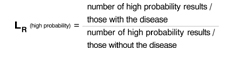
LRs greater than 1 tell us a test result is more likely to occur among patients with the disease than among those without the disease; LRs less than 1 tell us a result is less likely to occur among patients with the disease than among patients without the disease. LRs of 10 or more usually “rule in” disease; LRs of 0.1 or less usually “rule out” disease. An LR of 1 is completely useless in ruling disease in or out.
In the example above, if LR(high probability) is 10, this means that a high probability result is 10 times more likely to occur among people with the disease than among people without it.
Advantages of the likelihood ratio. Sensitivity and specificity can be used only with test results reported as positive or negative (dichotomous results). Likelihood ratios can be used with tests that have any number of outcomes. They can also be used in one form of Bayes’ theorem, as illustrated below, which has application to the Applied Evidence article on open-angle glaucoma in this issue.
Applying the likelihood ratio in this issue
On page 119 of this issue, Aref and Schmidt discuss the risk factors and diagnosis of open-angle glaucoma (OAG). Consider a 70-year-old African American woman who has difficulty seeing in the dark and has lost some peripheral vision in both eyes. Her sister has recently received a diagnosis of OAG. The patient’s risk factors and family history make a diagnosis of OAG likely.
How can a likelihood ratio help here? Direct ophthalmoscopy is warranted to determine if the patient has an elevated cup-disc ratio (>0.6). How useful would such an examination be in this case? In general, Bayes’ theorem tells us that new information should be interpreted in light of what is already known. The form of Bayes’ theorem applicable to diagnostic tests is the following:
Posttest odds of disease = Pretest odds of disease × likelihood ratio
First, calculate pretest odds. The relationship of odds to probability is fairly simple. Let’s assume, based on the patient’s history alone, we believe there is a 33% chance she has OAG. In other words, her pretest probability of OAG is 33%. We convert this probability to odds: Odds = probability/ 1 − probability = 0.33/ 1 − 0.33 = 1/2
Her odds of disease is therefore “1 in favor to 2 against.”
Next, find posttest odds. Now let’s assume that ophthalmoscopy reveals a cup-disc ratio of 0.8. According to Aref and Schmidt’s article, a cup-disc ratio of >0.6 (ie, a “positive” ophthalmoscopic examination) has an LR+ of 16; a cup-disc ratio of <0.6 has an LR− of 0.375 (or 3/8). Since the patient has a “positive” test result, we obtain:
Posttest odds of OAG = 1/2 × 16 = 16/2 = 8/1
Translating back to probability. To make things easier, we can convert this posttest odds of 8/1 to a probability:
Probability = odds in favor/odds in favor + odds against = 8/8+1 = 0.89 (or 89%).
After combining our ophthalmoscopic examination with the history, we can conclude that the patient has an 89% chance of having OAG.
If the result of the ophthalmoscopic examination was negative,
Posttest odds of OAG = 1/2 × 3/8 = 3/16.
The corresponding probability is roughly 0.16 (or 16%).
When likelihood ratios are most useful
In general, diagnostic tests of any kind are most useful for patients like the one described— those who have an intermediate pre-test probability of disease (usually 20%–60%). Very high or very low pretest probabilities of disease are less likely to influence post-test probability of disease.
You may be concerned that the value of pretest probability we chose is subjective. Bear in mind that much of our thinking in medicine is subjective, but based upon clinical experience and knowledge. Evidence-based medicine is a complement to, not a substitute for, clinical experience. Combining the objectivity of likelihood ratios with subjective pretest probabilities using Bayes’ theorem is consistent with the principles of evidence-based medicine.
Corresponding author
Goutham Rao, MD. E-mail: raog@upmc.edu.
Like sensitivity and specificity, a likelihood ratio (LR) can be used to express the usefulness of diagnostic tests. A likelihood ratio is a ratio of 2 proportions: the subset of people with a particular test result among all those who have a specific disease, divided by the subset of people with the same test result among all those without the disease. The mathematical expression of this is:
![]()
What ratio results mean
Consider a study to assess the usefulness of a new blood test for colon cancer. Results of the blood test are reported as high probability of cancer, intermediate probability, or low probability.
All patients in the study undergo the blood test and colonoscopy, the gold standard for identifying colon cancer. The likelihood ratio of a “high probability” result is calculated thus:
LRs greater than 1 tell us a test result is more likely to occur among patients with the disease than among those without the disease; LRs less than 1 tell us a result is less likely to occur among patients with the disease than among patients without the disease. LRs of 10 or more usually “rule in” disease; LRs of 0.1 or less usually “rule out” disease. An LR of 1 is completely useless in ruling disease in or out.
In the example above, if LR(high probability) is 10, this means that a high probability result is 10 times more likely to occur among people with the disease than among people without it.
Advantages of the likelihood ratio. Sensitivity and specificity can be used only with test results reported as positive or negative (dichotomous results). Likelihood ratios can be used with tests that have any number of outcomes. They can also be used in one form of Bayes’ theorem, as illustrated below, which has application to the Applied Evidence article on open-angle glaucoma in this issue.
Applying the likelihood ratio in this issue
On page 119 of this issue, Aref and Schmidt discuss the risk factors and diagnosis of open-angle glaucoma (OAG). Consider a 70-year-old African American woman who has difficulty seeing in the dark and has lost some peripheral vision in both eyes. Her sister has recently received a diagnosis of OAG. The patient’s risk factors and family history make a diagnosis of OAG likely.
How can a likelihood ratio help here? Direct ophthalmoscopy is warranted to determine if the patient has an elevated cup-disc ratio (>0.6). How useful would such an examination be in this case? In general, Bayes’ theorem tells us that new information should be interpreted in light of what is already known. The form of Bayes’ theorem applicable to diagnostic tests is the following:
Posttest odds of disease = Pretest odds of disease × likelihood ratio
First, calculate pretest odds. The relationship of odds to probability is fairly simple. Let’s assume, based on the patient’s history alone, we believe there is a 33% chance she has OAG. In other words, her pretest probability of OAG is 33%. We convert this probability to odds: Odds = probability/ 1 − probability = 0.33/ 1 − 0.33 = 1/2
Her odds of disease is therefore “1 in favor to 2 against.”
Next, find posttest odds. Now let’s assume that ophthalmoscopy reveals a cup-disc ratio of 0.8. According to Aref and Schmidt’s article, a cup-disc ratio of >0.6 (ie, a “positive” ophthalmoscopic examination) has an LR+ of 16; a cup-disc ratio of <0.6 has an LR− of 0.375 (or 3/8). Since the patient has a “positive” test result, we obtain:
Posttest odds of OAG = 1/2 × 16 = 16/2 = 8/1
Translating back to probability. To make things easier, we can convert this posttest odds of 8/1 to a probability:
Probability = odds in favor/odds in favor + odds against = 8/8+1 = 0.89 (or 89%).
After combining our ophthalmoscopic examination with the history, we can conclude that the patient has an 89% chance of having OAG.
If the result of the ophthalmoscopic examination was negative,
Posttest odds of OAG = 1/2 × 3/8 = 3/16.
The corresponding probability is roughly 0.16 (or 16%).
When likelihood ratios are most useful
In general, diagnostic tests of any kind are most useful for patients like the one described— those who have an intermediate pre-test probability of disease (usually 20%–60%). Very high or very low pretest probabilities of disease are less likely to influence post-test probability of disease.
You may be concerned that the value of pretest probability we chose is subjective. Bear in mind that much of our thinking in medicine is subjective, but based upon clinical experience and knowledge. Evidence-based medicine is a complement to, not a substitute for, clinical experience. Combining the objectivity of likelihood ratios with subjective pretest probabilities using Bayes’ theorem is consistent with the principles of evidence-based medicine.
Corresponding author
Goutham Rao, MD. E-mail: raog@upmc.edu.
The Problem with Ps
Most studies include a measure of the significance of treatment effects such as a P value or confidence interval (CI). CIs (Journal of family practice, December 2003, 53:970) are usually preferred to P values, which have notable limitations.
1. P values are easily misinterpreted
A P value is the probability of obtaining a result (usually a difference between treatments) as large or larger than that observed in a study if the null hypothesis (ie, no difference exists between treatments) is true. Differences in treatment effects can be expressed as absolute differences or as odds ratios. No difference, for example, corresponds to an absolute difference of zero or an odds ratio of 1.0.
Consider a recent primary care study from the UK comparing the effectiveness of different lipid-lowering drugs to simvastatin.1 The odds ratio for achieving a cholesterol level ≤5 mmol/L with pravastatin compared with simvastatin was 0.58, with a P value of .003 (ie, simvastatin superior to pravastatin). This means that if there is no difference between pravastatin and simvastatin (ie, null hypothesis is true), the probability of getting an odds ratio of 0.58 or less is just .003 (0.3%).
A P<.05 (sometimes <.01) is usually considered to be sufficient evidence to reject the null hypothesis. This is not intuitively obvious and does not appear to provide useful information.
Many clinicians misinterpret the P value “backwards” as the probability of the null hypothesis assuming the results. In the example above, the misinterpretation would be that there is a 0.3% probability of there being no difference between simvastatin and pravastatin based on the results. Misinterpreting the P value in this way is serious, since the true probability of the null hypothesis based on the results is often much greater than the P value.
2. P values tell us nothing about the magnitude of a significant difference
In the example above, the odds ratio of 0.58, P=.003 has a 95% CI of 0.400.83. The confidence interval, unlike the P value, provides a measure of the precision of the odds ratio.
3. P values are very sensitive to sample size
A small difference between 2 treatments that is clinically insignificant (eg, 1-week difference in mean life expectancy between 2 lipid-lowering treatments) may have a statistically significant P value (ie, <.05) if the sample size is large enough. P values, therefore, can exaggerate the significance of results.
Correspondence
Goutham Rao, MD, 3518 Fifth Avenue, Pittsburgh, PA 15361. E-mail: grao@upmc.edu.
REFERENCE
1. Hippisley-Cox J, Cater R, Pringle M, Coupland C. Cross sectional survey of effectiveness of lipid lowering drugs in reducing serum cholesterol concentration in patients in 17 general practices. BMJ 2003;326:689-693.
Most studies include a measure of the significance of treatment effects such as a P value or confidence interval (CI). CIs (Journal of family practice, December 2003, 53:970) are usually preferred to P values, which have notable limitations.
1. P values are easily misinterpreted
A P value is the probability of obtaining a result (usually a difference between treatments) as large or larger than that observed in a study if the null hypothesis (ie, no difference exists between treatments) is true. Differences in treatment effects can be expressed as absolute differences or as odds ratios. No difference, for example, corresponds to an absolute difference of zero or an odds ratio of 1.0.
Consider a recent primary care study from the UK comparing the effectiveness of different lipid-lowering drugs to simvastatin.1 The odds ratio for achieving a cholesterol level ≤5 mmol/L with pravastatin compared with simvastatin was 0.58, with a P value of .003 (ie, simvastatin superior to pravastatin). This means that if there is no difference between pravastatin and simvastatin (ie, null hypothesis is true), the probability of getting an odds ratio of 0.58 or less is just .003 (0.3%).
A P<.05 (sometimes <.01) is usually considered to be sufficient evidence to reject the null hypothesis. This is not intuitively obvious and does not appear to provide useful information.
Many clinicians misinterpret the P value “backwards” as the probability of the null hypothesis assuming the results. In the example above, the misinterpretation would be that there is a 0.3% probability of there being no difference between simvastatin and pravastatin based on the results. Misinterpreting the P value in this way is serious, since the true probability of the null hypothesis based on the results is often much greater than the P value.
2. P values tell us nothing about the magnitude of a significant difference
In the example above, the odds ratio of 0.58, P=.003 has a 95% CI of 0.400.83. The confidence interval, unlike the P value, provides a measure of the precision of the odds ratio.
3. P values are very sensitive to sample size
A small difference between 2 treatments that is clinically insignificant (eg, 1-week difference in mean life expectancy between 2 lipid-lowering treatments) may have a statistically significant P value (ie, <.05) if the sample size is large enough. P values, therefore, can exaggerate the significance of results.
Correspondence
Goutham Rao, MD, 3518 Fifth Avenue, Pittsburgh, PA 15361. E-mail: grao@upmc.edu.
REFERENCE
1. Hippisley-Cox J, Cater R, Pringle M, Coupland C. Cross sectional survey of effectiveness of lipid lowering drugs in reducing serum cholesterol concentration in patients in 17 general practices. BMJ 2003;326:689-693.
REFERENCE
1. Hippisley-Cox J, Cater R, Pringle M, Coupland C. Cross sectional survey of effectiveness of lipid lowering drugs in reducing serum cholesterol concentration in patients in 17 general practices. BMJ 2003;326:689-693.
Remembering the meanings of sensitivity, specificity, and predictive values
Sensitivity, specificity, positive predictive value (PPV), and negative predictive value (NPV) —collectively known as “test charac-teristics” —are important ways to express the usefulness of diagnostic tests. The 2 x 2 tables from which these terms are derived are familiar to some physicians ( Table ).
Sensitivity is the “true positive rate,” equivalent to a/a+c. Specificity is the “true negative rate,” equivalent to d/b+d. PPV is the proportion of people with a positive test result who actually have the disease (a/a+b); NPV is the proportion of those with a negative result who do not have the disease (d/c+d).
Sensitivity and specificity are fixed for a particular type of test. PPV and NPV for a particular type of test depend upon the prevalence of a disease in a population. For example, though current screening tests for HIV have high sensitivity and specificity, the low prevalence of HIV in the general population cannot justify universal screening since the majority of positive tests would be falsely positive (ie, low PPV).
TABLE
2 x 2 table for diagnostic test results
| Disease present (+) | Disease absent (−) | Totals | |
|---|---|---|---|
| Test result positive (+) | a | b | a + b |
| Test result negative (−) | c | d | c + d |
| Totals | a + c | b + d | — |
How to Remember these Terms
Begin by assuming that you have 4 patients. For the first 2 you know only their disease status; for the last 2 patients you know only their test result.
You know your patient’s disease status:
- Sensitivity: “I know my patient has the dis-ease. What is the chance that the test will show that my patient has it?”
- Specificity: “I know my patient doesn’t have the disease. What is the chance that the test will show that my patient doesn’t have it?”
- PPV: “I just got a positive test result back on my patient. What is the chance that my patient actually has the disease?”
- For NPV: “I just got a negative test result back on my patient. What is the chance that my patient actually doesn’t have the disease?”
Keeping these 4 questions in mind as you run across these frequently used terms will help you interpret diagnostic tests accurately and efficiently, without having to think about more awkward 2 x 2 tables.
Correspondence
Goutham Rao, MD, 3518 Fifth Avenue, Pittsburgh, PA 15261. E-mail: raog@msx.upmc.edu.