User login
Cancer patients’ complaints about care
A new study suggests cancer patients may be more concerned with the human aspects of care than the technical ones.
Researchers studied complaints made by outpatients (or proxies) to a cancer institute over a 2-year period.
A majority of the complaints were management-related issues (48%), such as finance and billing problems, or relationship-related (41%), such as patient-staff dialogue.
Only 11% of the complaints were related to clinical issues, such as errors in diagnosis. However, these complaints were frequently of higher severity than others.
Jennifer W. Mack, MD, of Dana-Farber Cancer Institute in Boston, Massachusetts, and her colleagues reported these findings in The Joint Commission Journal on Quality and Patient Safety.
The researchers looked at complaints made to the Patient/Family Relations Office at the Dana-Farber Cancer Institute from January 2013 through December 2014.
There were 78,668 outpatients treated during this time, and 266 complaints were filed. Most complaints were filed by the patient (73%), 17% by the patient’s spouse/partner, 3% by a parent, 12% by another family member, 0.4% by a friend, 2% by the referring provider, and 1% by a social worker.
The complaints were placed in 3 categories—management, relationship, and clinical issues.
For 48% of the complaints, “management” was the primary category. This encompassed complaints related to:
- Service issues—15%
- Delays—13%
- Finance and billing—10%
- Access and admission—6%
- Bureaucracy—2%
- Environment—2%
- Referrals—0.4%.
For 41% of the complaints, “relationship” was the primary category, which encompassed:
- Communication breakdown—15%
- Respect, dignity, caring—15%
- Patient-staff dialogue—5%
- Staff attitudes—3%
- Confidentiality—2%
- Incorrect information—1%.
For 11% of the complaints, “clinical” was the primary category, which encompassed complaints related to:
- Quality of care—4%
- Skills and conduct—2%
- Patient journey—2%
- Treatment—1%
- Errors in diagnosis—1%
- Safety incidents—1%
- Examinations—0.4%.
Fifty-seven percent of clinical complaints were considered high severity, as were 28% of relationship complaints and 7% of management complaints
Overall, most (64%) complaints were classified as low severity, 16% were moderate, and 20% were high severity.
The following aspects raised the severity level of a complaint:
- Involvement of a prescribing oncologist—27%
- Strong affect of the complainant, including anger—15%
- Allegation of a medical error or suboptimal care—6%
- Request or desire to transfer care to another provider (12%) or institution (5%)
- Mention of malpractice or a desire to pursue legal action—1%.
The researchers said this study provides insight into patient and family values when it comes to cancer care, suggesting they prioritize high-quality relationships and communication. And a systematic review of complaints could reveal areas where care fails to meet patient and family needs. ![]()
How APL cells evade the immune system
New research has revealed a way in which acute promyelocytic leukemia (APL) cells evade destruction by the immune system.
The study showed how group 2 innate lymphoid cells (ILC2s) are recruited by leukemic cells to suppress an essential anticancer immune response.
Researchers believe this newly discovered immunosuppressive axis likely holds sway in other cancers, and it might be disrupted by therapies already in use to treat other diseases.
Camilla Jandus, MD, PhD, of the Ludwig Institute for Cancer Research in Lausanne, Switzerland, and her colleagues described this research in Nature Communications.
“ILCs are not very abundant in the body, but, when activated, they secrete large amounts of immune factors,” Dr Jandus said. “In this way, they can dictate whether a response will be pro-inflammatory or anti-inflammatory.”
ILC1, 2, and 3 have been shown to play a role in inflammation and autoimmune diseases. However, their role in cancer has remained unclear.
To address that question, Dr Jandus and her colleagues began with the observation that one subtype of the cells, ILC2s, are abnormally abundant and hyperactivated in patients with APL.
The researchers examined ILC2 immunology in patients with active APL and compared it to that of APL patients in remission.
“Our analyses suggest that, in patients with this leukemia, ILC2s are at the beginning of a novel immunosuppressive axis, one that is likely to be active in other types of cancer as well,” Dr Jandus said.
She and her colleagues found that APL cells secrete large quantities of PGD2 and express high levels of B7H6 on their surface. Both of these molecules bind to receptors on ILC2s—CRTH2 and NKp30, respectively—activating the ILC2s and prompting them to secrete interleukin-13 (IL-13).
The IL-13 switches on and expands the population of monocytic myeloid-derived immune cells (M-MDSCs). These cells suppress immune responses and allow leukemic cells to evade immune system attack.
The researchers tested these findings in a mouse model of APL. Like patients, mice with APL displayed abnormal activation of ILC2s and M-MDSCs.
However, interfering with all the signals of the immunosuppressive axis restored anti-cancer immunity and prolonged survival in the mice.
Treating mice with a PGD2 inhibitor, an NKp30-blocking antibody, and an anti-IL-13 antibody resulted in reduced APL cell engraftment and a decrease in PGD2, ILC2s, and M-MDSCs. These mice also had significantly longer survival than untreated control mice (P<0.05).
Dr Jandus and her colleagues noted that antibodies against IL-13 and inhibitors of PGD2 are already in clinical use for other diseases, and antibodies that interfere with NKp30-B7H6 binding are in clinical development.
“We also found that this immunosuppressive axis may be operating in other types of cancer; in particular, prostate cancer,” Dr Jandus said. “We believe that some ILCs, like ILC2s, might suppress immune responses, while others might stimulate them. That’s what we are investigating in other types of tumors now.”
This research was supported by the Novartis Foundation for Medical-Biological Research, Ludwig Cancer Research, the Swiss National Science Foundation, Fondazione San Salvatore, ProFemmes UNIL, Fondation Pierre Mercier pour la Science, the Swiss Cancer League, and the Foundation for the Fight against Cancer. ![]()
EC expands approval of obinutuzumab in FL
The European Commission (EC) has expanded the marketing authorization for obinutuzumab (Gazyvaro).
The drug is now approved for use in combination with chemotherapy to treat patients with previously untreated, advanced follicular lymphoma (FL). Patients who respond to this treatment can then receive obinutuzumab maintenance.
This is the third EC approval for obinutuzumab.
The drug was first approved by the EC in 2014 to be used in combination with chlorambucil to treat patients with previously untreated chronic lymphocytic leukemia and comorbidities that make them unsuitable for full-dose fludarabine-based therapy.
In 2016, the EC approved obinutuzumab in combination with bendamustine, followed by obinutuzumab maintenance, in FL patients who did not respond to, or who progressed during or up to 6 months after, treatment with rituximab or a rituximab-containing regimen.
The EC’s latest approval of obinutuzumab is based on results of the phase 3 GALLIUM trial, which were presented at the 2016 ASH Annual Meeting.
The study enrolled 1401 patients with previously untreated, indolent non-Hodgkin lymphoma, including 1202 with FL.
Half of the FL patients (n=601) were randomized to receive obinutuzumab plus chemotherapy (followed by obinutuzumab maintenance for up to 2 years), and half were randomized to rituximab plus chemotherapy (followed by rituximab maintenance for up to 2 years).
The different chemotherapies used were CHOP (cyclophosphamide, doxorubicin, vincristine, and prednisolone), CVP (cyclophosphamide, vincristine, and prednisolone), and bendamustine.
Patients who received obinutuzumab had significantly better progression-free survival than patients who received rituximab. The 3-year progression-free survival rate was 73.3% in the rituximab arm and 80% in the obinutuzumab arm (hazard ratio [HR]=0.66, P=0.0012).
There was no significant difference between the treatment arms with regard to overall survival. The 3-year overall survival was 92.1% in the rituximab arm and 94% in the obinutuzumab arm (HR=0.75, P=0.21).
The overall incidence of adverse events (AEs) was 98.3% in the rituximab arm and 99.5% in the obinutuzumab arm. The incidence of serious AEs was 39.9% and 46.1%, respectively.
The incidence of grade 3 or higher AEs was higher among patients who received obinutuzumab.
Grade 3 or higher AEs occurring in at least 5% of patients in either arm (rituximab and obinutuzumab, respectively) included neutropenia (67.8% and 74.6%), leukopenia (37.9% and 43.9%), febrile neutropenia (4.9% and 6.9%), infections and infestations (3.7% and 6.7%), and thrombocytopenia (2.7% and 6.1%). ![]()
Combination ‘sets new standard’ for GVHD prophylaxis
A 2-drug combination sets a new standard for prevention of acute graft-versus-host disease (GVHD), according to an author of a new study.
The combination—sirolimus and KY1005—completely protected nonhuman primates from acute GVHD and significantly prolonged survival in the animals.
The combination controlled the expansion of effector T cells (Teffs) while augmenting the proportion of regulatory T cells (Tregs) in the primates’ bloodstreams, allowing transplanted stem cells to reconstitute the animals’ immune systems.
Researchers reported these results in Science Translational Medicine. The work was funded by Kymab, the company developing KY1005.
“KY1005, in combination with sirolimus, sets a new standard for [acute] GVHD prevention,” said study author Leslie Kean, MD, PhD, of Seattle Children’s Research Institute in Washington.
“These results in the complex and clinically relevant animal model suggest this regimen is an exceptional candidate for clinical translation.”
Dr Kean and her colleagues noted that no existing treatments for GVHD can successfully strike the delicate balance between controlling Teffs and maintaining the protective function of Tregs.
So the team decided to combine 2 treatments that partially suppress Teffs—sirolimus and KY1005, a monoclonal antibody that blocks a T-cell receptor ligand called OX40L.
The researchers tested the combination in rhesus macaques undergoing allogeneic hematopoietic stem cell transplant (HSCT). The team compared the combination to each agent alone, as well as to no prophylaxis.
KY1005 was given at a dose of 10 mg/kg, starting 2 days before HSCT and continuing once weekly until planned discontinuation on day 54. Sirolimus was given daily for the entire study period as an intramuscular formulation, with doses adjusted to achieve a serum trough concentration of 5 to 15 ng/mL.
Animals treated with both sirolimus and KY1005 survived—free from GVHD—for more than 100 days after HSCT, which was significantly longer than any other group (P<0.01).
In comparison, untreated animals succumbed to GVHD within 8 days of HSCT. And the median GVHD-free survival times were 14 days for the sirolimus group and 19.5 days for the KY1005 group.
The researchers also noted that untreated animals experienced “a rapid decline” in Tregs over the study period. They had a significant decrease in the ratio of Tregs to conventional T cells (Tconv)—2.0 ± 0.4 before HSCT and 0.6 ± 0.1 at last analysis (P<0.001).
When given alone, both KY1005 and sirolimus protected animals from this drop in the Treg/Tconv ratio.
But the combination regimen significantly augmented the Treg/Tconv ratio—1.30 ± 0.30 before HSCT and 1.82 ± 0.43 at last analysis (P<0.05).
Because sirolimus is already used as GVHD prophylaxis and KY1005 is in phase 1 testing as a psoriasis treatment, the researchers believe the combination is a strong candidate for clinical testing. ![]()
SCD drug receives rare pediatric disease designation
The US Food and Drug Administration (FDA) has granted rare pediatric disease designation to Altemia™ soft gelatin capsules for the treatment of children with sickle cell disease (SCD).
Altemia (formerly SC411) is being developed by Sancilio Pharmaceuticals Company, Inc. (SPCI) to treat SCD patients between the ages of 5 and 17 years.
Altemia consists of a mixture of fatty acids, primarily in the form of Ethyl Cervonate™ (a proprietary blend of docosahexaenoic acid and other omega-3 fatty acids), and surface active agents formulated using Advanced Lipid Technologies®.
According to SPCI, Advanced Lipid Technologies are proprietary formulation and manufacturing techniques used to create lipophilic drug products capable of increased bioavailability, avoidance of the first pass effect, and elimination of the food effects commonly associated with oral administration.
Altemia is designed to replenish the lipids destroyed by sickle hemoglobin. The product is intended to be taken once daily to reduce vaso-occlusive crises, anemia, organ damage, and other complications of SCD.
Altemia also has orphan drug designation from the FDA.
SPCI is currently conducting a phase 2 trial of Altemia. In this randomized, double-blind, placebo-controlled trial, researchers are evaluating the efficacy and safety of Altemia in pediatric patients with SCD.
The company plans to report top-line results from the study, known as the SCOT trial, early in the fourth quarter of this year.
About rare pediatric disease designation
Rare pediatric disease designation is granted to drugs that show promise to treat diseases affecting fewer than 200,000 patients in the US, primarily patients age 18 or younger.
The designation provides incentives to advance the development of drugs for rare disease, including access to the FDA’s expedited review and approval programs.
Under the FDA’s Rare Pediatric Disease Priority Review Voucher Program, if a drug with rare pediatric disease designation is approved, the drug’s developer may qualify for a voucher that can be redeemed to obtain priority review for any subsequent marketing application. ![]()
Antibiotic could help treat CML
The antibiotic tigecycline may enhance the treatment of chronic myeloid leukemia (CML), according to research published in Nature Medicine.
Using cells isolated from CML patients, researchers showed that treatment with tigecycline, an antibiotic used to treat bacterial infection, is effective in killing CML stem cells when used in combination with the tyrosine kinase inhibitor (TKI) imatinib.
The study also suggested the combination can stave off relapse in animal models of CML.
“We were very excited to find that, when we treated CML cells with both the antibiotic tigecycline and the TKI drug imatinib, CML stem cells were selectively killed,” said study author Vignir Helgason, PhD, of the University of Glasgow in Scotland.
“We believe that our findings provide a strong basis for testing this novel therapeutic strategy in clinical trials in order to eliminate CML stem cells and provide cure for CML patients.”
The researchers said they found that, in primitive CML stem and progenitor cells, mitochondrial oxidative metabolism is crucial for the production of energy and anabolic precursors. This suggested that restraining mitochondrial functions might have a therapeutic benefit in CML.
The team knew that, in addition to inhibiting bacterial protein synthesis, tigecycline inhibits the synthesis of mitochondrion-encoded proteins, which are required for the oxidative phosphorylation machinery.
So the researchers tested tigecycline, alone or in combination with imatinib, in CML cells. Both treatments (tigecycline monotherapy and the combination) “strongly impaired” the proliferation of primary CD34+ CML cells.
However, imatinib alone had “a moderate effect.” The researchers said this is in line with the preferential effect of imatinib on differentiated CD34− cells.
Each drug alone decreased the number of short-term CML colony-forming cells (CFCs), and the combination eliminated colony formation entirely. This correlated with an increase in cell death.
Neither monotherapy nor the combination had a significant effect on non-leukemic CFCs.
The researchers then turned to a xenotransplantation model of human CML. Starting 6 weeks after transplant, mice received daily doses of vehicle, tigecycline (escalating doses of 25–100 mg per kg body weight), imatinib (100 mg per kg body weight), or both drugs. All treatment was given for 4 weeks.
The team said there were no signs of toxicity in any of the mice.
Compared to controls, tigecycline-treated mice had a marginal decrease in the total number of CML-derived CD45+ cells in the bone marrow, and imatinib-treated mice had a significant decrease in these cells. But the CML burden decreased even further with combination treatment.
The researchers noted that imatinib alone marginally decreased the number of CD45+CD34+CD38− CML cells, but combination treatment eliminated 95% of these cells.
Finally, the team tested each drug alone and in combination (as well as vehicle control) in additional cohorts of mice with CML. After receiving treatment for 4 weeks, mice were left untreated for either 2 weeks or 3 weeks.
Mice that received imatinib alone showed signs of relapse at 2 and 3 weeks, as they had similar numbers of leukemic cells as vehicle-treated mice. However, most of the mice treated with the combination had low numbers of leukemic stem cells in the bone marrow.
“Our work in this study demonstrates, for the first time, that CML stem cells are metabolically distinct from normal blood stem cells, and this, in turn, provides opportunities to selectively target them,” said study author Eyal Gottlieb, PhD, of the Cancer Research UK Beatson Institute in Glasgow.
“It’s exciting to see that using an antibiotic alongside an existing treatment could be a way to keep this type of leukemia at bay and potentially even cure it,” added Karen Vousden, PhD, Cancer Research UK’s chief scientist.
“If this approach is shown to be safe and effective in humans too, it could offer a new option for patients who, at the moment, face long-term treatment with the possibility of relapse.”
This research was funded by AstraZeneca, Cancer Research UK, The Medical Research Council, Scottish Government Chief Scientist Office, The Howat Foundation, Friends of the Paul O’Gorman Leukaemia Research Centre, Bloodwise, The Kay Kendall Leukaemia Fund, Lady Tata International Award, and Leuka. ![]()
Midostaurin approved to treat AML, SM in Europe
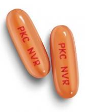
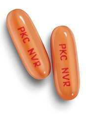
The European Commission has approved the multi-targeted kinase inhibitor midostaurin (Rydapt®) to treat acute myeloid leukemia (AML) and 3 types of advanced systemic mastocytosis (SM).
Midostaurin is approved to treat adults with newly diagnosed acute myeloid leukemia (AML) who are FLT3 mutation-positive. In these patients, midostaurin can be used in combination with standard daunorubicin and cytarabine induction, followed by high-dose cytarabine consolidation. Patients who achieve a complete response can then receive midostaurin as maintenance therapy.
Midostaurin is also approved as monotherapy for adults with aggressive SM (ASM), SM with associated hematological neoplasm (SM-AHN), and mast cell leukemia (MCL).
Midostaurin in AML
The approval of midostaurin in AML is based on results from the phase 3 RATIFY trial, which were published in NEJM last month.
In RATIFY, researchers compared midostaurin plus standard chemotherapy to placebo plus standard chemotherapy in 717 adults younger than age 60 who had FLT3-mutated AML.
The median overall survival was significantly longer in the midostaurin arm than the placebo arm—74.7 months and 25.6 months, respectively (hazard ratio=0.77, P=0.016).
And the median event-free survival was significantly longer in the midostaurin arm than the placebo arm—8.2 months and 3.0 months, respectively (hazard ratio=0.78, P=0.004).
The most frequent adverse events (AEs) in the midostaurin arm (occurring in at least 20% of patients) were febrile neutropenia, nausea, vomiting, mucositis, headache, musculoskeletal pain, petechiae, device-related infection, epistaxis, hyperglycemia, and upper respiratory tract infection.
The most frequent grade 3/4 AEs (occurring in at least 10% of patients) were febrile neutropenia, device-related infection, and mucositis. Nine percent of patients in the midostaurin arm stopped treatment due to AEs, as did 6% in the placebo arm.
Midostaurin in advanced SM
The approval of midostaurin in advanced SM is based on results from a pair of phase 2, single-arm studies, hereafter referred to as Study 2 and Study 3. Data from Study 2 were published in NEJM in June 2016, and data from Study 3 were presented at the 2010 ASH Annual Meeting.
Study 2 included 116 patients, 115 of whom were evaluable for response.
The overall response rate (ORR) was 17% in the entire cohort, 31% among patients with ASM, 11% among patients with SM-AHN, and 19% among patients with MCL. The complete response rates were 2%, 6%, 0%, and 5%, respectively.
Study 3 included 26 patients with advanced SM. In 3 of the patients, the subtype of SM was unconfirmed.
Among the 17 patients with SM-AHN, there were 10 responses (ORR=59%), including 1 partial response and 9 major responses. In the 6 patients with MCL, there were 2 responses (ORR=33%), which included 1 partial response and 1 major response.
In both studies combined, there were 142 adults with ASM, SM-AHN, or MCL.
The most frequent AEs (excluding laboratory abnormalities) that occurred in at least 20% of these patients were nausea, vomiting, diarrhea, edema, musculoskeletal pain, abdominal pain, fatigue, upper respiratory tract infection, constipation, pyrexia, headache, and dyspnea.
The most frequent grade 3 or higher AEs (excluding laboratory abnormalities) that occurred in at least 5% of patients were fatigue, sepsis, gastrointestinal hemorrhage, pneumonia, diarrhea, febrile neutropenia, edema, dyspnea, nausea, vomiting, abdominal pain, and renal insufficiency.
Serious AEs occurred in 68% of patients, most commonly infections and gastrointestinal disorders.
Twenty-one percent of patients discontinued treatment due to AEs, the most frequent of which were infection, nausea or vomiting, QT prolongation, and gastrointestinal hemorrhage. ![]()
The European Commission has approved the multi-targeted kinase inhibitor midostaurin (Rydapt®) to treat acute myeloid leukemia (AML) and 3 types of advanced systemic mastocytosis (SM).
Midostaurin is approved to treat adults with newly diagnosed acute myeloid leukemia (AML) who are FLT3 mutation-positive. In these patients, midostaurin can be used in combination with standard daunorubicin and cytarabine induction, followed by high-dose cytarabine consolidation. Patients who achieve a complete response can then receive midostaurin as maintenance therapy.
Midostaurin is also approved as monotherapy for adults with aggressive SM (ASM), SM with associated hematological neoplasm (SM-AHN), and mast cell leukemia (MCL).
Midostaurin in AML
The approval of midostaurin in AML is based on results from the phase 3 RATIFY trial, which were published in NEJM last month.
In RATIFY, researchers compared midostaurin plus standard chemotherapy to placebo plus standard chemotherapy in 717 adults younger than age 60 who had FLT3-mutated AML.
The median overall survival was significantly longer in the midostaurin arm than the placebo arm—74.7 months and 25.6 months, respectively (hazard ratio=0.77, P=0.016).
And the median event-free survival was significantly longer in the midostaurin arm than the placebo arm—8.2 months and 3.0 months, respectively (hazard ratio=0.78, P=0.004).
The most frequent adverse events (AEs) in the midostaurin arm (occurring in at least 20% of patients) were febrile neutropenia, nausea, vomiting, mucositis, headache, musculoskeletal pain, petechiae, device-related infection, epistaxis, hyperglycemia, and upper respiratory tract infection.
The most frequent grade 3/4 AEs (occurring in at least 10% of patients) were febrile neutropenia, device-related infection, and mucositis. Nine percent of patients in the midostaurin arm stopped treatment due to AEs, as did 6% in the placebo arm.
Midostaurin in advanced SM
The approval of midostaurin in advanced SM is based on results from a pair of phase 2, single-arm studies, hereafter referred to as Study 2 and Study 3. Data from Study 2 were published in NEJM in June 2016, and data from Study 3 were presented at the 2010 ASH Annual Meeting.
Study 2 included 116 patients, 115 of whom were evaluable for response.
The overall response rate (ORR) was 17% in the entire cohort, 31% among patients with ASM, 11% among patients with SM-AHN, and 19% among patients with MCL. The complete response rates were 2%, 6%, 0%, and 5%, respectively.
Study 3 included 26 patients with advanced SM. In 3 of the patients, the subtype of SM was unconfirmed.
Among the 17 patients with SM-AHN, there were 10 responses (ORR=59%), including 1 partial response and 9 major responses. In the 6 patients with MCL, there were 2 responses (ORR=33%), which included 1 partial response and 1 major response.
In both studies combined, there were 142 adults with ASM, SM-AHN, or MCL.
The most frequent AEs (excluding laboratory abnormalities) that occurred in at least 20% of these patients were nausea, vomiting, diarrhea, edema, musculoskeletal pain, abdominal pain, fatigue, upper respiratory tract infection, constipation, pyrexia, headache, and dyspnea.
The most frequent grade 3 or higher AEs (excluding laboratory abnormalities) that occurred in at least 5% of patients were fatigue, sepsis, gastrointestinal hemorrhage, pneumonia, diarrhea, febrile neutropenia, edema, dyspnea, nausea, vomiting, abdominal pain, and renal insufficiency.
Serious AEs occurred in 68% of patients, most commonly infections and gastrointestinal disorders.
Twenty-one percent of patients discontinued treatment due to AEs, the most frequent of which were infection, nausea or vomiting, QT prolongation, and gastrointestinal hemorrhage.
ESC working groups publish consensus on DVT
Experts in Europe have published a consensus document on the diagnosis and management of acute deep vein thrombosis (DVT).
The document provides advice on DVT diagnosis, initial management (first 5–21 days), long-term management (first 3–6 months), extended management (beyond 6 months), and special situations such as cancer, pregnancy, and DVT at unusual sites.
The document, published in the European Heart Journal, was produced by the European Society of Cardiology (ESC) Working Group on Aorta and Peripheral Vascular Diseases and Working Group on Pulmonary Circulation and Right Ventricular Function.
First, the document highlights the importance of clinical assessment and imaging with venous ultrasound for DVT diagnosis.
“The signs and the symptoms of DVT differ from one patient to another and are unspecific but are still very important and recommended for the initial evaluation of patients with suspected DVT,” said lead author of the document Lucia Mazzolai, MD, PhD, of Lausanne University Hospital in Switzerland.
“Ultrasound is recommended as the first-line diagnostic tool when lower or upper limb DVT is suspected. We also propose venous ultrasound in patients with confirmed PE [pulmonary embolism] as initial reference in case of DVT recurrence or further patient stratification in selected individuals.”
For initial and long-term management, the document includes advice according to the location of DVT, which can be proximal (popliteal, femoral, or iliac veins) or isolated distal (in the calf veins only).
Anticoagulation for patients with isolated distal DVT is under debate, and the document’s authors said patients should be stratified based on their individual risk.
Regarding the type of anticoagulant therapy to use in the first line of initial and long-term management, Dr Mazzolai said there has been a paradigm shift in recent years.
“We propose direct oral anticoagulants as first-line treatment for non-cancer patients,” she said. “We also recommend catheter-directed thrombolysis as an adjuvant treatment only in select patients.”
For the extended management phase, the authors recommend personalized decisions on the continuation of anticoagulation, based on risk/benefit balance.
In addition, a venous ultrasound should be performed when anticoagulation is discontinued. This can serve as a baseline comparative exam in case DVT recurs.
Regarding special situations, the authors said that, after 6 months, patients with cancer need personalized management. The decision on whether or not to continue anticoagulation, and with which drug, should be based on the patient’s risk/benefit ratio, tolerability, preference, and cancer activity.
Pregnant patients with suspected lower limb DVT should have venous ultrasound as the first-line diagnostic imaging test. Low-molecular-weight heparin is recommended for initial and long-term treatment of DVT during pregnancy.
“This is the first European document that addresses all aspects of modern DVT management because it discusses diagnosis, treatment, and follow-up, plus special situations like cancer, pregnancy, and unusual DVT sites,” Dr Mazzolai said.
“Together with the 2014 ESC guidelines on PE, clinicians now have comprehensive recommendations on the management of VTE [venous thromboembolism].”
FDA grants RMAT designation to HSCT adjunct
The US Food and Drug Administration (FDA) has granted Regenerative Medicine Advanced Therapy (RMAT) designation to ATIR101™, which is intended to be used as an adjunct to haploidentical hematopoietic stem cell transplant (HSCT).
ATIR101 is a personalized T-cell immunotherapy—a donor lymphocyte preparation selectively depleted of host-alloreactive T cells through the use of photo-dynamic therapy.
Recipient-reactive T cells from the donor are activated in a unidirectional mixed-lymphocyte reaction. The cells are then treated with TH9402 (a rhodamide-like dye), which is selectively retained in activated T cells.
Subsequent light exposure eliminates the activated recipient-reactive T cells but preserves the other T cells.
The final product is infused after CD34-selected haploidentical HSCT with the goal of preventing infectious complications, graft-versus-host disease (GVHD), and relapse.
About RMAT designation
The RMAT pathway is analogous to the breakthrough therapy designation designed for traditional drug candidates and medical devices. RMAT designation was specifically created by the US Congress in 2016 in the hopes of getting new cell therapies and advanced medicinal products to patients earlier.
Just like breakthrough designation, RMAT designation allows companies developing regenerative medicine therapies to interact with the FDA more frequently in the clinical testing process. In addition, RMAT-designated products may be eligible for priority review and accelerated approval.
A regenerative medicine is eligible for RMAT designation if it is intended to treat, modify, reverse, or cure a serious or life-threatening disease or condition, and if preliminary clinical evidence indicates the treatment has the potential to address unmet medical needs for such a disease or condition.
“To receive the RMAT designation from the FDA is an important milestone for Kiadis Pharma and a recognition by the FDA of the significant potential for ATIR101 to help patients receive safer and more effective bone marrow transplantations,” said Arthur Lahr, CEO of Kiadis Pharma, the company developing ATIR101.
“We are now going to work even closer with the FDA to agree a path to make this cell therapy treatment available for patients in the US as soon as possible. In Europe, ATIR101 was filed for registration in April 2017, and we continue to prepare the company for the potential European launch in 2019.”
ATIR101 trials
Results of a phase 2 trial of ATIR101 were presented at the 42nd Annual Meeting of the European Society of Blood and Marrow Transplantation in 2016.
Patients who received ATIR101 after haploidentical HSCT had significant improvements in transplant-related mortality and overall survival when compared to historical controls who received a T-cell-depleted haploidentical HSCT without ATIR101.
None of the patients who received ATIR101 developed grade 3-4 GVHD, but a few patients did develop grade 2 GVHD.
A phase 3 trial of ATIR101 is now underway. The trial is expected to enroll 200 patients with acute myeloid leukemia, acute lymphoblastic leukemia, or myelodysplastic syndrome.
The patients will receive a haploidentical HSCT with either a T-cell-depleted graft and adjunctive treatment with ATIR101 or a T-cell-replete graft and post-transplant cyclophosphamide.
Team creates guidelines on CAR T-cell-related toxicity
Researchers say they have created guidelines for managing the unique toxicities associated with chimeric antigen receptor (CAR) T-cell therapy.
The guidelines focus on cytokine release syndrome (CRS); neurological toxicity, which the researchers have dubbed “CAR-T-cell-related encephalopathy syndrome (CRES);” and adverse effects related to these syndromes.
“The toxicities are unique, and every member of the care team needs to be trained to recognize them and act accordingly,” said Sattva Neelapu, MD, of University of Texas MD Anderson Cancer Center in Houston.
Dr Neelapu and his colleagues described the toxicities and related recommendations in Nature Reviews Clinical Oncology.
The team’s guidelines include supportive-care considerations for patients receiving CAR T‑cell therapy. For example, they recommend:
- Baseline brain MRI to rule out central nervous system disease
- Cardiac monitoring starting on the day of CAR T‑cell infusion
- Assessing a patient’s vital signs every 4 hours after CAR T-cell infusion
- Assessing and grading CRS at least twice daily and whenever the patient’s status changes
- Assessing and grading CRES at least every 8 hours.
CRS
One section of the guidelines is dedicated to CRS, with subsections on pathophysiology, precautions and supportive care, the use of corticosteroids and IL‑6/IL‑6R antagonists, and grading CRS.
The researchers noted that CRS typically manifests with constitutional symptoms, such as fever, malaise, anorexia, and myalgias. However, CRS can affect any organ system in the body.
The team recommends managing CRS according to grade. For example, patients with grade 1 CRS should typically receive supportive care. However, physicians should consider giving tocilizumab or siltuximab to grade 1 patients who have a refractory fever lasting more than 3 days.
The researchers also noted that CRS can evolve into fulminant hemophagocytic lymphohistiocytosis (HLH), also known as macrophage-activation syndrome (MAS).
The team said HLH/MAS encompasses a group of severe immunological disorders characterized by hyperactivation of macrophages and lymphocytes, proinflammatory cytokine production, lymphohistiocytic tissue infiltration, and immune-mediated multi-organ failure.
The guidelines include diagnostic criteria for CAR T‑cell-related HLH/MAS and recommendations for managing the condition.
CRES
One section of the guidelines is dedicated to the grading and treatment of CRES, which typically manifests as a toxic encephalopathy.
The researchers said the earliest signs of CRES are diminished attention, language disturbance, and impaired handwriting. Other symptoms include confusion, disorientation, agitation, aphasia, somnolence, and tremors.
Patients with severe CRES (grade >2) may experience seizures, motor weakness, incontinence, mental obtundation, increased intracranial pressure, papilledema, and cerebral edema.
Therefore, the guidelines include recommendations for the management of status epilepticus and raised intracranial pressure after CAR T‑cell therapy.
The researchers also devised an algorithm, known as CARTOX-10, for identifying neurotoxicity. (An existing general method didn’t effectively quantify the neurological effects caused by CAR T-cell therapies.)
CARTOX-10 is a 10-point test in which patients are asked to do the following:
- Name the current month (1 point) and year (1 point)
- Name the city (1 point) and hospital they are in (1 point)
- Name the president/prime minister of their home country (1 point)
- Name 3 nearby objects (3 points)
- Write a standard sentence (1 point)
- Count backward from 100 by tens (1 point).
A perfect score indicates normal cognitive function. A patient has mild to severe impairment depending on the number of questions or activities missed.
Dr Neelapu and his colleagues believe their recommendations will be applicable to other types of cell-based immunotherapy as well, including CAR natural killer cells, T-cell receptor engineered T cells, and combination drugs that use an antibody to connect T cells to targets on cancer cells.
Researchers involved in this work have received funding from companies developing/marketing CAR T-cell therapies.


