User login
Combo proves ‘beneficial’ for ‘unfit’ CLL patients
STOCKHOLM—Obinutuzumab plus chlorambucil (G-Clb) is a “valid and beneficial” frontline treatment option for “unfit” patients with chronic lymphocytic leukemia (CLL), according to a speaker at the 23rd Congress of the European Hematology Association (EHA).
Final results from the CLL11 study have revealed additional benefits of G-Clb over rituximab plus chlorambucil (R-Clb) in patients with previously untreated CLL and comorbidities.
Prior results from this study showed that G-Clb produced higher response rates and prolonged progression-fee survival (PFS) compared to R-Clb.
Now, with a median follow-up of 5 years, researchers have found that G-Clb prolongs overall survival (OS) and time to next treatment (TTNT) as well.
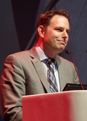
Valentin Goede, MD, of the University Hospital Cologne in Germany, presented these results during the Presidential Symposium of the EHA Congress (abstract S151).
The study was sponsored by Hoffmann-La Roche.
CLL11 enrolled patients with previously untreated CLL and coexisting medical conditions. They were randomized to receive six 28-day cycles of Clb alone, G-Clb, or R-Clb.
In stage 1, researchers compared G-Clb (n=238) to Clb alone (n=118) and R-Clb (n=233) to Clb alone (n=118). In stage 2, they compared G-Clb (n=333) and R-Clb (n=330).
“The treatment arms were well-balanced, not just with regard to patient [characteristics] but also with disease characteristics,” Dr Goede said.
Overall, the median age was 73 (range, 39-90), the median Cumulative Illness Rating Scale score was 8, and the median creatinine clearance was 62 mL/min.
Efficacy: G-Clb vs Clb
The median observation time for G-Clb vs Clb was 62.5 months.
The median PFS was 31.1 months in the G-Clb arm and 11.1 months in the Clb arm. The 5-year PFS rates were 25% and 2%, respectively. The hazard ratio (HR) was 0.21 (P<0.0001).
The median OS was not reached in the G-Clb arm and was 66.7 months in the Clb arm. The 5-year OS rates were 66% and 53%, respectively. The HR was 0.68 (P=0.0196).
Thirty-nine percent of the G-Clb arm died, as did 49% of the Clb arm. The main causes of death were adverse events (AEs) and disease progression.
Efficacy: G-Clb vs R-Clb
The median observation time for G-Clb vs R-Clb was 59.4 months.
The median PFS was 28.9 months in the G-Clb arm and 15.7 months in the R-Clb arm. The 5-year PFS rates were 23% and 9%, respectively. The HR was 0.49 (P<0.0001).
“The median PFS was nearly doubled, from approximately 15 months in the rituximab arm to almost 30 months in the obinutuzumab arm,” Dr Goede said. “And this translated into a clinically meaningful prolongation of time to next treatment.”
The median TTNT was 56.4 months in the G-Clb arm and 34.9 months in the R-Clb arm. At 5 years, TTNT rates were 49% and 32%, respectively. The HR was 0.58 (P<0.0001).
“In the rituximab arm, the median time to next treatment was a little greater than 2.5 years, and, in the obinutuzumab arm, it was almost 5 years,” Dr Goede said. “From a clinical perspective, I would consider treatment-free intervals of that duration as highly relevant and beneficial in an elderly population.”
The median OS was not reached in the G-Clb arm and was 73.1 months in the R-Clb arm. The 5-year OS rates were 66% and 57%, respectively. The HR was 0.76 (P=0.0245).
“This difference is clinically meaningful, and it is also remarkable in the context of the long follow-up, given the fact that approximately half of the patients have received at least 1 salvage treatment in the meantime,” Dr Goede said.
In all, 37% of the G-Clb arm died, as did 45% of the R-Clb arm. Again, the main causes of death were AEs and disease progression.
Safety
Dr Goede said no new safety signals or late-onset toxicities were detected.
“Adverse events of any grade, but particularly grade 3-5 and serious adverse events, were more frequent in the obinutuzumab arm compared to the other 2 arms,” he noted. “[This] was mainly driven by more infusion reactions and some greater hematological toxicity.”
“Importantly, the rate of fatal adverse events, during treatment but also during follow-up, was not higher in the obinutuzumab arm. And the most common fatal adverse events were second malignancies.”
G-Clb vs Clb
Ninety-five percent of patients in the G-Clb arm and 83% of those in the Clb arm had at least 1 AE. The rates of grade 3-5 AEs were 74% and 51%, respectively. The rates of serious AEs were 47% and 39%, respectively. The rates of fatal AEs were 8% and 11%, respectively.
Seventeen percent of patients in the G-Clb arm and 11% of those in the Clb arm had late-onset neutropenia. The rates of prolonged neutropenia were 3% and 9%, respectively.
Fourteen percent of patients in the G-Clb arm and 7% of those in the Clb arm had second malignancies (starting 6 months after treatment initiation). The most common of these were squamous cell carcinoma (2% vs 0%) and basal cell carcinoma (2% vs <1%).
G-Clb vs R-Clb
Ninety-four percent of patients in the G-Clb arm and 90% of those in the R-Clb arm had at least 1 AE. The rates of grade 3-5 AEs were 72% and 60%, respectively. The rates of serious AEs were 45% and 39%, respectively. The rates of fatal AEs were 7% and 10%, respectively.
Fifteen percent of patients in the G-Clb arm and 12% of those in the R-Clb arm had late-onset neutropenia. The rates of prolonged neutropenia were 2% and 4%, respectively.
Eleven percent of patients in the G-Clb arm and 10% of those in the Clb arm had second malignancies. Squamous cell carcinoma occurred in 2% of patients in both arms. Basal cell carcinoma occurred in 2% of G-Clb recipients and 1% of R-Clb recipients.
STOCKHOLM—Obinutuzumab plus chlorambucil (G-Clb) is a “valid and beneficial” frontline treatment option for “unfit” patients with chronic lymphocytic leukemia (CLL), according to a speaker at the 23rd Congress of the European Hematology Association (EHA).
Final results from the CLL11 study have revealed additional benefits of G-Clb over rituximab plus chlorambucil (R-Clb) in patients with previously untreated CLL and comorbidities.
Prior results from this study showed that G-Clb produced higher response rates and prolonged progression-fee survival (PFS) compared to R-Clb.
Now, with a median follow-up of 5 years, researchers have found that G-Clb prolongs overall survival (OS) and time to next treatment (TTNT) as well.
Valentin Goede, MD, of the University Hospital Cologne in Germany, presented these results during the Presidential Symposium of the EHA Congress (abstract S151).
The study was sponsored by Hoffmann-La Roche.
CLL11 enrolled patients with previously untreated CLL and coexisting medical conditions. They were randomized to receive six 28-day cycles of Clb alone, G-Clb, or R-Clb.
In stage 1, researchers compared G-Clb (n=238) to Clb alone (n=118) and R-Clb (n=233) to Clb alone (n=118). In stage 2, they compared G-Clb (n=333) and R-Clb (n=330).
“The treatment arms were well-balanced, not just with regard to patient [characteristics] but also with disease characteristics,” Dr Goede said.
Overall, the median age was 73 (range, 39-90), the median Cumulative Illness Rating Scale score was 8, and the median creatinine clearance was 62 mL/min.
Efficacy: G-Clb vs Clb
The median observation time for G-Clb vs Clb was 62.5 months.
The median PFS was 31.1 months in the G-Clb arm and 11.1 months in the Clb arm. The 5-year PFS rates were 25% and 2%, respectively. The hazard ratio (HR) was 0.21 (P<0.0001).
The median OS was not reached in the G-Clb arm and was 66.7 months in the Clb arm. The 5-year OS rates were 66% and 53%, respectively. The HR was 0.68 (P=0.0196).
Thirty-nine percent of the G-Clb arm died, as did 49% of the Clb arm. The main causes of death were adverse events (AEs) and disease progression.
Efficacy: G-Clb vs R-Clb
The median observation time for G-Clb vs R-Clb was 59.4 months.
The median PFS was 28.9 months in the G-Clb arm and 15.7 months in the R-Clb arm. The 5-year PFS rates were 23% and 9%, respectively. The HR was 0.49 (P<0.0001).
“The median PFS was nearly doubled, from approximately 15 months in the rituximab arm to almost 30 months in the obinutuzumab arm,” Dr Goede said. “And this translated into a clinically meaningful prolongation of time to next treatment.”
The median TTNT was 56.4 months in the G-Clb arm and 34.9 months in the R-Clb arm. At 5 years, TTNT rates were 49% and 32%, respectively. The HR was 0.58 (P<0.0001).
“In the rituximab arm, the median time to next treatment was a little greater than 2.5 years, and, in the obinutuzumab arm, it was almost 5 years,” Dr Goede said. “From a clinical perspective, I would consider treatment-free intervals of that duration as highly relevant and beneficial in an elderly population.”
The median OS was not reached in the G-Clb arm and was 73.1 months in the R-Clb arm. The 5-year OS rates were 66% and 57%, respectively. The HR was 0.76 (P=0.0245).
“This difference is clinically meaningful, and it is also remarkable in the context of the long follow-up, given the fact that approximately half of the patients have received at least 1 salvage treatment in the meantime,” Dr Goede said.
In all, 37% of the G-Clb arm died, as did 45% of the R-Clb arm. Again, the main causes of death were AEs and disease progression.
Safety
Dr Goede said no new safety signals or late-onset toxicities were detected.
“Adverse events of any grade, but particularly grade 3-5 and serious adverse events, were more frequent in the obinutuzumab arm compared to the other 2 arms,” he noted. “[This] was mainly driven by more infusion reactions and some greater hematological toxicity.”
“Importantly, the rate of fatal adverse events, during treatment but also during follow-up, was not higher in the obinutuzumab arm. And the most common fatal adverse events were second malignancies.”
G-Clb vs Clb
Ninety-five percent of patients in the G-Clb arm and 83% of those in the Clb arm had at least 1 AE. The rates of grade 3-5 AEs were 74% and 51%, respectively. The rates of serious AEs were 47% and 39%, respectively. The rates of fatal AEs were 8% and 11%, respectively.
Seventeen percent of patients in the G-Clb arm and 11% of those in the Clb arm had late-onset neutropenia. The rates of prolonged neutropenia were 3% and 9%, respectively.
Fourteen percent of patients in the G-Clb arm and 7% of those in the Clb arm had second malignancies (starting 6 months after treatment initiation). The most common of these were squamous cell carcinoma (2% vs 0%) and basal cell carcinoma (2% vs <1%).
G-Clb vs R-Clb
Ninety-four percent of patients in the G-Clb arm and 90% of those in the R-Clb arm had at least 1 AE. The rates of grade 3-5 AEs were 72% and 60%, respectively. The rates of serious AEs were 45% and 39%, respectively. The rates of fatal AEs were 7% and 10%, respectively.
Fifteen percent of patients in the G-Clb arm and 12% of those in the R-Clb arm had late-onset neutropenia. The rates of prolonged neutropenia were 2% and 4%, respectively.
Eleven percent of patients in the G-Clb arm and 10% of those in the Clb arm had second malignancies. Squamous cell carcinoma occurred in 2% of patients in both arms. Basal cell carcinoma occurred in 2% of G-Clb recipients and 1% of R-Clb recipients.
Umbralisib can revitalize ruxolitinib in MF
STOCKHOLM—The PI3K delta inhibitor umbralisib can “augment or resurrect” responses to ruxolitinib in patients with myelofibrosis (MF), according to a speaker at the 23rd Congress of the European Hematology Association (EHA).
Results of a phase 1 study showed that adding umbralisib to treatment with ruxolitinib could induce responses in MF patients who had a suboptimal or lost response to ruxolitinib.
Of the 23 patients who received the combination, 2 achieved a complete remission (CR), 11 had clinical improvement, and 8 had stable disease.
In addition, umbralisib plus ruxolitinib was considered well-tolerated. The most common adverse event (AE) was anemia.
Tamara K. Moyo, MD, PhD, of Vanderbilt University Medical Center in Nashville, Tennessee, presented these results at the EHA Congress as abstract S133. The research was sponsored by TG Therapeutics.
Patients
Dr Moyo reported results in 23 MF patients who had a suboptimal response, lost a response, or had no response while on a stable dose of ruxolitinib for at least 8 weeks. Their median age was 67 (range, 49-83), and 61% were male.
Patients had primary MF (30%), post-essential thrombocythemia (ET) MF (43%), or post-polycythemia vera (PV) MF (26%). Forty-three percent of patients had JAK2 V617F, 30% had CALR mutations, 17% had MPL mutations, and 13% were triple-negative. One patient had co-occurring CALR and MPL mutations.
Most patients had an ECOG performance score of 0 (39%) or 1 (52%). All had intermediate-1 (35%), intermediate-2 (35%), or high-risk disease (30%) according to DIPSS Plus.
Sixty-one percent of patients had splenomegaly.
Treatment
In stage 1, the patients received stable ruxolitinib and escalating umbralisib. In stage 2, patients received escalating ruxolitinib and umbralisib at the maximum tolerated dose (MTD) established from stage 1.
Patients could then proceed to expansion cohorts in which they would receive any dose of ruxolitinib and umbralisib at the MTD. The expansion cohorts include patients with treatment-naïve MF, PV, chronic myelomonocytic leukemia, and myelodysplastic syndromes/myeloproliferative neoplasms.
However, Dr Moyo reported only on the 23 ruxolitinib-experienced MF patients.
Safety
There were 2 dose-limiting toxicities of asymptomatic, grade 3 amylase/lipase elevations. One occurred in a patient receiving 800 mg of umbralisib daily and 10 mg of ruxolitinib twice daily. The other occurred in a patient receiving 800 mg of umbralisib daily and 15 mg of ruxolitinib twice daily.
Therefore, 600 mg daily was deemed the MTD of umbralisib.
Seventeen patients had at least 1 AE. There were 17 grade 3 or higher AEs in 13 patients.
AEs of any grade included anemia (n=10), neutrophil decrease (n=2), platelet decrease (n=5), AST increase (n=6), ALT increase (n=3), amylase increase (n=3), lipase increase (n=3), diarrhea (n=2), colitis (n=1), dyspnea (n=1), upper respiratory infection (n=2), pneumonia (n=4), other infections (n=6), and sepsis (n=1).
Grade 3 AEs included anemia (n=3), neutrophil decrease (n=2), amylase increase (n=2), lipase increase (n=2), diarrhea (n=2), colitis (n=1), dyspnea (n=1), pneumonia (n=1), and other infections (n=2). The case of sepsis was the only grade 4 AE.
Dr Moyo noted that anemia—the most common AE—was commonly attributed to disease rather than study treatment.
The case of colitis, which was grade 3, was deemed possibly related to treatment, so the patient was removed from the study.
Thirteen patients had discontinued study treatment at the time of analysis. Aside from the patient who discontinued due to colitis, 2 patients went off study due to dose-limiting toxicities, 3 due to progressive disease, 6 due to physician or patient decision, and 1 due to transplant.
Efficacy
Two patients could not be assessed for efficacy, and 8 had stable disease on umbralisib and ruxolitinib.
The combination produced clinical improvement—reduction in spleen volume, increase in hemoglobin, and improvement in MF-related symptoms—in 11 patients (48%).
And 2 patients (9%) achieved a CR. Dr Moyo said there were “few commonalities” between these 2 patients.
Both had intermediate-1-risk disease as well as persistent or progressive MF-related symptoms and thrombocytosis at baseline. However, 1 patient had post-ET MF, and 1 had post-PV MF.
The post-ET MF patient had an MPL driver mutation. She received ruxolitinib at 20 mg twice daily and umbralisib at 400 mg daily. The patient achieved a CR at cycle 15 and remained on study 2 years before proceeding to transplant. The patient is now about 1 year from her transplant with no evidence of disease.
The post-PV patient had a JAK2 V617F driver mutation. She received ruxolitinib at 15 mg twice daily and umbralisib at 600 mg daily. The patient achieved a CR at cycle 5 and remains on study, currently receiving cycle 12 of treatment.
Dr Moyo said these results suggest “the addition of umbralisib to ruxolitinib can augment or resurrect a response in MF patients who have had suboptimal or lost response to ruxolitinib alone, and this treatment combination warrants further investigation.”
Quizartinib can prolong OS in rel/ref, FLT3-ITD AML
STOCKHOLM—Phase 3 results suggest the FLT3 inhibitor quizartinib can prolong overall survival (OS) in patients with relapsed/refractory, FLT3-ITD acute myeloid leukemia (AML).
In the QuANTUM-R study, patients who received single-agent quizartinib had a significantly longer median OS than patients who received salvage chemotherapy.
There was a trend toward improved event-free survival (EFS) with quizartinib as well.
“QuANTUM-R represents the first study that shows a significant improvement in overall survival for a single agent—a FLT3 inhibitor or any other targeted agent—in this population of FLT3-mutated AML patients with refractory or relapsed disease . . .,” said study investigator Jorge Cortes, MD, of MD Anderson Cancer Center in Houston, Texas.
Dr Cortes presented results from QuANTUM-R at the 23rd Congress of the European Hematology Association (EHA). The research was selected as the best late-breaking abstract (LB2600).
The study was funded by Daiichi Sankyo, Inc., and Dr Cortes is a consultant for the company.
Patients and treatment
QuANTUM-R enrolled adults with FLT3-ITD AML (at least 3% FLT3-ITD allelic ratio) who had refractory disease or had relapsed within 6 months of their first complete remission. They had received at least 1 cycle of an induction regimen containing standard-dose anthracycline or mitoxantrone.
Patients were randomized to receive once-daily treatment with quizartinib (n=245) or a salvage chemotherapy regimen (n=122)—low-dose cytarabine (LoDAC, n=29); combination mitoxantrone, etoposide, and cytarabine (MEC, n=40); or combination fludarabine, cytarabine, and idarubicin (FLAG-IDA, n=53).
Responders could proceed to hematopoietic stem cell transplant (HSCT), and those in the quizartinib arm could resume quizartinib after HSCT.
Baseline characteristics were similar between the treatment arms. The median age was 55 (range, 19-81) for patients receiving quizartinib and 58 (range, 18-78) for those receiving chemotherapy.
Thirty-three percent of the quizartinib arm had refractory disease, and 67% had relapsed disease. Thirty-four percent of the chemotherapy arm had refractory disease, and 66% had relapsed disease.
The percentage of patients with a prior allogeneic HSCT was 25% in the quizartinib arm and 23% in the chemotherapy arm. Most patients in both arms had intermediate-risk cytogenetics—78% of the quizartinib arm and 66% of the chemotherapy arm.
In all, 241 patients received quizartinib, and 94 received salvage chemotherapy—LoDAC (n=22), MEC (n=25), and FLAG-IDA (n=47). Of the 28 patients in the chemotherapy group who were not treated, most withdrew consent.
The median treatment duration was 4 cycles (range, 1-3) in the quizartinib arm and 1 cycle (range, 1-2) for patients who received LoDAC, MEC, and FLAG-IDA.
The most common reason for discontinuation of chemotherapy was lack of response/progression (n=49), followed by death (n=6). Twenty-four patients completed salvage chemotherapy.
In the quizarinib arm, the most common reasons for treatment discontinuation were HSCT (n=79), relapse (n=60), or lack of response/progression (n=47).
Thirty-two percent of quizartinib-treated patients and 12% of the chemotherapy group went on to HSCT.
Results
The median follow-up was 23.5 months. The efficacy results include all randomized patients, and the safety results include only those who received their assigned treatment.
The study’s primary endpoint was OS. The median OS was 6.2 months in the quizartinib arm and 4.7 months in the chemotherapy arm (hazard ratio=0.76, P=0.0177). The 1-year OS rate was 27% and 20%, respectively.
The median EFS was 6.0 weeks in the quizartinib arm and 3.7 weeks in the chemotherapy arm (hazard ratio=0.90, P=0.1071). Dr Cortes noted that patients who did not receive treatment were censored on day 1, and partial responses were counted as failures in the EFS analysis.
The overall response rate was 69% in the quizartinib arm and 30% in the chemotherapy arm.
The composite complete response (CR) rate was 48% in the quizartinib arm and 27% in the chemotherapy arm. This includes the CR rate (4% and 1%, respectively), the rate of CR with incomplete platelet recovery (4% and 0%, respectively), and the rate of CR with incomplete hematologic recovery (40% and 26%, respectively). The rate of partial response was 21% and 3%, respectively.
Dr Cortes said rates of treatment-emergent adverse events (TEAEs) were similar between the treatment arms.
Grade 3 or higher hematologic TEAEs occurring in at least 5% of patients (in the quizartinib and chemotherapy groups, respectively) included thrombocytopenia (35% and 34%), anemia (30% and 29%), neutropenia (32% and 25%), febrile neutropenia (31% and 21%), and leukopenia (17% and 16%).
Grade 3 or higher nonhematologic TEAEs occurring in at least 5% of patients (in the quizartinib and chemotherapy groups, respectively) included fatigue (8% and 1%), hypokalemia (12% and 9%), sepsis/septic shock (16% and 18%), dyspnea (5% for both), hypophosphatemia (5% for both), and pneumonia (12% and 9%).
Three percent of patients in the quizartinib arm had grade 3 QTcF prolongation, but there were no grade 4 cases. Two patients discontinued quizartinib due to QTcF prolongation.
“The safety of this drug has remained constant across over 1600 patients that have been treated with quizartinib across a variety of studies,” Dr Cortes said.
He added that QuANTUM-R results open up the possibility that quizartinib could be used in other settings. Researchers are already testing standard chemotherapy with and without quizartinib in a phase 3 trial of patients with newly diagnosed, FLT-ITD AML (QuANTUM-First).
Promising results with expanded UCB product
SALT LAKE CITY—An expanded umbilical cord blood (UCB) product can produce favorable outcomes as a stand-alone graft, according to a presentation at the 2018 BMT Tandem Meetings.
The product, MGTA-456, provided “rapid and durable” engraftment in patients with hematologic malignancies, according to John E. Wagner, MD, of the University of Minnesota in Minneapolis.
He also said MGTA-456 “preserved the clinical benefits” of UCB transplant, including low rates of graft-vs-host disease (GVHD) and high overall survival (OS).
Dr Wagner presented these results as one of the “Best Abstracts” at this year’s BMT Tandem Meetings (abstract 4). The research was supported by Novartis and Magenta Therapeutics.
MGTA-456 is developed by dividing a UCB unit into a CD34- portion and a CD34+ portion, then expanding the CD34+ portion for 15 days via culture with an aryl hydrocarbon receptor antagonist (SR-1), stem cell factor, FLT3 ligand, interleukin-6, and thrombopoietin.
In a previous study,* MGTA-456 enhanced hematopoietic recovery when given as half of a double UCB transplant.
With the current research, Dr Wagner and his colleagues evaluated MGTA-456 as a stand-alone graft. The team conducted two phase 2 trials of MGTA-456, one in which patients received myeloablative conditioning (MAC) and one in which patients received non-myeloablative conditioning (NMAC).
Treatment
Each trial included 10 patients with a high-risk hematologic malignancy and a partially HLA-matched UCB unit. In each trial, 1 patient could not receive MGTA-456 due to low expansion.
So 9 patients received MAC—cyclophosphamide (CY) at 60 mg/kg/day on days -6 and -5, fludarabine (FLU) at 25 mg/m2/day on days -7 to -5, and total body irradiation (TBI) at 1320 cGy on days -4 to -1.
And 9 patients received NMAC—CY at 50 mg/kg on day -6, FLU at 40 mg/m2/day on days -6 to -2, and TBI at 200 cGy on day -1. Some patients who had not received recent chemotherapy also received antithymocyte globulin as part of their conditioning regimen.
For MAC recipients, the median expansion of CD34+ cells was 406-fold (range, 162-1643). The median CD34 cell dose they received was 16.2 x 10⁶/kg.
For NMAC recipients, the median expansion of CD34+ cells was 274-fold (range, 42-527). The median CD34 cell dose they received was 13.4 x 10⁶/kg.
All patients received cyclosporine and mycophenolate mofetil as GVHD prophylaxis.
Dr Wagner and his colleagues compared outcomes in these MGTA-456 recipients to outcomes in historical control subjects—151 patients who received MAC and 132 who received NMAC.
MAC recipients
The 9 MAC/MGTA-456 recipients had a median age of 25 (range, 15-53). Seven of the patients had acute leukemia, 1 had myelodysplastic syndrome (MDS), and 1 had lymphoma.
Eleven percent of patients had high-risk disease, 89% were cytomegalovirus seropositive, and 89% had a Karnofsky performance score of 90 to 100.
The only significant difference between the MGTA-456 recipients and historical controls was weight. The median weight was 93.8 kg (range, 41-107) for MGTA-456 recipients and 66.7 kg (range, 11-136) for controls (P=0.04).
The MGTA-456 recipients had superior hematopoietic recovery compared to historical controls.
The rate of neutrophil engraftment was 100% for MGTA-456 recipients and 89% for controls. The median time to neutrophil engraftment was 14 days and 23 days, respectively (P<0.01).
The rate of platelet engraftment was 89% for MGTA-456 recipients and 71% for controls. The median time to platelet engraftment was 46 days and 64 days, respectively (P=0.01).
There was no significant difference between MGTA-456 recipients and historical controls when it came to GVHD or OS.
The incidence of grade 3-4 acute GVHD at 100 days was 22% for MGTA-456 recipients and 24% for controls (P=0.78). The incidence of chronic GVHD at 1 year was 11% and 21%, respectively (P=0.48).
The 2-year OS rate was 67% in MGTA-456 recipients and 55% in controls (P=0.59).
NMAC recipients
There were significant differences between the 9 NMAC/MGTA-456 recipients and the 132 NMAC historical controls when it came to age (P=0.03), disease type (P<0.01), and disease status (P=0.03).
The median age was 65 (range, 29-70) for MGTA-456 recipients and 53 (range, 6-72) for historical controls. The median weights were 93.4 kg (range, 55-111) and 81.4 kg (range, 22-145), respectively.
Diagnoses among MGTA-456 recipients included acute leukemia (n=1), MDS (n=4), chronic leukemia (n=1), lymphoma (n=1), and “other” (n=2). Diagnoses among historical controls included acute leukemia (n=61), MDS (n=25), chronic leukemia (n=9), lymphoma (n=35), and “other” (n=2).
Eighty-nine percent of MGTA-456 recipients and 49% of historical controls had high-risk disease. Sixty-seven percent and 64%, respectively, were cytomegalovirus seropositive. Sixty-seven percent and 85%, respectively, had a Karnofsky performance score of 90 to 100.
NMAC recipients who received MGTA-456 had superior neutrophil recovery but platelet recovery that was comparable to that of historical controls.
The rate of neutrophil engraftment was 100% in MGTA-456 recipients and 95% in historical controls. The median time to neutrophil engraftment was 7 days and 15 days, respectively (P<0.01).
The rate of platelet engraftment was 56% for MGTA-456 recipients and 77% for historical controls. The median time to platelet engraftment was 107 days and 47 days, respectively (P=0.19).
There was no significant difference between MGTA-456 recipients and historical controls when it came to GVHD or OS.
The incidence of grade 3-4 acute GVHD at 100 days was 43% for MGTA-456 recipients and 15% for controls (P=0.11). The incidence of chronic GVHD at 1 year was 0% and 19%, respectively (P=0.17).
The 2-year OS rate was 44% in MGTA-456 recipients and 49% in controls (P=0.80).
*Wagner JE Jr et al; Phase I/II Trial of StemRegenin-1 Expanded Umbilical Cord Blood Hematopoietic Stem Cells Supports Testing as a Stand-Alone Graft. Cell Stem Cell 2016; 18(1):144-155.
Outcomes appear similar with MAC and RIC
SALT LAKE CITY—New research suggests outcomes may be similar whether patients receive myeloablative conditioning (MAC) or reduced-intensity conditioning (RIC) prior to haploidentical peripheral blood stem cell transplant (haploPBSCT) with post-transplant cyclophosphamide (PTCy).
Results from 2 parallel, phase 2 trials showed that MAC-PTCy-haploPBSCT and RIC-PTCy-haploPBSCT produced comparable rates of engraftment, acute and chronic graft-versus host disease (GVHD), relapse, and non-relapse mortality (NRM).
Rates of overall survival (OS) and event-free survival (EFS) were significantly higher in the MAC group. However, the fact that some RIC recipients had received prior allogeneic transplants—and none of the MAC recipients had—appeared to play a role in survival outcomes.
Junichi Sugita, MD, PhD, of Hokkaido University in Sapporo, Japan, presented these results at the 2018 BMT Tandem Meetings (abstract 50*).
To compare MAC and RIC in the context of PTCy-haploPBSCT, Dr Sugita and his colleagues conducted 2 parallel studies—JSCT Haplo 14 MAC and JSCT Haplo 14 RIC.
Patients
There were 50 patients in the MAC trial and 77 in the RIC trial. They had median ages of 36 (range, 17 to 60) and 58 (range, 22 to 65), respectively (P<0.01). There was a greater percentage of male patients among MAC recipients (82% vs 62%, P=0.028).
Diagnoses were similar between the groups and included:
- Acute myeloid leukemia—23 MAC, 34 RIC
- Acute lymphoblastic leukemia—11 MAC, 14 RIC
- Myelodysplastic syndromes/myeloproliferative neoplasms—6 MAC, 12 RIC
- Lymphoma—6 MAC, 14 RIC
- “Other”—4 MAC, 3 RIC.
Forty-eight percent (n=24) of MAC recipients and 58% (n=45) of RIC recipients were not in remission at transplant (P=0.48). There were no significant differences in disease risk index (P=0.34).
Thirty-nine percent of RIC recipients (n=30) had a history of allogeneic transplant, but none of the MAC recipients did (P<0.01).
Conditioning and prophylaxis
There were 2 MAC regimens. One consisted of fludarabine (Flu, 30 mg/m2/day on days -6 to -4) plus total body irradiation (TBI, 12 Gy on days -3 to -1). The other consisted of Flu (30 mg/m2/day on days -6 to -2), busulfan (BU, 3.2 mg/kg/day on days -6 to -3), and TBI (4 Gy on day -1).
The RIC regimen consisted of Flu (30 mg/m2/day on days -6 to -2), BU (3.2 mg/kg/day on days -4 to -3), and TBI (4 Gy on day -1).
All patients received GVHD prophylaxis consisting of cyclophosphamide (50 mg/kg/day on days 3 and 4), tacrolimus (days 5 to 180), and mycophenolate mofetil (days 5 to 60).
Graft
Siblings were the most common donors for MAC recipients (50%, n=25), followed by parents (28%, n=14), children (16%, n=8), and “other” donors (6%, n=3).
Children were the most common donors for RIC recipients (60%, n=46), followed by siblings (33%, n=25), and parents (8%, n=6).
There was no significant difference between MAC and RIC recipients when it came to human leukocyte antigen matching, cytomegalovirus serostatus, or CD34 cell dose.
However, there was a significant difference in donor-recipient gender matching (P=0.033).
Fifty-two percent (n=26) of MAC recipients and 62% (n=48) of RIC recipients had a donor-recipient gender match. Forty-two percent (n=21) and 22% (n=17), respectively, had female donor to male recipient.
Engraftment and GVHD
“Hematopoietic recovery was similar between MAC and RIC,” Dr Sugita said.
The cumulative incidence of neutrophil engraftment was 98% in MAC recipients and 94% in RIC recipients. The median time to neutrophil engraftment was 17 days and 18 days, respectively (P=0.10).
The cumulative incidence of platelet engraftment was 84% in the MAC recipients and 74% in the RIC recipients. The median time to platelet engraftment was 31 days and 37 days, respectively (P=0.32).
“Complete chimerism was achieved in all engrafted patients,” Dr Sugita noted.
There was no significant difference between MAC and RIC recipients when it came to acute or chronic GVHD.
At day 100, the cumulative incidence of grade 2-4 acute GVHD was 18% in the MAC group and 14% in the RIC group (P=0.52). Grade 3-4 acute GVHD was 8% and 5%, respectively (P=0.52).
At 2 years, the cumulative incidence of all-grade chronic GVHD was 36% in the MAC group and 27% in the RIC group (P=0.24). Moderate to severe chronic GVHD was 20% in both groups (P=1.0).
Relapse and survival
There was no significant between-group difference in NRM or relapse.
The cumulative incidence of NRM at 2 years was 20% in the RIC group and 10% in the MAC group (P=0.15). The cumulative incidence of relapse at 2 years was 45% and 36%, respectively (P=0.32).
Survival was superior in the MAC recipients. The 2-year OS was 68% in the MAC group and 44% in the RIC group (P=0.02). The 2-year EFS was 54% and 35%, respectively (P=0.04).
However, survival appeared to be affected by history of allogeneic transplant.
“Patients with a history of prior allogenic SCT have significantly worse overall survival and event-free survival,” Dr Sugita said.
Two-year OS was 31% in RIC recipients with a history of transplant and 52% in RIC recipients without a history of transplant (P=0.04). The OS was 68% in MAC recipients, all of whom had no history of transplant.
Two-year EFS was 21%, 44%, and 54%, respectively (P=0.02 for difference between 2 RIC groups).
In a multivariate analysis, conditioning regimen was not a significant predictor of NRM. The hazard ratio (HR) for RIC was 1.13 (P=0.85).
Likewise, conditioning regimen was not a significant predictor of relapse (HR=0.81, P=0.53), OS (HR=0.85, P=0.66), or EFS (HR=0.73, P=0.34).
“Our results indicate that both MAC and RIC are valid options for PTCy-haplo,” Dr Sugita said in closing.
“Ideally, a more precise comparison of MAC and RIC should be studied further in the setting of, if possible, a randomized trial.”
*Data in the abstract differs from the presentation.
HSCT approach provides ‘excellent’ survival in FA
SALT LAKE CITY—A “risk-adjusted” approach leads to “excellent” survival in patients with Fanconi anemia (FA) undergoing alternative donor hematopoietic stem cell transplant (HSCT), according to a speaker at the 2018 BMT Tandem Meetings.
All FA patients who received personalized doses of busulfan in place of total body irradiation (TBI) were alive and disease-free after undergoing HSCT for bone marrow failure or myelodysplastic syndrome (MDS).
None of the patients developed graft-vs-host disease (GVHD), and the most common toxicity was viral infection.
Parinda A. Mehta, MD, of Cincinnati Children’s Hospital Medical Center in Ohio, presented these results at this year’s BMT Tandem Meetings as abstract 109.*
“We all know that inherent chemotherapy and radiation sensitivity makes transplant for patients with Fanconi anemia quite challenging,” Dr Mehta began. “In our recently published, prospective, multi-institutional study, we showed excellent outcomes of alternative donor transplant in patients with Fanconi anemia without using radiation.”
“In that study,** TBI was replaced by pharmacokinetically adjusted busulfan. It proved that, yes, we can do alternative donor transplant successfully without radiation by showing an overall survival of 80% for a total of 45 patients. We were quite ecstatic to see these numbers.”
The study also showed that younger patients fared better with this regimen, and younger patients did best with the lowest dose of busulfan tested (0.6 mg/kg vs 0.8 to 1.0 mg/kg). In addition, patients who underwent HSCT for bone marrow failure had better outcomes than patients who had MDS.
This led Dr Mehta and her colleagues to hypothesize that adjusting busulfan dosing based on a patient’s age and disease status at HSCT could minimize toxicity and improve outcomes.
Patients
The researchers tested their theory in 22 FA patients. They had a median age of 7 (range, 4-27), and most (n=13) were female.
Twelve patients had pancytopenia, 6 had severe single-lineage cytopenia, 3 had low-grade MDS, and 1 patient had acute myeloid leukemia (AML).
Eighteen patients had a history of transfusions, and 3 had a history of androgen use.
Treatment
The preparative regimen consisted of 4 doses of busulfan (every 12 hours on day -7 to -6), followed by cyclophosphamide at 10 mg/kg/day (on day -5 to -2), fludarabine at 35 mg/m2/day (on day -5 to -2), and rabbit antithymocyte globulin at 2.5 mg/kg/day (on day -5 to -2).
Busulfan doses were adjusted according to age and disease status.
Children (age 18 and younger) with bone marrow failure received busulfan at 0.6 to 0.8 mg/kg. Children with MDS/AML received busulfan at 0.8 to 1.0 mg/kg. Adults (19 and older) received the lowest dose of busulfan—0.4 mg/kg—regardless of disease status.
“At the first sight, this will look counterintuitive . . . ,” Dr Mehta said. “However, based on our previous experience, in general and also from results of our previous study, this was specifically designed to avoid upfront TRM [transplant-related mortality] for these adult patients.”
All 22 patients received CD34-selected, T-cell-depleted peripheral blood stem cells from unrelated donors. Eleven patients received a fully matched graft (10/10), 8 patients had a 9/10 match, and 3 had an 8/10 match.
The median number of CD34+ cells/kg was 23.9 x 10⁶ (range, 4.9-76.6), and the median number of CD3 cells/kg was 1 x 10⁴ (range, 0.003-3.1).
T-cell depletion was the only GVHD prophylaxis used.
Patients with MDS/AML could receive azacitidine at day 42 after HSCT, an option intended to prevent relapse in these patients.
Toxicity
There were no cases of acute or chronic GVHD.
Toxicities included infections (n=24), oral mucositis (n=14), hyperbilirubinemia (n=2), pulmonary hemorrhage (n=1), and sinusoidal obstruction syndrome (n=1).
There were 20 viral infections, 4 bacterial infections, and no fungal infections. Viral infections included BK virus (n=7), cytomegalovirus (n=6), Epstein-Barr virus (n=6), and adenovirus (n=1).
Dr Mehta noted that viral infections are “not unexpected in a T-cell-depleted graft setting.”
“Because we know this complication [can occur], and we worry about our patients, one of the things that, in recent years, we have done is, we manufacture viral-specific CTLs [cytotoxic T lymphocytes] for all of these patients ahead of time whenever possible,” she said.
“To give you an example, 19 out of these 20 patients’ viral infections—or rather, viremias—are completely under control with the use of either antivirals or donor-specific CTLs, including a third-party CTL in one of the patients.”
Response and survival
All 22 patients engrafted. The median time to neutrophil engraftment was 9 days (range, 8-10), and the median time to platelet engraftment was 16 days (range, 11-40).
Twenty-one of the 22 patients (95%) were alive and disease-free at last follow-up. The median follow-up was 21 months (range, 6-44).
The single AML patient achieved remission but died of post-transplant lymphoproliferative disorder (PTLD) on day 202 after HSCT. Dr Mehta said this was due to partial loss of follow-up and noncompliance with medical recommendations during PTLD treatment.
The AML patient also had “significant upfront toxicity” but “recovered very nicely,” according to Dr Mehta. He had severe mucositis, herpetic stomatitis, and sinusoidal obstruction syndrome that responded to defibrotide.
“Overall, we are quite excited to see 95% overall survival for this cohort and conclude that the current risk-adjusted approach leads to excellent overall survival and disease-free survival in patients undergoing alternative donor transplant either for marrow failure or MDS/AML,” Dr Mehta said.
“Enrollment is ongoing, and we hope to see continued success in patients with MDS/AML as well as in adult patients.”
*Data in the abstract differ from the presentation.
**Mehta PA et al. Radiation-free, alternative donor HCT for Fanconi anemia patients: results from a prospective multi-institutional study. Blood 2017; doi: https://doi.org/10.1182/blood-2016-09-743112.
Study confirms higher risk of infection with CB transplant
SALT LAKE CITY—Results of a large, retrospective analysis support the notion that patients who receive cord blood (CB) transplants have a higher risk of infection than other hematopoietic stem cell transplant (HSCT) recipients.
Investigators found that CB recipients had a significantly higher risk of bacterial, viral, and fungal infections in the early post-transplant period than patients who received peripheral blood (PB) or bone marrow (BM) transplants.
In addition, CB recipients had longer hospital stays, higher inpatient costs, and greater inpatient mortality than PB and BM recipients.
Amandeep Godara, MD, of Tufts Medical Center in Boston, Massachusetts, presented these results at the 2018 BMT Tandem Meetings (abstract 30*).
“Infections are considered more common in cord blood transplant recipients based on some prior retrospective analyses,” Dr Godara noted. “But there is limited data comparing these infectious complications between cord blood transplant and peripheral blood/bone marrow stem cell transplants during the inpatient stay for the stem cell transplant.”
With this in mind, Dr Godara and his colleagues analyzed data from the Healthcare Cost and Utilization Project National Inpatient Sample. This database covers 46 US states and contains data from more than 7 million hospital stays each year.
The investigators searched the database for hospital admissions for HSCT from 2002 to 2014. They identified 2979 CB transplants and 56,845 PB or BM transplants.
The CB recipients had a median age of 48, and 55% were male. Fifty-nine percent were white, 18% Hispanic, 13% black, 5% Asian, and 5% “other.” Sixty-six percent of patients had acute leukemia, 18% non-Hodgkin lymphoma, 5% Hodgkin lymphoma, and 11% “other” diseases.
The PB/BM recipients had a median age of 45, and 58% were male. Seventy-nine percent were white, 8% Hispanic, 6% black, 3% Asian, and 4% “other.” Sixty-one percent of patients had acute leukemia, 16% non-Hodgkin lymphoma, 4% Hodgkin lymphoma, and 19% “other” diseases.
Results
Dr Godara and his colleagues compared the rates and types of infection from the time of HSCT to hospital discharge in CB and PB/BM recipients. The team also compared early inpatient mortality, the cost of hospitalization, and the length of hospital stay.
“[W]e observed a higher risk for infections in cord blood transplant patients compared to peripheral blood and bone marrow stem cell transplant patients, and this risk for infection extended through a wide spectrum of pathogens,” Dr Godara said.
“We also observed a higher all-cause inpatient mortality in cord blood transplant compared to peripheral blood and bone marrow transplant, especially in patients who had bacterial sepsis or invasive fungal infection.”
The rate of bacterial sepsis was 34.87% in CB recipients and 20.20% in PB/BM recipients (P<0.0001). Rates of viral infection were 20.05% and 8.19%, respectively (P<0.0001). And rates of invasive fungal infection were 12.87% and 7.89% (P<0.0001).
There was a similar distribution of bacterial infections in CB and PB/BM recipients. The most common was pneumonia (47% and 41%, respectively), followed by abdominal infections (29% and 31%, respectively), urinary tract infections (17% and 21%, respectively), central line-associated bloodstream infections (4% and 6%, respectively), and acute sinusitis (3% and 1%, respectively).
The rate of Clostridium difficile infection was significantly higher in CB recipients than PB/BM recipients—11.75% and 8.90%, respectively (P=0.02). However, there was no significant difference in mortality related to C. difficile—14% and 10%, respectively (P=0.3).
On the other hand, all-cause inpatient mortality was significantly higher in CB recipients than PB/BM recipients—16% and 7%, respectively (P<0.0001).
Inpatient mortality rates were significantly higher for CB recipients with bacterial sepsis (33% vs 23%, P=0.001) and invasive fungal infections (29% vs 16%, P=0.0045) but not viral infections (19% vs 17%, P=0.5).
The median length of hospital stay was 36 days for CB recipients and 25 days for PB/BM recipients. The mean inpatient charges were $448,892 and $250,437 respectively.
*Data in the abstract differ from the presentation.
Team identifies biomarkers for cGVHD in kids
SALT LAKE CITY—Researchers say they have identified prognostic biomarkers for chronic graft-vs-host disease (cGVHD) in children.
The group found that recent thymic emigrants (RTEs) and regulatory T cells expressing CD31 and CD45RA (Treg RTEs) were significantly lower at day 100 after transplant in patients who developed cGVHD.
Because prior research indicated that RTEs are higher in adults with cGVHD, the researchers concluded that thymic function may play a bigger role in cGVHD development for children than for adults.
Geoff D.E. Cuvelier, MD, of CancerCare Manitoba in Winnipeg, Canada, presented these findings at the 2018 BMT Tandem Meetings (abstract 104).
“Our group became interested in these populations of recent thymic emigrants when 2 adult studies1,2 . . . showed that higher proportions of recent thymic emigrants at day 100 were prognostic for the development of chronic graft-vs-host disease,” Dr Cuvelier said.
This led his group to evaluate whether RTEs (CD4+CD45RA+CD31+ T cells), as well as Treg RTEs—Tregs (CD4+CD25+CD127Low) that co-express naïve and recently emigrated markers (CD45RA+CD31+)—are prognostic for pediatric cGVHD at day 100 after allogeneic hematopoietic stem cell transplant (allo-HSCT).
The researchers’ study enrolled patients younger than 18 years of age who underwent allo-HSCT to treat malignant and non-malignant diseases. There were 144 patients who had 1 year of follow-up after allo-HSCT.
Thirty-seven of the patients (25.7%) had cGVHD, and 34 (23.6%) had late acute GVHD (post-day 100). The remaining 73 patients (50.7%) had neither cGVHD nor late acute GVHD, so they served as controls.
Twenty cases of cGVHD were severe, 12 were moderate, and 5 were mild. cGVHD was initially diagnosed at a mean of 192 days post-transplant. The median number of organ systems involved was 3 (range, 1-6). Seventeen patients (46%) had overlap syndrome.
The researchers found that RTEs as a percentage of CD4 T cells were significantly lower at day 100 in patients with cGVHD than in controls—7.8% and 13.5%, respectively (P=0.007). The difference between controls and patients with late acute GVHD was not significant—13.5% and 9.4%, respectively (P=0.08).
Treg RTEs as a percentage of all Tregs were also significantly lower at day 100 in patients with cGVHD than in controls—6.4% and 13.2%, respectively (P=0.002). Again, the difference between controls and patients with late acute GVHD was not significant—13.2% and 9.1%, respectively (P=0.06).
However, further analysis revealed that percentages of RTEs and Treg RTEs were significantly lower at day 100 in patients who developed any type of GVHD after day 100.
The percentage of RTEs was 13.5% in controls and 8.5% in patients with any GVHD after day 100 (P=0.009). The percentage of Treg RTEs was 13.2% and 7.6%, respectively (P=0.004).
“Both recent thymic emigrants and Treg RTEs at day 100 are prognostic biomarkers for chronic graft-vs-host disease in children in this large, multi-institutional, prospective trial,” Dr Cuvelier said in closing.
“Unlike adults, in children, RTEs are proportionally lower, not higher, in patients that later develop chronic GVHD compared to no chronic GVHD. This may suggest that thymic dysfunction and thymic output may play a greater role in chronic GVHD development in children compared to adults.”
To expand upon this research, Dr Cuvelier and his colleagues are planning to begin model building using prognostic cellular and plasma biomarkers at day 100 to determine high-risk profiles for cGVHD.
1. Greinix HT et al; CD19+CD21low B Cells and CD4+CD45RA+CD31+ T Cells Correlate with First Diagnosis of Chronic Graft-versus-Host Disease. BBMT 2015; 21(2):250-258.
2. Li AM et al. An Early Naïve T Cell Population Lacking PD1 Expression at Day 100 As A Prognostic Biomarker of Chronic GVHD. TSS 2017; 210.6.
Expanded UCB product can stand alone
SALT LAKE CITY—The expanded umbilical cord blood (UCB) product NiCord can be used as a stand-alone graft, according to research presented at the 2018 BMT Tandem Meetings.
Researchers found that a single NiCord unit provided “robust” engraftment in a phase 1/2 study of patients with high-risk hematologic malignancies.
NiCord recipients had quicker neutrophil and platelet engraftment than matched control subjects who received standard myeloablative UCB transplant (single or double).
Mitchell Horwitz, MD, of the Duke University Medical Center in Durham, North Carolina, presented these results at the meeting as abstract 49.* The research was sponsored by Gamida Cell, the company developing NiCord.
“[NiCord] is an ex vivo expanded cell product that’s derived from an entire unit of umbilical cord blood,” Dr Horwitz explained. “It’s manufactured starting with a CD133-positive selection, which is the progenitor cell population that’s cultured, and a T-cell containing CD133-negative fraction that is provided also at the time of transplant.”
“The culture system contains nicotinamide—that’s the active ingredient in the culture. And that’s supplemented with cytokines—thrombopoietin, IL-6, FLT-3 ligand, and stem cell factor. The culture is 21 days.”
Previous research showed that double UCB transplant including a NiCord unit could provide benefits over standard double UCB transplant. This led Dr Horwitz and his colleagues to wonder if NiCord could be used as a stand-alone graft.
So the team evaluated the safety and efficacy of NiCord alone in 36 adolescents/adults with high-risk hematologic malignancies.
Patients had acute myelogenous leukemia (n=17), acute lymphoblastic leukemia (n=9), myelodysplastic syndrome (n=7), chronic myelogenous leukemia (n=2), and Hodgkin lymphoma (n=1).
Most patients had intermediate (n=15) or high-risk (n=13) disease. They had a median age of 44 (range, 13-63) and a median weight of 75 kg (range, 41-125).
Treatment
For conditioning, 19 patients received thiotepa, busulfan, and fludarabine. Fifteen patients received total body irradiation and fludarabine with or without cyclophosphamide or thiotepa. And 2 patients received clofarabine, fludarabine, and busulfan.
Most patients had a 4/6 human leukocyte antigen (HLA) match (n=26), 8 had a 5/6 HLA match, and 2 had a 6/6 HLA match.
The median total nucleated cell dose was 2.4 x 10⁷/kg prior to expansion of the UCB unit and 3.7 x 10⁷/kg after expansion. The median CD34+ cell dose was 0.2 x 10⁶/kg and 6.3 x 10⁶/kg, respectively.
“CD34 cells were expanded 33-fold in the 3-week culture system,” Dr Horwitz noted. “That translated to a median CD34 dose of 6.3 x 10⁶/kg, a dose comparable to what would be obtained from an adult donor graft.”
Engraftment
There was 1 case of primary graft failure and 2 cases of secondary graft failure. One case of secondary graft failure was associated with an HHV-6 infection, and the other was due to a lethal adenovirus infection.
Of those patients who engrafted, 97% achieved full donor chimerism, and 3% had mixed chimerism.
Dr Horwitz and his colleagues compared engraftment results in the NiCord recipients to results in a cohort of patients from the CIBMTR registry who underwent UCB transplants from 2010 to 2013. They had similar characteristics as the NiCord patients—age, conditioning regimen, disease status, etc.
In total, there were 148 CIBMTR registry patients, 20% of whom received a single UCB unit.
The median time to neutrophil engraftment was 11.5 days (range, 6-26) with NiCord and 21 days in the CIBMTR matched control cohort (P<0.001). The cumulative incidence of neutrophil engraftment was 94.4% and 89.7%, respectively.
The median time to platelet engraftment was 34 days (range, 25-96) with NiCord and 46 days in the CIBMTR controls (P<0.001). The cumulative incidence of platelet engraftment was 80.6% and 67.1%, respectively.
“There’s a median 10-day reduction in neutrophil recovery [and] 12-day reduction in time to platelet recovery [with NiCord],” Dr Horwitz noted. “There is evidence of robust and durable engraftment with a NiCord unit, with one patient now over 7 years from his first transplant on the pilot trial.”
Relapse, survival, and GVHD
Dr Horwitz reported other outcomes in the NiCord recipients without making comparisons to the CIBMTR matched controls.
The estimated 2-year rate of non-relapse mortality in NiCord recipients was 23.8%, and the estimated 2-year incidence of relapse was 33.2%.
The estimated disease-free survival was 49.1% at 1 year and 43.0% at 2 years. The estimated overall survival was 51.2% at 1 year and 2 years.
At 100 days, the rate of grade 2-4 acute GVHD was 44.0%, and the rate of grade 3-4 acute GVHD was 11.1%.
The estimated 1-year rate of mild to severe chronic GVHD was 40.5%, and the estimated 2-year rate of moderate to severe chronic GVHD was 9.8%.
Dr Horwitz said these “promising results” have led to the launch of a phase 3 registration trial in which researchers are comparing NiCord to standard single or double UCB transplant. The trial is open for accrual.
*Information in the abstract differs from the presentation.
Minihaplo-BMT can cure severe hemoglobinopathies
SALT LAKE CITY—Cure of severe hemoglobinopathies is now possible for most patients, according to a speaker at the 2018 BMT Tandem Meetings.
Results of a small study suggest that nonmyeloablative haploidentical bone marrow transplant (minihaplo-BMT), when used with 400 cGy total body irradiation and post-transplant cyclophosphamide, can cure sickle cell disease (SCD) and beta-thalassemia while conferring limited toxicity.
“This is our second study showing that you can certainly get rid of these conditions using haploidentical donors and, also, that you can cure these conditions with a nonmyeloablative regimen,” said Javier Bolaños-Meade, MD, of Johns Hopkins University in Baltimore, Maryland.
“What we saw [in the current study] is that increasing the total body irradiation from 200 cGy to 400 cGy can overcome the very high rate of graft failure we observed in our prior study.”
However, the results also suggest that major ABO incompatible donors should be avoided.
Dr Bolaños-Meade presented these findings as a late-breaking abstract (LBA3) at this year’s BMT Tandem Meetings.
He began the presentation by pointing out that allogeneic BMT is the only potential cure for SCD and beta-thalassemia. Unfortunately, most of these patients have been excluded from transplant due to a lack of matched donors, concerns over graft-vs-host disease (GVHD), and/or compromised end organ function precluding them from myeloablative conditioning.
With a prior study, Dr Bolaños-Meade and his colleagues found that minihaplo-BMT with post-transplant cyclophosphamide expanded the donor pool and was well tolerated in patients with SCD. However, the rate of secondary graft failure was high, at 43%.
“We hypothesized that increasing the amount of radiation that was given to these patients, making the regimen absolutely more intensive, will help us to overcome this rate of graft failure and retain the tolerability of the regimen,” Dr Bolaños-Meade said.
He and his colleagues tested this theory in 12 patients with SCD and 5 with beta-thalassemia.
The SCD patients had a median age of 26 (range, 6-31), and most (n=8) were female. Ten were African-American, and 2 were of Arab descent.
All SCD patients had at least 2 hospital admissions in the last 2 years, 5 had osteonecrosis, 3 were transfusion-dependent, 2 had a history of acute chest syndrome, and 1 patient had changes in brain magnetic resonance imaging.
The thalassemia patients had a median age of 7 (range, 6-16). Four were female, and all 5 were of Arab descent. All patients were transfusion-dependent from infancy.
Treatment
All patients received antithymocyte globulin (rabbit) at 0.5 mg/kg on day -9 and 2 mg/kg on days -8 and -7, fludarabine at 30 mg/m2 on days -6 to -2, cyclophosphamide at 14.5 mg/kg on days -6 and -5, and total body irradiation at 400 cGy on day -1.
Patients received unmanipulated bone marrow that was collected and infused on day 0. Donors included 1 aunt, 4 brothers, 3 sisters, 5 mothers, and 4 fathers. There were 2 major ABO incompatible pairs, 5 minor ABO incompatible pairs, and 10 ABO compatible pairs.
For GVHD prophylaxis, patients received 50 mg/kg/day of cyclophosphamide on days 3 and 4, sirolimus (levels of 5-10 ng/dl) from days 5 to 365, and mycophenolate mofetil at 1 g every 8 hours from days 5 to 35.
Efficacy
All 17 patients are still alive at a median follow-up of 15 months (range, 3-34).
“Of all the patients transplanted, we only observed 1 graft failure,” Dr Bolaños-Meade said. “This is in sharp contrast to the 40% graft failure in our prior study.”
Thirteen patients achieved full donor chimerism, and 3 had mixed chimerism.
Four of the 5 beta-thalassemia patients achieved full donor chimerism, and 1 had mixed chimerism. All 5 patients became transfusion-independent.
Eleven of the 12 SCD patients engrafted, and 9 achieved full donor chimerism.
All but one of the engrafted SCD patients became transfusion-independent. The exception was a mixed chimeric patient who had a major ABO mismatched donor. The patient remains transfusion-dependent more than 30 months after BMT.
“This is the main problem we have seen in all our sickle cell and hemoglobinopathy experiences,” Dr Bolaños-Meade said. “If you have a major ABO incompatibility, and there’s mixed chimerism, you’re asking for trouble.”
Toxicity
All SCD patients had sickle cell crises after antithymocyte globulin. Other toxicities included sirolimus-induced diabetes in 1 patient, worsening of Meniere’s disease in 1 patient, and BK cystitis in 1 patient.
Two patients developed grade 2 acute GVHD, and 1 developed grade 3 acute GVHD. Three patients had chronic GVHD, 2 mild and 1 moderate.
However, all cases of GVHD resolved. And, of the 8 engrafted patients with more than 1 year of follow-up, 6 are off systemic immunosuppression.